Click here to view cell apoptosis assay products

α-SMA Polyclonal Antibody

  • Cat.No.:E-AB-34268

  • Host: Rabbit
  • Reactivity: H,M,R
  • Applications: WB,IHC-p,IF,ELISA

To Purchase E-AB-34268

Size:
  • 20μL
  • 60μL
  • 120μL
  • 200μL
Price: $73
Qty:

Test Application

  • Verified Samples

    Reactivity Application
    Human WB
    (Hela,)

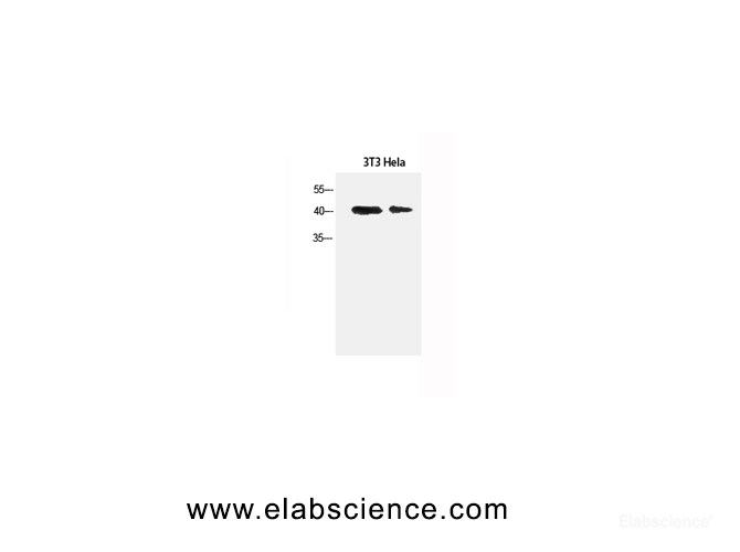
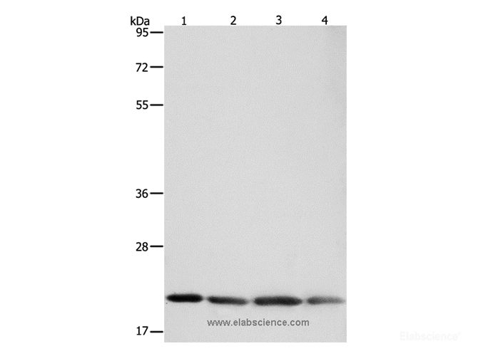
    Western Blot analysis of 3T3 cells and Hela cells using α-SMA Polyclonal Antibody at dilution of 1:1500.

    Rat IHC
    (lung,)

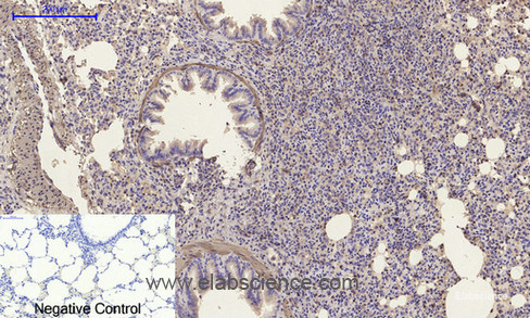
    Immunohistochemistry of paraffin-embedded Rat lung tissue using α-SMA Polyclonal Antibody at dilution of 1:200.

    Mouse WB
    (3T3,)

    Western Blot analysis of 3T3 cells and Hela cells using α-SMA Polyclonal Antibody at dilution of 1:1500.

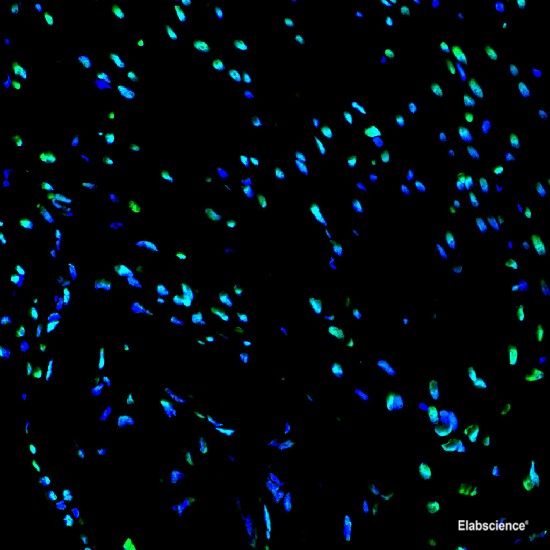
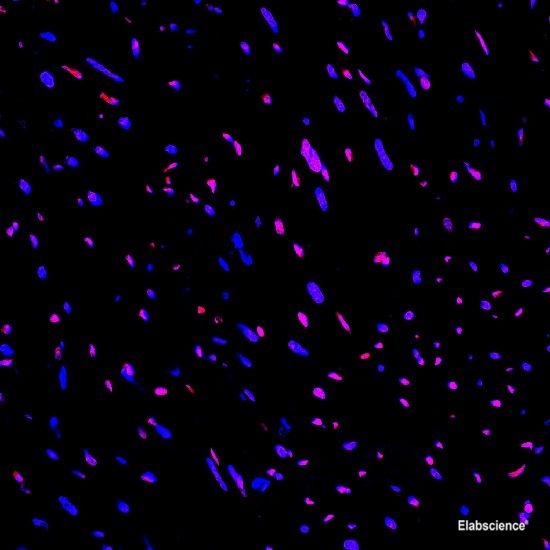
    IF
    (heart,)

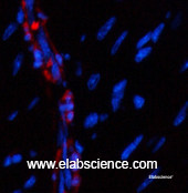
    Immunofluorescence analysis of Mouse heart tissue using α-SMA Polyclonal Antibody at dilution of 1:200.

  • Dilution

    WB 1:500-2000
    IHC 1:50-300
    IF 1:50-1:200
    ELISA 1:10000-20000

Preparation of protein samples

1.Protein extraction

1)For tissue sample
a. Take the samples, wash the tissue thoroughly with pre-cooled PBS (0.01 M, pH=7.4)(Cat# E-BC-R187) to remove the surface blood and internal debris.
b. Weigh and smash the tissue, add an appropriate ratio of RIPA Lysis Buffer (Cat# E-BC-R327)(add 10 μL PMSF and 10 μL Na3VO4 to each 1 mL RIPA Lysis) and homogenizely lyse the tissue.
It is recommended to homogenize according to the ratio of tissue weight: RIPA volume = 3:10. For example, add 1 mL RIPA Lysis Buffer to 0.3 g tissue sample, the specific volume can be adjusted according to experimental requirements.
c. Shake and lyse on the ice for 30 min after homogenization. And then sonicate the sample for 1 min (under ice water bath conditions) with 2 s’ sonication and 2 s’ intervals to make cells fully lysis and reduce the viscosity of sample.
d. Centrifuge at 12,000 rpm for 10 min at 4℃.
e. Take the supernatant and measure the protein concentration mentioned in step2.

2)For cell sample
a. Collect the cells, wash them thoroughly with pre-cooled PBS (0.01 M, pH=7.4) to remove the medium off (it is generally recommended to wash 3 times).
b. Add an appropriate ratio of RIPA Lysate Buffer (10 μL PMSF and 10 μL Na3VO4 in each 1 mL RIPA Lysis) and lyse on the ice for 30 min.
It is recommended to add 0.1 mL of RIPA Lysis Buffer to each well of a 6-well plates (the protein content in different cells may vary, and the volume of the lysate added can be appropriately adjusted).
c. Sonicate the sample for 1 min (under ice water bath conditions) with 2 s’ sonication and 2 s’ intervals to make cells fully lyse and reduce viscosity of sample.
d. Centrifuge at 12,000 rpm for 10 min at 4℃.
e. Take the supernatant and measure the protein concentration mentioned in step2.

2.Measurement of protein concentration
By the BCA method (see the Total Protein Colorimetric Assay Kit (Cat# E-BC-K318) instructions).

3.Boiling the samples
Adjust the protein concentration with PBS Buffer. Add 5 × SDS Loading Buffer (Cat# E-BC-R288) with the ratio of the protein sample: 5 × SDS Loading Buffer = 4:1 and boil the mixture for 10 min. Centrifuge at 12,000 rpm for 2 min and collect the supernatant. The denatured protein can be employed to Western Blot experiments or stored at -20℃ or -80℃.

Note: It is recommended that the total protein loading amount of test sample is about 50 μg in each well. Try to make the loading volume of each sample close to 10 μL.

Electrophoresis

1.According to the molecular weight of the target protein, prepare 0% separation gel. Add the test sample to each well, and add 5 μL of Pre-stained Protein Marker (Cat# E-BC-R273)to a reserved well in order to verify the target molecular weight and the extent of membrane transfer. Add Electrophoresis Buffer ( Cat# E-BC-R331) and start electrophoresis.

2.Electrophoresis at 80v when the samples are in stacking gel, then convert to 120v when the blue flow into the separating gel. Electrophoresis time is about 2-3 h till bromophenol blue reaches the bottom of the gel.

Transfer Membrane

1.Choose the PVDF Membrane (Cat# E-BC-R266) with a pore size of μm according to the molecular weight of the target protein. Soak the PVDF Membrane in methanol for 1 min to activate it, and then soak the PVDF Membrane in the Transmembrane Buffer (Cat# E-BC-R333), the filter paper and fiber mat must be soaked in the Transmembrane Buffer for use too.

2.Follow manufacture instructions of Transfer System for wet, semi-dry, or dry transfer.

Incubation of antibodies

1.Soak the PVDF Membrane with TBST Buffer (Cat# E-BC-R335) containing 5% Skim Milk Powder as blocking buffer and block the membrane at room temperature for .

2.According to the recommended primary antibody dilution ratio, use the TBST Buffer containing 5% Skim Milk Powder to dilute the α-SMA Antibody at , soak the PVDF Membrane in the primary antibody working solution, incubate overnight at 4 ℃, and gently shake.

3.Wash the PVDF Membrane with TBST Buffer for .

4.According to the recommended secondary antibody dilution ratio, use a TBST Buffer solution containing 2% Skim Milk Powder to dilute Goat Anti-Rabbit IgG (H+L) (peroxidase/HRP conjugated) (Cat# E-AB-1003) at . Incubate at room temperature for 1 h on a shaker.

5.Wash the PVDF Membrane with TBST Buffer for .

Detection

1.Mix A and B in the Excellent Chemiluminescent Substrate Detection kit (Cat# E-BC-R347) at the ratio of 1:1 as working solution.

2.Take out the PVDF Membrane from TBST Buffer and absorb the liquid with the filter paper. Pave the PVDF Membrane on the detection machine, add ECL working solution continuously on the PVDF Membrane, discharge the bubble and detect the result.

3.Adjust the contrast and the exposure time to get the best image.

Appendix

Product Details

Clonality Polyclonal
Isotype IgG
Concentration 1 mg/mL
Storage Store at -20℃. Avoid freeze / thaw cycles.
Buffer PBS with 0.02% sodium azide, 0.5% protective protein and 50% glycerol, pH7.4
Purification Method Affinity purification
Research Areas Cancer, Cardiovascular, Developmental Biology, Signal Transduction, Stem Cells
Conjugation Unconjugated

Immunogen Details

Immunogen Synthesized peptide derived from α-SMA
Abbre α-SMA
Synonyms a actin,AAT6,ACTA,ACTA2,Actin alpha 2 smooth muscle aorta,Actin aortic smooth muscle,Actin,aortic smooth muscle,ACTSA,ACTVS,Alpha 2 actin,Alpha actin 2,Alpha cardiac actin,alpha sma,Alpha-actin-2,Cell growth inhibiting gene 46 protein,Cell growth-inhibiting gene 46 protein,GIG46,Growth inhibiting gene 46,MYMY5,ACTC,ACTC,ACTC1,Actin alpha cardiac muscle 1,Actin,alpha cardiac muscle 1,Alpha-cardiac actin,ASD5,CMD1R,CMH11,LVNC4,a actin,Actin alpha skeletal muscle,Actin,actin,alpha 1,skeletal muscle 1,actin,alpha 1,skeletal muscle,Actin,alpha skeletal muscle,actina,actine,ACTS,aktin,Alpha Actin 1,,ASMA,CFTD,CFTD1,CFTDM,MPFD,NEM1,NEM2,NEM3,nemaline myopathy type 3
Swissprot P62736,P68133,P63267
Calculated MW 42kDa
Observed MW 42kDa
Cellular Localization Cytoskeleton,actin filament,cytoskeleton,filamentous actin,Cytosol,Extracellular region or secreted,blood microparticle,extracellular exosome,extracellular matrix,extracellular space,Nucleus,Plasma Membrane,Other locations:dense body,focal adhesion,membrane,myelin sheath,myofibril,phagocytic vesicle

Background

Actins are highly conserved proteins that are involved in various types of cell motility and are ubiquitously expressed in all eukaryotic cells. ACTA2 (Actin, Alpha 2, Smooth Muscle, Aorta) is a Protein Coding gene. Diseases associated with ACTA2 include Multisystemic Smooth Muscle Dysfunction Syndrome and Moyamoya Disease 5. Among its related pathways are ICos-ICosL Pathway in T-Helper Cell and GPCR Pathway. GO annotations related to this gene include protein kinase binding. An important paralog of this gene is ACTG2. ACTA1 (Actin, Alpha 1, Skeletal Muscle) is a Protein Coding gene. Diseases associated with ACTA1 include Nemaline Myopathy 3, Autosomal Dominant Or Recessive and Myopathy, Congenital, With Fiber-Type Disproportion. Among its related pathways are ICos-ICosL Pathway in T-Helper Cell and GPCR Pathway. GO annotations related to this gene include structural constituent of cytoskeleton and myosin binding. An important paralog of this gene is ACTC1. ACTG2 (Actin, Gamma 2, Smooth Muscle, Enteric) is a Protein Coding gene. Diseases associated with ACTG2 include Visceral Myopathy and Chronic Intestinal Pseudoobstruction. Among its related pathways are ICos-ICosL Pathway in T-Helper Cell and GPCR Pathway. An important paralog of this gene is ACTA2.

Citations

  1. Nature Communications (2022) IF: 14.919
    ADAMTS4-specific MR probe to assess aortic aneurysms in vivo using synthetic peptide libraries

    DOI: 10.1038/s41467-022-30464-8

    PMID: 35606349

    Sample: aortic wall
  2. INTERNATIONAL JOURNAL OF BIOLOGICAL MACROMOLECULES (2022) IF: 8.025
    Bioengineering strategies for 3D bioprinting of tubular construct using tissue-specific decellularized extracellular matrix

    DOI: 10.1016/j.ijbiomac.2022.11.064

  3. INTERNATIONAL JOURNAL OF MOLECULAR SCIENCES (2023) IF: 6.208
    Human Colonoid–Myofibroblast Coculture for Study of Apical Na+/H+ Exchangers of the Lower Cryptal Neck Region

    DOI: 10.3390/ijms24054266

    Sample: colonoids
  4. LIFE SCIENCES (2022) IF: 6.78
    Thymol protects against bleomycin-induced pulmonary fibrosis via abrogation of oxidative stress, inflammation, and modulation of miR-29a/TGF-β and PI3K/Akt signaling in mice

    DOI: 10.1016/j.lfs.2022.121256

    Sample: lung tissues
  5. LIFE SCIENCES (2022) IF: 6.78
    Femtosecond laser attenuates oxidative stress, inflammation, and liver fibrosis in rats: Possible role of PPARγ and Nrf2/HO-1 signaling

    DOI: 10.1016/j.lfs.2022.120877

    PMID: 35963297

    Sample: liver
  6. Journal of Inflammation Research (2021) IF: 6.922
    Silenced SNHG1 Inhibited Epithelial-Mesenchymal Transition and Inflammatory Response of ARPE-19 Cells Induced by High Glucose

    DOI: 10.2147/JIR.S299010

    Sample: ARPE-19 cell
  7. Cells (2019) IF: 5.656
    Development of a Stromal Microenvironment Experimental Model Containing Proto-Myofibroblast Like Cells and Analysis of Its Crosstalk with Melanoma Cells: A New Tool to Potentiate and Stabilize Tumor Suppressor Phenotype of Dermal Myofibroblasts

    DOI: 10.3390/cells8111435

    PMID: 31739477

    Sample: Cell lysate,Tissue homogenate
  8. JOURNAL OF DAIRY SCIENCE (2023) IF: 4.225
    Liver fibrosis is a common pathological change in the liver of dairy cows with fatty liver

    DOI: 10.3168/jds.2022-22021

    Sample: liver
  9. Foods (2022) IF: 4.35
    Changes in Histological Structure, Interleukin 12, Smooth Muscle Actin and Nitric Oxide Synthase 1. and 3. Expression in the Liver of Running and Non-Running Wistar Rats Supplemented with Bee Pollen or Whey Protein

    DOI: 10.3390/foods11081131

    PMID: 35454718

    Sample: liver
  10. Cancer Medicine (2022) IF: 4.711
    HEG1 as a novel potential biomarker for the prognosis of lung adenocarcinoma

    DOI: 10.1002/cam4.5081

    Sample: Lung cancer tissue
  11. Bioengineered (2021) IF: 3.269
    Plumbagin attenuates traumatic tracheal stenosis in rats and inhibits lung fibroblast proliferation and differentiation via TGF-β1/Smad and Akt/mTOR pathways

    DOI: 10.1080/21655979.2021.1954580

    PMID: 34304701

    Sample: tracheal,fibroblasts
  12. JOURNAL OF PHARMACOLOGY AND EXPERIMENTAL THERAPEUTICS (2018) IF: 3.706
    A Combination of Chitosan, Cellulose, and Seaweed Polysaccharide Inhibits Postoperative Intra-abdominal Adhesion in Rats

    DOI: 10.1124/jpet.117.244400

    PMID: 29263242

    Sample: Tissue homogenate
  13. JOURNAL OF ETHNOPHARMACOLOGY (2017) IF: 2.981
    Protective effect of ethyl acetate fraction of Drynaria quercifolia against CCl4 induced rat liver fibrosis via Nrf2/ARE and NFκB signalling pathway

    DOI: 10.1016/j.jep.2017.11.015

    Sample: Liver tissue
  14. Experimental and Therapeutic Medicine (2020) IF: 1.785
    Lung histomorphological alterations in rats exposed to cigarette smoke and electronic cigarette vapour

    DOI: 10.3892/etm.2020.8530

    Sample: lung
Show AllShorten

Reviews/Q&A

  • Show all
  • Reviews
  • Q&A
... Show All Show Less

People Also Bought

Apply for 10 Test Free Trial FCM Antibody
*Product Name:
*Catalog Number:
*Name:
*Email:
*Message:
*Country:
*When will you use it?

*Captcha: