Click here to view cell apoptosis assay products

beta Tubulin Monoclonal Antibody

  • Cat.No.:E-AB-20033

  • Host: Mouse
  • Reactivity: H,M,R,Mk,Ch,Dg,Hm,Rb,Sh,Insect,Yeast
  • Applications: WB,IHC-p,IF

To Purchase E-AB-20033

Size:
  • 30μL
  • 60μL
  • 120μL
  • 200μL
Price: $73
Qty:

Test Application

  • Verified Samples

    Reactivity Application
    Human WB
    (A549,)

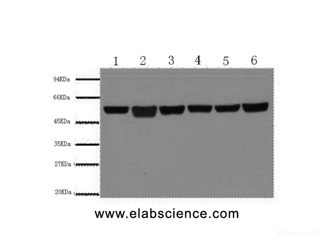
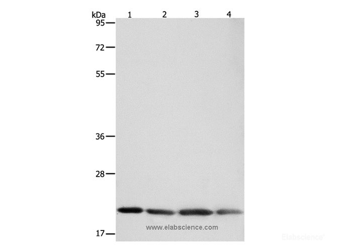
    Western Blot analysis of A549 cells, Rat brain, Mouse brain, Chicken lung, Rabbit testis and Sheep muscle using beta Tubulin Monoclonal Antibody at dilution of 1:5000.

    IF
    (Hela,)

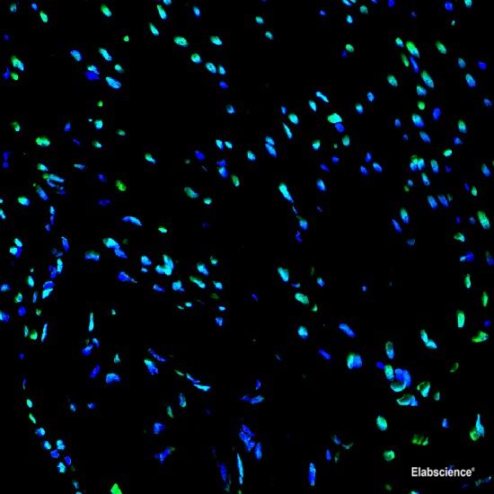
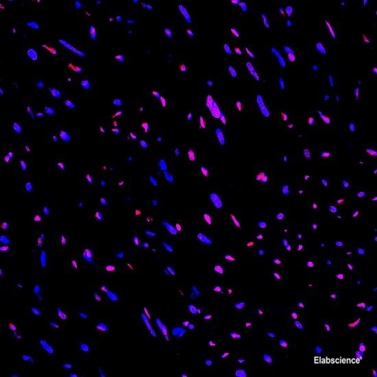
    Immunofluorescence analysis of Hela tissue using beta Tubulin Monoclonal Antibody at dilution of 1:100.

    Rat WB
    (brain,)

    Western Blot analysis of A549 cells, Rat brain, Mouse brain, Chicken lung, Rabbit testis and Sheep muscle using beta Tubulin Monoclonal Antibody at dilution of 1:5000.

    Mouse WB
    (brain,)

    Western Blot analysis of A549 cells, Rat brain, Mouse brain, Chicken lung, Rabbit testis and Sheep muscle using beta Tubulin Monoclonal Antibody at dilution of 1:5000.

    IHC
    (testis,)

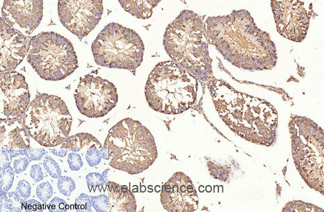
    Immunohistochemistry of paraffin-embedded Mouse testis tissue using beta Tubulin Monoclonal Antibody at dilution of 1:200.

    Others WB
    (Chicken lung,Rabbit testis,Sheep muscle,)

    Western Blot analysis of A549 cells, Rat brain, Mouse brain, Chicken lung, Rabbit testis and Sheep muscle using beta Tubulin Monoclonal Antibody at dilution of 1:5000.

    Western Blot analysis of A549 cells, Rat brain, Mouse brain, Chicken lung, Rabbit testis and Sheep muscle using beta Tubulin Monoclonal Antibody at dilution of 1:5000.

    Western Blot analysis of A549 cells, Rat brain, Mouse brain, Chicken lung, Rabbit testis and Sheep muscle using beta Tubulin Monoclonal Antibody at dilution of 1:5000.

  • Dilution

    WB 1:5000-1:10000
    IHC 1:100-1:300
    IF 1:100-1:300

Preparation of protein samples

1.Protein extraction

1)For tissue sample
a. Take the samples, wash the tissue thoroughly with pre-cooled PBS (0.01 M, pH=7.4)(Cat# E-BC-R187) to remove the surface blood and internal debris.
b. Weigh and smash the tissue, add an appropriate ratio of RIPA Lysis Buffer (Cat# E-BC-R327)(add 10 μL PMSF and 10 μL Na3VO4 to each 1 mL RIPA Lysis) and homogenizely lyse the tissue.
It is recommended to homogenize according to the ratio of tissue weight: RIPA volume = 3:10. For example, add 1 mL RIPA Lysis Buffer to 0.3 g tissue sample, the specific volume can be adjusted according to experimental requirements.
c. Shake and lyse on the ice for 30 min after homogenization. And then sonicate the sample for 1 min (under ice water bath conditions) with 2 s’ sonication and 2 s’ intervals to make cells fully lysis and reduce the viscosity of sample.
d. Centrifuge at 12,000 rpm for 10 min at 4℃.
e. Take the supernatant and measure the protein concentration mentioned in step2.

2)For cell sample
a. Collect the cells, wash them thoroughly with pre-cooled PBS (0.01 M, pH=7.4) to remove the medium off (it is generally recommended to wash 3 times).
b. Add an appropriate ratio of RIPA Lysate Buffer (10 μL PMSF and 10 μL Na3VO4 in each 1 mL RIPA Lysis) and lyse on the ice for 30 min.
It is recommended to add 0.1 mL of RIPA Lysis Buffer to each well of a 6-well plates (the protein content in different cells may vary, and the volume of the lysate added can be appropriately adjusted).
c. Sonicate the sample for 1 min (under ice water bath conditions) with 2 s’ sonication and 2 s’ intervals to make cells fully lyse and reduce viscosity of sample.
d. Centrifuge at 12,000 rpm for 10 min at 4℃.
e. Take the supernatant and measure the protein concentration mentioned in step2.

2.Measurement of protein concentration
By the BCA method (see the Total Protein Colorimetric Assay Kit (Cat# E-BC-K318) instructions).

3.Boiling the samples
Adjust the protein concentration with PBS Buffer. Add 5 × SDS Loading Buffer (Cat# E-BC-R288) with the ratio of the protein sample: 5 × SDS Loading Buffer = 4:1 and boil the mixture for 10 min. Centrifuge at 12,000 rpm for 2 min and collect the supernatant. The denatured protein can be employed to Western Blot experiments or stored at -20℃ or -80℃.

Note: It is recommended that the total protein loading amount of test sample is about 50 μg in each well. Try to make the loading volume of each sample close to 10 μL.

Electrophoresis

1.According to the molecular weight of the target protein, prepare 0% separation gel. Add the test sample to each well, and add 5 μL of Pre-stained Protein Marker (Cat# E-BC-R273)to a reserved well in order to verify the target molecular weight and the extent of membrane transfer. Add Electrophoresis Buffer ( Cat# E-BC-R331) and start electrophoresis.

2.Electrophoresis at 80v when the samples are in stacking gel, then convert to 120v when the blue flow into the separating gel. Electrophoresis time is about 2-3 h till bromophenol blue reaches the bottom of the gel.

Transfer Membrane

1.Choose the PVDF Membrane (Cat# E-BC-R266) with a pore size of μm according to the molecular weight of the target protein. Soak the PVDF Membrane in methanol for 1 min to activate it, and then soak the PVDF Membrane in the Transmembrane Buffer (Cat# E-BC-R333), the filter paper and fiber mat must be soaked in the Transmembrane Buffer for use too.

2.Follow manufacture instructions of Transfer System for wet, semi-dry, or dry transfer.

Incubation of antibodies

1.Soak the PVDF Membrane with TBST Buffer (Cat# E-BC-R335) containing 5% Skim Milk Powder as blocking buffer and block the membrane at room temperature for .

2.According to the recommended primary antibody dilution ratio, use the TBST Buffer containing 5% Skim Milk Powder to dilute the β-Tubulin Antibody at , soak the PVDF Membrane in the primary antibody working solution, incubate overnight at 4 ℃, and gently shake.

3.Wash the PVDF Membrane with TBST Buffer for .

4.According to the recommended secondary antibody dilution ratio, use a TBST Buffer solution containing 2% Skim Milk Powder to dilute Goat Anti-Rabbit IgG (H+L) (peroxidase/HRP conjugated) (Cat# E-AB-1003) at . Incubate at room temperature for 1 h on a shaker.

5.Wash the PVDF Membrane with TBST Buffer for .

Detection

1.Mix A and B in the Excellent Chemiluminescent Substrate Detection kit (Cat# E-BC-R347) at the ratio of 1:1 as working solution.

2.Take out the PVDF Membrane from TBST Buffer and absorb the liquid with the filter paper. Pave the PVDF Membrane on the detection machine, add ECL working solution continuously on the PVDF Membrane, discharge the bubble and detect the result.

3.Adjust the contrast and the exposure time to get the best image.

Appendix

Product Details

Clonality Monoclonal
Isotype IgG
Concentration 1 mg/mL
Storage Store at -20℃. Avoid freeze / thaw cycles.
Buffer PBS with 0.02% sodium azide and 50% glycerol pH 7.4.
Purification Method Protein A purification
Research Areas Cancer, Neuroscience, Signal Transduction, Tags and Cell Markers
Clone No. Clone:8B2
Conjugation Unconjugated

Immunogen Details

Immunogen Synthetic Peptide
Abbre β-Tubulin
Synonyms beta 3 tubulin,beta-4,CDCBM,CDCBM1,CFEOM3,CFEOM3A,FEOM3,M(beta)3,M(beta)6,MC1R,Neuron specific beta III Tubulin,Neuron-specific class III beta-tubulin,QccE-11995,QccE-15186,TBB3,Tubb 3,TUBB3,TUBB4,Tubulin beta 3,Tubulin beta 3 chain,Tubulin beta 4,Tubulin beta III,Tubulin beta-3 chain,Tubulin beta-4 chain,Tubulin beta-III
Swissprot Q13509
Calculated MW 50kDa
Observed MW 55kDa

Western blotting is a method for detecting a certain protein in a complex sample based on the specific binding of antigen and antibody. Different proteins can be divided into bands based on different mobility rates. The mobility is affected by many factors, which may cause the observed band size to be inconsistent with the expected size. The common factors include:

1. Post-translational modifications: For example, modifications such as glycosylation, phosphorylation, methylation, and acetylation will increase the molecular weight of the protein.

2. Splicing variants: Different expression patterns of various mRNA splicing bodies may produce proteins of different sizes.

3. Post-translational cleavage: Many proteins are first synthesized into precursor proteins and then cleaved to form active forms, such as COL1A1.

4. Relative charge: the composition of amino acids (the proportion of charged amino acids and uncharged amino acids).

5. Formation of multimers: For example, in protein dimer, strong interactions between proteins can cause the bands to be larger. However, the use of reducing conditions can usually avoid the formation of multimers.

If a protein in a sample has different modified forms at the same time, multiple bands may be detected on the membrane.

Cellular Localization Cytoplasm>cytoskeleton.
Tissue Specificity Expression is primarily restricted to central and peripheral nervous system.

Background

There are five tubulins in human cells: alpha, beta, gamma, delta, and epsilon. Tubulins are conserved across species. They form heterodimers, which multimerize to form a microtubule filament. An alpha and beta tubulin heterodimer is the basic structural unit of microtubules. The heterodimer does not come apart, once formed. The alpha and beta tubulins, which are each about 55 kDa MW, are homologous but not identical. Alpha, beta, and gamma tubulins have all been used as loading controls. Tubulin expression may vary according to resistance to antimicrobial and antimitotic drugs.

Citations

  1. INTERNATIONAL JOURNAL OF BIOLOGICAL MACROMOLECULES (2022) IF: 8.025
    Thermal treatment of magnesium particles in polylactic acid polymer films elicits the expression of osteogenic differentiation markers and lipidome profile remodeling in human adipose stem cells

    DOI: 10.1016/j.ijbiomac.2022.11.005

  2. JOURNAL OF STEROID BIOCHEMISTRY AND MOLECULAR BIOLOGY (2022) IF: 5.011
    Postnatal developmental expression of apelin receptor proteins and its role in juvenile mice testis

    DOI: 10.1016/j.jsbmb.2022.106178

    PMID: 36108814

    Sample: testis sample
  3. HUMAN MUTATION (2020) IF: 4.124
    Mandibular‐pelvic‐patellar syndrome is a novel PITX1‐related disorder due to alteration of PITX1 transactivation ability

    DOI: 10.1002/humu.24070

    Sample: Cell culture supernatant
  4. INTERNATIONAL JOURNAL OF MOLECULAR SCIENCES (2019) IF: 4.183
    CB2 Receptor Stimulation and Dexamethasone Restore the Anti-Inflammatory and Immune-Regulatory Properties of Mesenchymal Stromal Cells of Children with Immune Thrombocytopenia

    DOI: 10.3390/ijms20051049

    PMID: 30823385

    Sample: Cell culture supernate
  5. JOURNAL OF STEROID BIOCHEMISTRY AND MOLECULAR BIOLOGY (2021) IF: 4.294
    Co-treatment of testosterone and estrogen mitigates heat-induced testicular dysfunctions in a rat model

    DOI: 10.1016/j.jsbmb.2021.106011

    PMID: 34688845

    Sample: testicular tissue
  6. PHARMACOLOGICAL RESEARCH (2018) IF: 4.897
    Bortezomib and endocannabinoid/endovanilloid system: a synergism in osteosarcoma

    DOI: 10.1016/j.phrs.2018.09.017

    Sample: cell lines
  7. INVESTIGATIVE OPHTHALMOLOGY & VISUAL SCIENCE (2020) IF: 3.47
    Indoleamine 2,3-Dioxygenase Regulates Macrophage Recruitment, Polarization and Phagocytosis in Aspergillus Fumigatus Keratitis

    DOI: 10.1167/iovs.61.8.28

    PMID: 32692841

    Sample: Tissue homogenate
  8. IMMUNOLOGY AND CELL BIOLOGY (2020) IF: 3.764
    Dimethyl itaconate protects against fungal keratitis by activating the Nrf2/HO‐1 signaling pathway

    DOI: 10.1111/imcb.12316

    Sample: Cell culture supernatant,Tissue homogenate
  9. JOURNAL OF STEROID BIOCHEMISTRY AND MOLECULAR BIOLOGY (2019) IF: 3.785
    Vitamin D3 mediated regulation of steroidogenesis mitigates testicular activity in an aged rat model

    DOI: 10.1016/j.jsbmb.2019.03.016

    PMID: 30923019

    Sample: Tissue homogenate
  10. CYTOKINE (2021) IF: 3.861
    Expression and localization of apelin and its receptor in the testes of diabetic mice and its possible role in steroidogenesis

    DOI: 10.1016/j.cyto.2021.155554

    PMID: 33962842

    Sample: Tissue homogenate
  11. ANDROLOGIA (2022) IF: 2.532
    Evaluation of a single dose of intra-testicular insulin treatment in heat-stressed mice model

    DOI: 10.1111/and.14603

    PMID: 36156807

    Sample: testicular tissue
  12. THERIOGENOLOGY (2021) IF: 2.74
    Synthetic leptin c-fragment peptide minimises heat-induced impairment of spermatogenesis in mice via Stat3 signalling

    DOI: 10.1016/j.theriogenology.2021.10.028

    PMID: 34763177

  13. MOLECULAR REPRODUCTION AND DEVELOPMENT (2020) IF: 2.823
    Effect of metformin on testicular expression and localization of leptin receptor and levels of leptin in the diabetic mice

    DOI: 10.1002/mrd.23342

    Sample: testes
  14. JOURNAL OF PEDIATRIC GASTROENTEROLOGY AND NUTRITION (2020) IF: 2.937
    The Role of Cannabinoid Receptor Type 2 in the Bone Loss Associated With Pediatric Celiac Disease

    DOI: 10.1097/MPG.0000000000002863

    PMID: 33093370

    Sample: Cell lysate
  15. ANDROLOGIA (2020) IF: 1.951
    Heat‐induced changes in the expression and localisation of PGC‐1α in the mice testis

    DOI: 10.1111/and.13713

    Sample: Tissue homogenate
  16. Oncotarget (2020)
    Can Denosumab be used in combination with Doxorubicin in Osteosarcoma?

    DOI: 10.18632/oncotarget.27669

    Sample: MG63 cell,U-2 OS cell
  17. JOURNAL OF EVOLUTIONARY BIOCHEMISTRY AND PHYSIOLOGY (2021) IF: 0.444
    Diabetes Induced Changes in the Expression of Markers for Alveolar Epithelial Type I and II Cells in the Lung of the Albino Rat

    DOI: 10.1134/S0022093021060120

    Sample: lung tissue
Show AllShorten

Reviews/Q&A

  • Show all
  • Reviews
  • Q&A
... Show All Show Less

People Also Bought

Apply for 10 Test Free Trial FCM Antibody
*Product Name:
*Catalog Number:
*Name:
*Email:
*Message:
*Country:
*When will you use it?

*Captcha: