

When we accidentally cut our finger, or when a cold virus quietly slips into our respiratory tract, an invisible, time-critical rescue operation is already underway inside our body. And who is the first to rush past the warning line and dash straight to the scene?
It's not some mysterious cell from a distant organ. It's the tissue "sentry" that has long been quietly stationed throughout our body: the macrophage. Today, let's get to know these swift-response guardians that wear many hats. Where do they actually come from, and what powerful abilities do they harbor?
Macrophages are a class of white blood cells that reside in tissues, differentiating from monocytes, which in turn originate from precursor cells in the bone marrow. Both macrophages and monocytes belong to the phagocyte family and participate in both non-specific immunity (innate immunity) and specific immunity (cellular immunity) in vertebrates. Some macrophages are fixed residents in tissues, while others roam freely in a patrolling manner. Upon detecting cellular debris or invading pathogens, they swiftly engulf and digest them. At the same time, macrophages can activate other immune cells, prompting the body to mount a precise immune response. It can be said that macrophages are by no means mere "scavengers" that clean up debris; they are more like immune all-rounders that combine the roles of scout and commander. Precisely because their functions are so diverse and critical, macrophages have become important subjects of study in phagocytosis, cellular immunity, and molecular immunology.
Table of Contents
1. Macrophage phagocytosis: mechanisms and biological functions
2. Macrophage secretory functions: cytokine and chemokine-mediated immune communication
3. Macrophage polarization and immunoregulatory functions in the microenvironment
4. Methods for macrophage detection and phenotypic characterization
5. Emerging applications of macrophages in biomedical research and immunotherapy
01 Macrophage phagocytosis: mechanisms and biological functions
Phagocytosis is the process of engulfing and clearing particulate cells or cellular debris, including microorganisms, foreign substances, senescent cells, damaged cells, and mutated cells. As the primary phagocytes, macrophages possess powerful phagocytic and clearance capabilities for cellular debris.
1.1 Recognition
Precise target identification is the first step in the macrophage's immune defense. How does it distinguish between "enemies" (pathogens) and "garbage" (apoptotic cells)? It relies on capturing specific molecular "labels."
(1) Pathogens carry pathogen-associated molecular patterns (PAMPs), such as bacterial lipopolysaccharide (LPS), peptidoglycan, and others;
(2) Damaged or apoptotic cells expose damage-associated molecular patterns (DAMPs), such as HMGB1, ATP, necrotic DNA, and others. The macrophage surface is equipped with two types of "detectors":
Pattern Recognition Receptors (PRRs): These directly recognize PAMPs/DAMPs, including C-type lectins (such as the mannose receptor, which captures sugar molecules on pathogen surfaces), scavenger receptors (which recognize LPS or phosphatidylserine exposed on apoptotic cells), and certain Toll-like receptors.
Opsonin Receptors: When opsonins, such as antibodies (IgG) or complement molecules, first "tag" the target, macrophages deploy opsonin receptors (Fcγ receptors and complement receptors (CRs)) to bind these tags, efficiently triggering phagocytosis.
These receptors do not act alone; they cooperate to recruit and cluster together, ensuring precise targeting of "enemies" and "garbage," paving the way for subsequent engulfment and digestion.
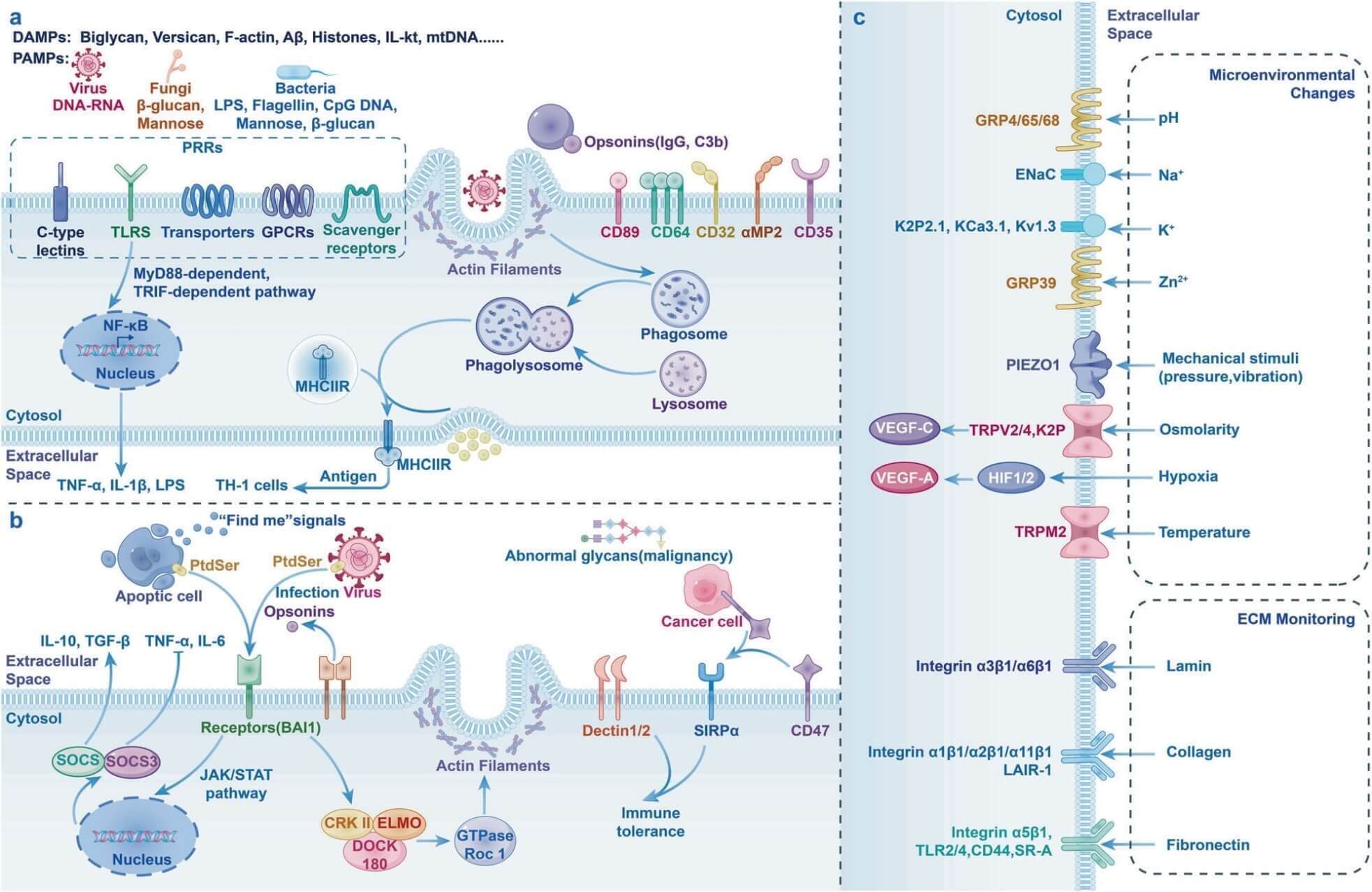
Fig. 1 Recognition Mechanisms of Macrophages[1].
1.2 Engulfment
Following recognition, a series of signaling pathways are activated, leading to the formation of phagocytic cups, pseudopods, and phagosomes. First, driven by the Arp2/3 complex, the cell membrane rapidly assembles a branched actin network, pushing the membrane forward to form a cup-like phagocytic cup. As the pseudopods continue to extend and embrace the target, BAR-domain-containing proteins guide actin polarization and recruit shear-related proteins such as dynamin, ultimately tightening and sealing the membrane opening to form a completely enclosed vesicle; this is the nascent phagosome.
1.3 Digestion
The nascent phagosome lacks killing capacity and must undergo a maturation journey. Through a "kiss-and-run" mechanism, it repeatedly undergoes rapid fusion and fission with endocytic organelles, gradually acquiring the acidifying enzymes and hydrolases required at each stage. Ultimately, the mature phagosome fuses with the lysosome, upgrading into a phagolysosome with potent degradative capability. Within this acidic "digestive workshop," pathogens or cellular debris are thoroughly broken down into harmless small molecules. After degradation is complete, the lysosome can be regenerated through complex fission and fusion processes for continued recycling.
02 Macrophage secretory functions: cytokine and chemokine-mediated immune communication
Beyond their phagocytic function, macrophages also serve as immune signal messengers, "calling out" by secreting cytokines and chemokines to recruit other immune cells.
2.1 Secretion of Cytokines
Macrophages are among the most important "cytokine factories" in the body, capable of rapidly synthesizing and releasing a variety of cytokines upon stimulation by microorganisms or their products (such as lipopolysaccharide, LPS).
· M1-type macrophages (classically activated) primarily secrete pro-inflammatory cytokines, including tumor necrosis factor-α (TNF-α), interleukin-1β (IL-1β), IL-6, IL-12, and IL-23, which activate immune cells and promote inflammatory responses.
· M2-type (alternatively activated) macrophages mainly secrete anti-inflammatory cytokines, such as IL-10 and transforming growth factor-β (TGF-β), which serve to suppress inflammation and promote tissue repair.
· Cytokines are exported out of the cell via the constitutive secretory pathway (via Golgi apparatus → recycling endosomes → cell membrane) or non-classical pathways (such as IL-1β released through pyroptosis-associated membrane pores).
2.2 Secretion of Chemokines
Macrophages can secrete a variety of chemokines, with the primary function of recruiting circulating leukocytes to migrate toward sites of inflammation or infection.
· Key chemokines produced by macrophages include: monocyte chemoattractant protein-1 (MCP-1/CCL2), MCP-2, MCP-3, macrophage inflammatory protein-1α (MIP-1α/CCL3), MIP-1β (CCL4), and RANTES (CCL5), among others.
· MCP-1 is the most representative chemokine. By binding to the CCR2 receptor on monocytes, it potently chemoattracts monocytes and memory T cells to infiltrate inflamed tissues, and upregulates integrin expression to facilitate monocyte transendothelial migration across vascular endothelium. Macrophages also secrete CXC chemokines such as IL-8 (CXCL8) and CXCL1/CXCL2, which primarily recruit neutrophils during the early stages of inflammation. At the same time, chemokines can also act back on macrophages themselves: for example, MCP-1 can enhance macrophage CD11b/CD11c expression and respiratory burst activity, forming a positive feedback regulatory network.
Overall, through the secretion of cytokines and chemokines, macrophages serve as a central hub that regulates both the innate and adaptive immune systems within the disease microenvironment.
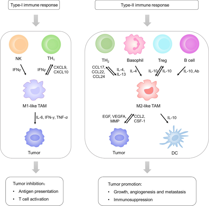
Fig. 2 Macrophages recruit other immune cells via secretion of cytokines and chemokines in the tumor microenvironment[2].
03 Macrophage polarization and immunoregulatory functions in the microenvironment
Macrophage polarization refers to the process by which macrophages dynamically differentiate into distinct functional phenotypes (such as pro-inflammatory M1 type or anti-inflammatory/repair-oriented M2 type) in response to microenvironmental signals. Through this flexible switching, macrophages act like the "chief steward" of the microenvironment: sometimes transforming into a combat mode to clear pathogens, other times shifting into a repair mode to promote healing, thereby precisely regulating local immunity and tissue homeostasis.
3.1 M1 Type (Classical Activation)
M1 macrophages are known as the "combat mode," activated by signals such as pathogens or interferon-γ. They release large amounts of pro-inflammatory cytokines, including TNF-α, IL-6, IL-1, IL-12, and inducible nitric oxide synthase (iNOS), while also secreting CXCL family chemokines. These substances effectively kill pathogens, block intracellular infections, promote Th1-type immune responses, and exert anti-tumor effects. M1 macrophages also serve as the first line of defense against pathogen invasion.
3.2 M2 Type (Alternative Activation)
M2 macrophages represent the "repair mode," involved in anti-inflammation, tissue repair, and angiogenesis. They highly express markers such as CD206, CD163, and the mannose receptor, and produce anti-inflammatory and repair-related substances including IL-10 and chitinase-like proteins. Based on functional differences, M2 macrophages are further subdivided into: M2a (promotes tissue repair), M2b (activates Th2 and modulates immunity), M2c (regulates phagocytosis and tissue repair), and M2d (also known as tumor-associated macrophages, TAMs, which can promote tumor progression and invasion).
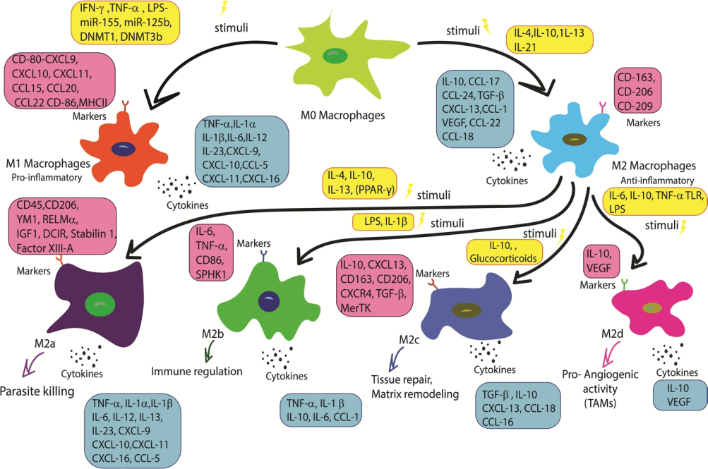
Fig. 3 Macrophage subtypes and their principal functions[3].
3.3 Functional Switching of Macrophages
The sophistication of macrophage polarization lies in its reversible functional switching. When signals such as pathogen-associated molecular patterns or interferon-γ are present, macrophages polarize toward the M1 phenotype, releasing pro-inflammatory factors to combat infection. Conversely, upon receiving anti-inflammatory signals like IL-4 and IL-13, they switch to the M2 phenotype, exerting tissue repair and anti-inflammatory effects. This switching is not a binary on/off process but rather a continuous spectrum: changes in the microenvironment can induce macrophage reprogramming and even give rise to mixed phenotypes. It is precisely this flexible switching capacity that enables macrophages to maintain a dynamic balance between inflammation and resolution, as well as between injury and repair, making them a key therapeutic target in various diseases, including diabetes, asthma, and cancer.
04 Methods for macrophage detection and phenotypic characterization
Due to the high plasticity of macrophage phenotypes and functions, accurate detection and identification of macrophages are essential for both fundamental immunological research and clinical disease diagnosis. Currently, the most commonly used methods for macrophage detection include the following aspects:
4.1 Immunohistochemistry (IHC) and Immunofluorescence (IF)
IHC and IF are the most classical methods for detecting macrophages in tissue sections. The principle involves using specific antibodies to recognize macrophage markers, followed by visualization through enzymatic chromogenic reactions (e.g., DAB yielding a brown color) or fluorescent labeling. The most commonly used pan-macrophage marker in human tissues is CD68. However, a single marker cannot identify all subpopulations: for example, in mice, F4/80 labels mature tissue-resident macrophages, but F4/80-negative, CD68-positive cells also exist; CD11b is often considered a marker for monocyte-derived macrophages, yet it is also positive on neutrophils.
4.2 Flow Cytometry
Flow cytometry is currently one of the most widely applied quantitative methods for macrophage detection, particularly suitable for phenotypic identification and functional analysis of single-cell suspensions. Macrophages lack a single definitive distinguishing antigen comparable to T cell markers such as CD4/CD8; therefore, a combination of markers must be used to identify macrophages. For mouse macrophages, commonly used combinations include F4/80, CD11b, Ly6C, and THY-1. Through multi-color fluorescent labeling and flow cytometric analysis, macrophages can be precisely identified and sorted from heterogeneous cell populations.
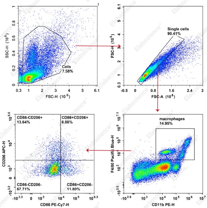
Fig. 4 Ascites samples were collected from C57BL/6J mice with peritoneal inflammation. Cells were stained with PE Anti-Mouse/Human/Monkey CD11b Antibody [M1/70] (E-AB-F1081D), Elab Fluor® Violet 450 Anti-Mouse F4/80 Antibody [CI:A3-1] (E-AB-F0995Q), PE/Cyanine7 Anti-Mouse CD86 Antibody [GL-1] (E-AB-F0994H), and APC Anti-Mouse CD206/MMR Antibody [C068C2] (E-AB-F1135E), and analyzed by flow cytometry for functional phenotyping of macrophages. CD11bhi/midF4/80hi/mid macrophages represent inflammatory-activated macrophages. Further analysis of CD206 and CD86 expression within this cell population was performed to assess macrophage functional status based on differential cell proportions and subpopulation distribution.
4.3 Quantitative Real-Time PCR (qRT-PCR) for Gene Expression Detection
qRT-PCR is a commonly used molecular biology method for detecting macrophage gene expression profiles. It can be used to detect the expression of transcription factors and marker genes associated with macrophage polarization. For example, M1-type macrophages highly express transcription factors such as STAT1, SOCS3, and IRF5, while M2-type macrophages highly express STAT6. In addition, cytokine profiling (e.g., M1 secretion of pro-inflammatory factors such as IL-6, IL-1β, and TNF-α; M2 secretion of anti-inflammatory factors such as IL-10 and CCL18) is also a commonly used method for functional confirmation.
4.4 Machine Learning Methods Based on Image Analysis
In recent years, approaches combining high-throughput image analysis of cell morphology with machine learning algorithms have gained attention. Studies have shown that by simply analyzing the morphological features of the nucleus (DAPI staining) and the actin cytoskeleton (Phalloidin staining), classifiers such as Random Forest can distinguish M1 and M2 macrophages with an accuracy exceeding 89%. However, its application remains limited by the high heterogeneity of macrophages.
4.5 Mass Spectrometry Imaging (MALDI MSI)
Matrix-assisted laser desorption/ionization mass spectrometry imaging (MALDI MSI) is a label-free, untargeted detection technique that can distinguish macrophage subtypes at the metabolite and lipid levels. Studies have utilized MALDI MSI to differentiate human M1, M2a, and M2c macrophages, revealing significant metabolic heterogeneity among monocytes derived from different donors, thus requiring large sample sizes to identify universal biomarkers.
4.6 Single-Cell RNA Sequencing (scRNA-seq)
Single-cell RNA sequencing (scRNA-seq) can resolve the transcriptional heterogeneity of macrophages at single-cell resolution, uncovering novel cell subpopulations not encompassed by the traditional M1/M2 binary classification. For example, studies using scRNA-seq in muscle regeneration and fibrosis models have identified new macrophage subpopulations, such as CD9⁺CD301b⁺MHCIIhi (regeneration-associated R1 cluster) and CD9hiCD301b⁻MHCII⁻IL36γ⁺ (pro-fibrotic F2 cluster). These findings challenge the applicability of the traditional M1/M2 classification in the in vivo context.
Table 1. Comparison of different detection methods for macrophages
|
Detection Method |
Product Name |
Main Limitations |
Common Markers/Indicators |
|
Immunohistochemistry / Immunofluorescence |
Allows observation of cell distribution, morphology, and spatial relationships in situ within tissues |
Low throughput; multiplex staining is technically challenging |
CD68, CD163、CD206, F4/80 (mouse), iNOS、Arginase-1 |
|
Flow Cytometry |
High throughput, quantitative, allows cell sorting, multiparameter simultaneous detection |
Requires preparation of single-cell suspensions; loss of spatial information |
CD45⁺CD64⁺F4/80hi(mouse); CD68, CD163, CD206, CD86 (human) |
|
qRT-PCR |
High sensitivity, allows quantitative measurement of mRNA expression levels |
Requires cell lysis; cannot distinguish spatial distribution of subpopulations |
STAT1, STAT6, SOCS3, IRF5, CD163, Mrc1, etc. |
|
Image Analysis / Machine Learning |
Automated, rapid, label‑free |
Requires large training data; limited accuracy for highly heterogeneous populations |
Cell area, nuclear/cytoplasmic ratio, actin texture, etc. |
|
MALDI Mass Spectrometry Imaging |
Label‑free, untargeted, simultaneous detection of multiple metabolites and lipids |
Lower sensitivity than LC‑MS; identification requires MS/MS validation |
Specific lipid/metabolite molecular ion peaks |
|
Single‑Cell RNA Sequencing |
Provides whole‑transcriptome information; enables discovery of novel subpopulations and differentiation trajectories |
High cost; complex data analysis |
Whole‑transcriptome gene expression profiles |
In summary, immunohistochemistry and flow cytometry remain the most commonly used mainstream methods for macrophage detection. qRT-PCR is widely employed as a complementary approach for gene expression analysis. Meanwhile, emerging technologies such as single‑cell RNA sequencing, MALDI mass spectrometry imaging, and machine learning are rapidly advancing. In practice, multiple methods are often combined based on specific research objectives to obtain a comprehensive understanding of macrophage phenotype and function.
05 Emerging applications of macrophages in biomedical research and immunotherapy
Macrophages are capable of not only phagocytosing pathogens and apoptotic cells but also secreting hundreds of cytokines to regulate inflammation and repair, while playing precise modulatory roles in tissue homeostasis and tumor immunity. The trinity of phagocytosis, secretion, and regulation makes macrophages ubiquitous in both health maintenance and disease progression. Currently, a growing body of research is focused on modulating macrophages for immunotherapeutic applications.
Example: M2→M1 Macrophage Reprogramming and CAR-M Therapy in Cancer Immunotherapy
Tumor-associated macrophages (TAMs) are among the most abundant immune cells in the tumor microenvironment, typically exhibiting a pro-tumor M2 phenotype that supports tumor progression through the secretion of immunosuppressive factors and promotion of angiogenesis. Reprogramming M2-type "accomplices" into M1-type "killers" has become an important strategy in cancer immunotherapy. Current major strategies include:
①Cytokine intervention: Utilizing IL-12, IL-21, and other cytokines to promote M1 polarization, or blocking M2 polarization-inducing factors such as IL-4, IL-6, and IL-10;
②Nanocarrier-targeted delivery: For example, using mannose-modified polyethylenimine (MPEI) nanocomplexes to deliver CAR and IFN-γ encoding genes to macrophages, enabling in situ conversion of M2 TAMs into CAR-M1 macrophages in vivo, thereby enhancing tumor-specific phagocytosis and anti-tumor immunity;
③CAR-M (Chimeric Antigen Receptor-Macrophage) therapy: Next-generation CAR-M incorporates the TIR domain of TLR4 in tandem with CD3ζ, enabling not only phagocytosis upon tumor antigen recognition but also sustained M1 polarization and M2 resistance driven through the NF-κB pathway, with significant inhibition of glioblastoma and hepatocellular carcinoma growth demonstrated in vivo.
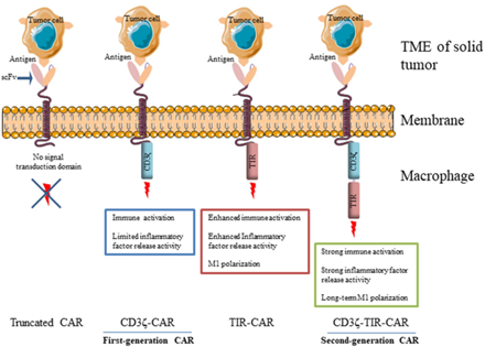
Fig. 5 Schematic representation of next-generation CAR-M[4].
In conclusion, macrophages silently protect the body's health. A thorough understanding of these cells is essential for us to better command the power of immunity.
Related Product Recommendations:
Table 2. Recommended Elabscience® reagents for macrophage phenotyping and cytokine quantification
|
Cat. No. |
Product Name |
|
XJM004 |
RAW 264.7 Polarized M1 Macrophage Induction and Identification Kit |
|
E-AB-F1081D |
PE Anti-Mouse/Human CD11b Antibody[M1/70] |
|
E-AB-F0995Q |
Elab Fluor® Violet 450 Anti-Mouse F4/80 Antibody[CI:A3-1] |
|
E-AB-F0994H |
PE/Cyanine7 Anti-Mouse CD86 Antibody[GL-1] |
|
E-AB-F1135E |
APC Anti-Mouse CD206/MMR Antibody[C068C2] |
|
CQM002 |
CellaQuant™ Mouse TNF-α (Tumor Necrosis Factor Alpha) ELISA Kit |
|
E-HSEL-H0001 |
High Sensitivity Human IL-1β (Interleukin 1 Beta) ELISA Kit |
|
E-EL-M0007 |
Mouse MIP-1α(Macrophage Inflammatory Protein 1 Alpha) ELISA Kit |
|
E-EL-M0032 |
Mouse GM-CSF(Granulocyte-Macrophage Colony Stimulating Factor) ELISA Kit |
|
E-EL-M0014 |
Mouse MDC/CCL22(Macrophage-Derived Chemokine) ELISA Kit |
References:
[1] Guan F, Wang R, Yi Z, et al. Tissue macrophages: origin, heterogeneity, biological functions, diseases and therapeutic targets. Signal Transduction and Targeted Therapy. 2025;10:93. doi:10.1038/s41392-025-02124-y
[2] Liu L, Li H, Wang J, et al. Leveraging macrophages for cancer theranostics. Advanced Drug Delivery Reviews. 2022;183:114136. doi:10.1016/j.addr.2022.114136
[3] Gharavi AT, Hanjani NA, Movahed E, et al. The role of macrophage subtypes and exosomes in immunomodulation. Cellular and Molecular Biology Letters. 2022;27:83. doi:10.1186/s11658-022-00384-y
[4] Lei A, Yu H, Lu S, et al. A second-generation M1-polarized CAR macrophage with antitumor efficacy. Nature Immunology. 2024;25:102-116. doi:10.1038/s41590-023-01687-8
[5] Khamehgir-Silz P, Schnitter F, Wagner AH, et al. Strategy for marker-based differentiation of pro- and anti-inflammatory macrophages using matrix-assisted laser desorption/ionization mass spectrometry imaging. The Analyst. 2018;143(18):4273-4282. doi:10.1039/c8an00659h
[6] Perciani CT, MacParland SA. Lifting the veil on macrophage diversity in tissue regeneration and fibrosis. Science Immunology. 2019;4(40). doi:10.1126/sciimmunol.aaz0749
[7] Mortezaee K, Majidpoor J. Roles for macrophage-polarizing interleukins in cancer immunity and immunotherapy. Cellular Oncology. 2022;45:333-353. doi:10.1007/s13402-022-00667-8
[8] Kang M, Lee SH, Kwon M, et al. Nanocomplex‐Mediated In Vivo Programming to Chimeric Antigen Receptor‐M1 Macrophages for Cancer Therapy. Advanced Materials. 2021;33(43). doi:10.1002/adma.202103258

