

Alveolar macrophages play a pivotal role in the onset and progression of acute pneumonia, making them a major focus of respiratory immunology research. However, researchers often encounter several practical challenges during experiments, such as how to establish a reliable acute pneumonia model, efficiently isolate alveolar macrophages from bronchoalveolar lavage fluid (BALF), and accurately identify and analyze these cells by flow cytometry. To address these common experimental challenges, this article outlines the workflow for isolating alveolar macrophages from BALF, describes flow cytometric analysis strategies, and compares changes in alveolar macrophages between healthy and acute pneumonia conditions, providing a practical reference for respiratory immunology research.
Table of Contents
1. Alveolar macrophages in acute pneumonia research
2. Bronchoalveolar Lavage Fluid macrophage isolation and flow cytometry workflow
3. Flow cytometry gating strategy and results
01 Alveolar macrophages in acute pneumonia research
Pneumonia and its associated acute lung injury (ALI)/acute respiratory distress syndrome (ARDS) are common acute critical illnesses in clinical practice. Their main pathological features include disruption of the alveolar-capillary barrier, pulmonary edema, and uncontrolled inflammatory responses, with a mortality rate as high as 30%–45%[1]. Despite continuous advancements in supportive measures such as mechanical ventilation, specific therapeutic interventions targeting the core pathological processes remain scarce[2].
Macrophages are the central innate immune cells in the lungs, playing a critical role in inflammatory regulation, tissue repair, and fibrotic processes[3]. Alveolar macrophages (AMs) constitute approximately 70% of alveolar immune cells and reside on the alveolar surface. They are often referred to as the "scavengers" of immune cells, capable of directly phagocytosing pathogens and harmful particulates inhaled into the lungs[4]. Under homeostatic conditions, AMs predominantly exhibit an M2 anti-inflammatory phenotype, maintaining tissue immune tolerance. Upon stimulation by pathogen-associated molecular patterns such as lipopolysaccharide (LPS), AMs rapidly polarize toward a pro-inflammatory M1 phenotype, releasing inflammatory cytokines such as TNF-α and IL-1β, which drive neutrophil recruitment and lung injury. Studies have shown that the imbalance of M1/M2 polarization is a key mechanism underlying the development and progression of ALI.
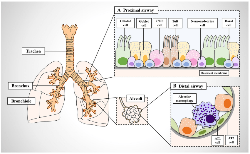
Fig. 1 Structure and cellular composition of the lung. A. The proximal airways are composed of ciliated cells, mucus-secreting goblet cells, secretory columnar cells, brush cells, undifferentiated basal cells, and neuroendocrine cells. B. Alveolar type 1 (AT1) and type 2 (AT2) epithelial cells constitute the distal airways. Resident alveolar macrophages continuously monitor the surrounding microenvironment to identify potentially harmful pathogens[3].
The intranasal instillation of LPS to induce an acute pneumonia model is a classic method for studying ALI. LPS is the main component of the cell wall of Gram-negative bacteria. After airway administration, it can effectively activate alveolar macrophages and trigger an acute pulmonary inflammatory response characterized by neutrophil infiltration and a cytokine storm, which closely mimics the pathological processes of human bacterial pneumonia and ALI[6].
This article primarily introduces how to isolate macrophages from bronchoalveolar lavage fluid (BALF) and use flow cytometry to detect phenotypic differences between normal and acute pneumonia states, aiming to provide experimental references for respiratory immunology research among the scientific community.
02 Bronchoalveolar Lavage Fluid macrophage isolation and flow cytometry workflow
Bronchoalveolar lavage fluid macrophages refer to the macrophage population obtained from the pulmonary luminal spaces via bronchoalveolar lavage technique, predominantly consisting of alveolar macrophages (AMs), which serve as the first immune barrier defending the lungs against pathogen invasion. In mouse models of pneumonia, detecting the number, phagocytic activity, cytokine secretion, and surface marker changes of BALF macrophages can provide real-time reflection of the local inflammatory status and immune response dynamics in lung tissues, enabling quantitative assessment of the degree of inflammatory injury and repair trends. Furthermore, it allows for early evaluation of the remodeling effects of anti-inflammatory drugs or interventions on the pulmonary microenvironment, thereby providing experimental evidence and theoretical guidance for clinical therapeutic strategies for pneumonia.
2.1 Preparation of the acute pneumonia mouse model
(1) Weigh an appropriate amount of LPS, dissolve it in sterile PBS to prepare a 1 mg/mL stock solution, and store at –20°C.
(2) Select C57BL/6J mice, hold the mouse in an upright position, and administer LPS intranasally at a dose of 20 mg/kg.
(3) Use a pipette to aspirate the LPS solution, and gently place a droplet at one nostril at a time, allowing the mouse to inhale it naturally.
(4) After instillation, keep the mouse in an upright position for approximately 30 s to 1 min to ensure that the liquid fully enters the lower respiratory tract, then continue to house the mice for 24 h.
2.2 Preparation of bronchoalveolar lavage fluid
(1) After anesthetizing normal mice and acute pneumonia mice separately, secure them in a supine position, fully expose the neck and chest areas, and disinfect the operating area.
(2) Use ophthalmic scissors to make an approximately 1 cm longitudinal incision at the midline of the mouse neck, and carefully separate the skin, subcutaneous tissue, and muscle layer by layer to expose the white trachea.
(3) Attach a 1 mL syringe to an endotracheal cannula (26G scalp needle), insert the needle parallel into the trachea, with an insertion depth of approximately 0.8–1 cm.
(4) Slowly inject PBS (containing 2 mM EDTA and 1% BSA) at a volume of 0.5–1 mL per instillation (can be flexibly adjusted according to mouse body weight).
(5) After instillation, pause for 5–10 s. Gently massage the mouse chest to facilitate thorough contact of the saline with the alveoli, then gently aspirate the lavage fluid.
(Note: The natural recovery rate of bronchoalveolar lavage fluid is relatively low, typically 50%–70%. The lavage procedure can be repeated 3–4 times. Throughout the entire process, keep the collected lavage fluid on ice to prevent sample deterioration that could affect subsequent detection.)
2.3 Flow cytometry detection
(1) Centrifuge the collected bronchoalveolar lavage fluid at 300 × g for 3–5 min. After centrifugation, discard the supernatant and retain the white pellet.
(2) Add Cell Staining Buffer to resuspend the cells and adjust the cell density to 1 × 10⁷/mL.
(3) Take 100 μL of the cell suspension, add CD16/32 blocking antibody, gently pipette to mix, and incubate at 4°C for 15 min for blocking.
(4) Then add flow cytometry detection antibodies, gently pipette to mix, and continue incubating at 4°C for 30 min. Add 1–2 mL of Cell Staining Buffer to resuspend the cells, gently pipette, and centrifuge at 150 × g for 3–5 min.
(5) Discard the supernatant, then add 100–300 μL of Cell Staining Buffer to resuspend the cells, and proceed to flow cytometry analysis.
03 Flow cytometry gating strategy and results
3.1 Gating strategy
(1) AMs (Alveolar Macrophages): Select live cells, then from the CD45⁺ population, gate on CD64⁺MerTK⁺ double-positive cells as AMs.
(2) cDC (Conventional Dendritic Cells): From the CD64⁻MerTK⁻ cell population (excluding AMs), further analyze and identify CD11c⁺MHC II⁺ cells as cDCs.
(3) Eos (Eosinophils): From the CD64⁻MerTK⁻ cell population, further analyze and identify Siglec-F⁺CD11c⁻ cells as Eos.
(4) Neu (Neutrophils): From the CD64⁻MerTK⁻ cell population, further analyze and identify CD11b⁺Gr-1⁺ cells as Neu.
Notes:
① It is recommended to set up isotype controls for MHC II/CD64/MerTK.
② The use of CD16/32 blocking can affect CD64 staining. To avoid this, stain for CD64 first, then add CD16/32 blocking, and subsequently stain for other markers.
3.2 Flow cytometry detection results of alveolar macrophages in bronchoalveolar lavage fluid from normal mice
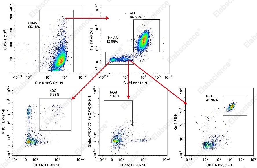
Fig. 2 Flow cytometry detection results of alveolar macrophages in bronchoalveolar lavage fluid from C57BL/6J mice.
Bronchoalveolar lavage fluid samples were collected from C57BL/6J mice and stained with mouse CD45, CD64, MerTK, CD11c, MHC II, Siglec-F, CD11b, and Gr-1 flow cytometry antibodies. The functional subsets of macrophages were then analyzed using flow cytometry.
As shown in the figure above, in the bronchoalveolar lavage fluid of normal mice, alveolar macrophages (AMs), characterized by high MerTK expression, accounted for 84.58% of CD45⁺ cells, while the non-alveolar macrophage population (Non-AM) accounted for 13.85% of CD45⁺ cells. Subsequent subset analysis of the Non-AM population identified conventional dendritic cells (cDCs) comprising 6.53% of Non-AM cells, eosinophils (EOS) comprising 1.40% of Non-AM cells, and neutrophils (NEU) comprising 42.98% of Non-AM cells. This figure illustrates the predominance of macrophages in the bronchoalveolar lavage fluid of normal mice.
3.3 Comparison of flow cytometry detection results of alveolar macrophages in bronchoalveolar lavage fluid between normal and acute pneumonia mice
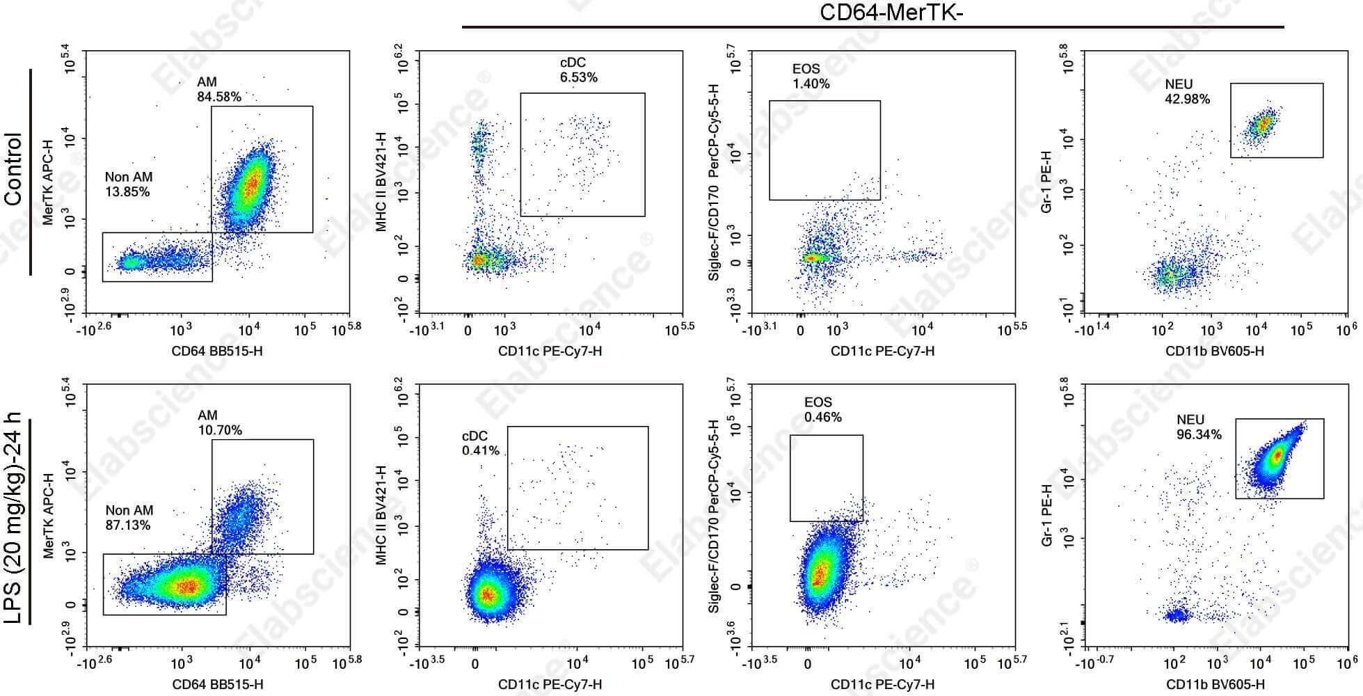
Fig. 3 Comparison of flow cytometry detection results of alveolar macrophages in bronchoalveolar lavage fluid between normal and LPS-induced acute pneumonia mice.
Bronchoalveolar lavage fluid samples were collected from normal mice and LPS-induced acute pneumonia mice, stained with mouse CD45, CD64, MerTK, CD11c, MHC II, Siglec-F, CD11b, and Gr-1 flow cytometry antibodies, and analyzed for macrophage functional subsets using flow cytometry.
As shown in the figure above, compared with the healthy control group, the LPS-induced acute pneumonia mice exhibited a significant decrease in the proportion of macrophages in the bronchoalveolar lavage fluid, dropping from 84.58% to 10.70%; cDCs also decreased from 6.53% to 0.41%; eosinophils decreased from 1.40% to 0.46%; while the proportion of neutrophils within the Non-AM population increased dramatically, rising from 42.98% to 96.34%.
This visually demonstrates the dramatic remodeling of the immune cell population in bronchoalveolar lavage fluid (BALF) during the LPS-induced acute lung injury model. At 24 hours post-LPS stimulation, the pulmonary microenvironment underwent a fundamental shift from "homeostatic maintenance" to "acute inflammatory outburst." Neutrophils replaced the originally resident cells and became the absolutely dominant population in BALF, while the proportion of alveolar macrophages showed a precipitous decline.
Elabscience® Recommended Products:
Table 1. Antibody panel for flow cytometric analysis of immune cells in mouse BALF
|
Cat. No. |
Product Name |
|
AN00871E |
APC Anti-Mouse MERTK Antibody[2B10C42] |
|
E-AB-F09832E |
APC Rat IgG2a, κ Isotype Control[2A3] |
|
E-AB-F0990Q2 |
Elab Bright™ Violet 421 Anti-Mouse MHC II (I-A/I-E) Antibody[M5/114] |
|
AN00821Q2 |
Elab Bright™ Violet 421 Rat IgG2b, κ Isotype Control[R35-38] |
|
E-AB-F1136S |
Elab Fluor® Red 780 Anti-Mouse CD45 Antibody[30-F11] |
|
E-AB-F1081T |
Elab Fluor® Violet 610 Anti-Mouse/Human/Monkey CD11b Antibody[M1/70] |
|
E-AB-F1186C |
FITC Anti-Mouse CD64/FcγRI Antibody[X54-5/7.1] |
|
E-AB-F09792C |
FITC Mouse IgG1, κ Isotype Control[MOPC-21] |
|
E-AB-F1120D |
PE Anti-Mouse Ly-6G/Ly-6C (Gr-1) Antibody[RB6-8C5] |
|
E-AB-F0991H |
PE/Cyanine7 Anti-Mouse CD11c Antibody[N418] |
|
AN00629J |
PerCP/Cyanine5.5 Anti-Mouse Siglec-F/CD170 Antibody[S17007L] |
|
E-AB-F0997A |
Purified Anti-Mouse CD16/32 Antibody[2.4G2] |
References:
[1] Wu X, Wu L, Wu Y, et al. Heme oxygenase-1 ameliorates endotoxin-induced acute lung injury by modulating macrophage polarization via inhibiting TXNIP/NLRP3 inflammasome activation. Free Radical Biology and Medicine. 2023;194:12-22.
[2] Cheng P, Li S, Chen H. Macrophages in Lung Injury, Repair, and Fibrosis. Cells. 2021;10(2):436
[3] Pyung YJ, Park DJ, Kim CG, Yun CH. Remodeling and Restraining Lung Tissue Damage Through the Regulation of Respiratory Immune Responses. Tissue Engineering and Regenerative Medicine. 2023;20(3):329-339.
[4] Wang L, Li D, Yang K, et al. The role of immunometabolism in macrophage polarization and its impact on acute lung injury/acute respiratory distress syndrome[J]. Frontiers in Immunology, 2023, 14: 1117548.
[5] Lu HL, Huang XY, Luo YF, et al. Activation of M1 macrophages plays a critical role in the initiation of acute lung injury. Bioscience Reports. 2018;38(2).
[6] Duan M, Steinfort DP, Smallwood D, et al. CD11b immunophenotyping identifies inflammatory profiles in the mouse and human lungs. Mucosal Immunology. 2016;9(2):550-563.

