

B cells serve as key orchestrators of adaptive immunity. They not only act as the exclusive producers of high-affinity antibodies, but also function as essential regulators of immune homeostasis via antigen presentation, cytokine secretion, and direct intercellular interactions. The dual ability of B cells to mediate humoral immune protection through plasma cells and sustain immune tolerance via regulatory B cell subsets highlights their multifaceted functions in both physiological and pathological immune processes.
This review systematically elaborates on the molecular mechanisms underlying high-affinity antibody production by B cells, the pivotal role of B cells in antigen presentation to CD4+ T cells, the profile and immunoregulatory effects of B cell-derived cytokines, the inhibitory mechanisms of regulatory B cells in immune responses, the protective functions of memory B cells in long-term immunological memory, and the functional specialization of plasma cells as professional antibody-secreting effector cells.
Table of Contents
1. How Do B Cells Produce High-Affinity Antibodies?
2. The Role of B Cells in Antigen Presentation to CD4+ T Cells
3. Cytokines Secreted by B Cells and Their Immunoregulatory Functions
4. How Regulatory B Cells Suppress Immune Responses
5. Functions of Memory B Cells in Long-Term Immune Protection
6. Plasma Cells as Specialized Antibody-Secreting Effector Cells
01 How Do B Cells Produce High-Affinity Antibodies?
Humoral immunity B cells generate high-affinity antibodies via a sophisticated, iterative Darwinian cascade termed affinity maturation, which primarily takes place within germinal centers (GCs), specialized microstructures residing in secondary lymphoid organs. This process couples targeted genetic diversification of immunoglobulin variable-region genes with strict antigen- and T cell–dependent selection, continuously enriching B cell clones whose receptors exhibit improved antigen-binding kinetics, most notably slower antigen dissociation rates. This cascade underpins the capacity of the adaptive immune system to establish long-lasting humoral immunity and serves as a fundamental foundation for effective vaccination[1].
The process initiates when naive B cells encounter cognate antigens and receive co-stimulatory signals from CD4+ T cells, driving B cell activation and follicular entry to form nascent GCs. Within GCs, B cells establish a unique transcriptional program governed by the master transcription factor Bcl6 and segregate into two functionally distinct compartments: the dark zone (DZ) and light zone (LZ). Such spatial compartmentalization is indispensable for the cyclic progression of affinity maturation and supports the diverse B cell functions required for effective immune priming and antibody optimization. In the DZ, GC B cells known as centroblasts undergo robust clonal expansion. During proliferation, these cells express activation-induced cytidine deaminase (AID), an enzyme that initiates somatic hypermutation (SHM) through cytosine deamination within immunoglobulin variable gene segments. This enzymatic reaction generates U:G DNA mismatches, which are subsequently processed by error-prone base excision repair and mismatch repair machineries to introduce point mutations, as well as sporadic insertions and deletions across complementarity-determining regions. These mutagenic events yield a heterogeneous pool of B cell clones with broad antigen-binding affinities and specificities; some clones acquire enhanced binding capacity, whereas others carry deleterious mutations or gain self-reactivity[2].
After acquiring somatic mutations, mutated B cells migrate to the LZ to compete for survival under stringent selective pressure. Here, centrocytes must capture native antigens presented on the surface of follicular dendritic cells (FDCs). The magnitude of B cell receptor (BCR) signaling and the abundance of captured antigen jointly determine the efficiency of peptide–MHC II complex processing and presentation to T follicular helper (Tfh) cells. Tfh cells supply limited survival cues, including CD40L–CD40 ligation and cytokines such as Interleukin-21 (IL-21), which exclusively license the highest-competence B cells to survive and proliferate. B cells that fail to capture sufficient antigen or receive insufficient Tfh co-stimulation undergo apoptosis and are eliminated by tingible body macrophages, thereby retaining only high-affinity variants. Repeated shuttling between the DZ (for mutagenesis) and LZ (for selection) iteratively refines antibody affinity over weeks to months[2,3].
Accumulating evidence demonstrates that the clonal diversity of GC founder B cells and the availability of antigen profoundly shape the final outcome of affinity maturation. Computational modeling indicates that GCs frequently maintain a broad repertoire of B cell clones with variable affinities rather than being rapidly dominated by a single high-affinity clone, enabling parallel affinity maturation across multiple independent lineages. Moreover, GC selection is not dictated solely by intrinsic antigen-binding affinity; it is also modulated by dynamic Tfh cell availability and interclonal competitive pressure within the GC niche. The transcription factor NF-κB c-Rel has also been characterized as a core selection regulator, whose intracellular abundance controls GC B cell maintenance and proliferative potential[2,3,4].
Beyond driving affinity maturation, AID orchestrates class switch recombination (CSR), a rearrangement event that converts antibody isotypes from IgM to IgG, IgA or IgE without altering antigen-binding specificity. Isotype switching remodels antibody Fc-domain effector functions, such as complement activation and opsonization, and thereby augments the overall potency of humoral immune responses. High-affinity GC B cells ultimately differentiate into two major effector subsets: long-lived plasma cells that home to the bone marrow to constitutively secrete abundant high-affinity antibodies, and memory B cells that circulate persistently and re-enter GCs upon antigen re-exposure for further affinity refinement[5].
The robustness and durability of GC-dependent humoral responses are well exemplified by clinical studies of SARS-CoV-2 mRNA vaccines, which sustain human GC activity for months post-immunization. Persistent GC reactions support continuous SHM-driven affinity maturation, generating memory B cells that produce antibodies with expanded neutralization breadth and elevated potency against emerging viral variants. Nevertheless, this mutagenic process carries intrinsic risks: aberrant SHM and CSR induce off-target oncogenic mutations that promote B cell lymphomagenesis, or generate pathogenic high-affinity autoantibodies that fuel autoimmune disorders. To counteract such adverse outcomes, multiple regulatory circuits, including T follicular regulatory (Tfr) cells, suppress the outgrowth of self-reactive B cell clones, balancing protective immunity and immune tolerance. In addition, recent mechanistic work reveals that RNA-binding proteins TIA1 and TIAL1 confer apoptotic resistance to GC B cells by boosting the translation of pro-survival factors, underscoring the intricate post-transcriptional regulatory network required for productive affinity maturation[6].
In summary, high-affinity antibody production by adaptive immunity B cells constitutes a tightly controlled, multi-stage cascade featuring AID-mediated somatic hypermutation in the DZ and rigorous Tfh-dependent clonal selection in the GC LZ. Repeated cycling between these two compartments selects B cell clones with optimal antigen-binding properties, which subsequently differentiate into long-lived plasma cells and memory B cells to confer potent, sustained humoral immunity.
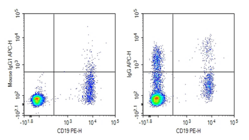
Fig. 1 Detection and analysis of IgG expression in human peripheral blood B lymphocytes. Human peripheral blood mononuclear cells (PBMCs) were stained with 0.2 μg purified anti-human IgG antibody (clone G18-145, right) or 0.2 μg mouse IgG1 κ isotype control antibody (left). Cells were subsequently incubated with APC-conjugated goat anti-mouse IgG secondary antibody, followed by staining with PE-conjugated anti-human CD19 monoclonal antibody. (The data are provided by Elabscience.)
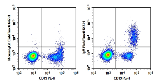
Fig. 2 Detection and analysis of IgM expression in human peripheral blood B lymphocytes. Human peripheral blood lymphocytes were stained with 0.2 μg Purified Anti-Human IgM Antibody[MHM-88] (Right) and 0.2 μg Mouse IgG1, κ Isotype Control (Left), followed by Elab Fluor® 647-conjugated Goat Anti-Mouse IgG Secondary Antibody, then anti-Human CD19 PE-conjugated Monoclonal Antibody. (The data are provided by Elabscience.)
Elabscience® Quick Overview of Popular Products:
Table 1. Reagents for research on high-affinity antibody generation by humoral immune B cells
|
Product Name |
Cat. No. |
|
Purified Anti-Human IgM Antibody[MHM-88] |
E-AB-F11720P |
|
PE/Cyanine5.5 Anti-Human IgM Antibody[MHM-88] |
E-AB-F1172I |
|
Elab Fluor® 700 Anti-Human IgM Antibody[MHM-88] |
E-AB-F1172M1 |
|
Purified Anti-Human IgG Antibody[G18-145] |
AN010450P |
|
PE Anti-Mouse CD19 Antibody[1D3] |
E-AB-F0986D |
|
PE Anti-Human/Monkey CD19 Antibody[CB19] |
E-AB-F1004D |
|
PE Anti-Mouse CD40 Antibody[FGK4.5/FGK45] |
E-AB-F1028D |
|
FITC Anti-Human CD40 Antibody[3A8] |
E-AB-F1037C |
|
Elab Fluor® 647 Anti-Human CD154 (CD40L) Antibody[24-31] |
AN00341M |
|
Mouse IgG(Immunoglobulin G) ELISA Kit |
E-EL-M0692 |
|
Human IgG(Immunoglobulin G) ELISA Kit |
E-EL-H0169 |
|
Porcine IgG(Immunoglobulin G) ELISA Kit |
E-EL-P0004 |
|
QuicKey Pro Mouse IgA(Immunoglobulin A) ELISA Kit |
E-OSEL-M0007 |
|
Rat IgA(Immunoglobulin A) ELISA Kit |
E-EL-R3015 |
|
QuicKey Pro Human IgA(Immunoglobulin A) ELISA Kit |
E-OSEL-H0009 |
|
Human PBMC Separation Solution(P 1.077) |
E-CK-A103 |
|
10×ACK Lysis Buffer |
E-CK-A105 |
|
10× RBC Lysis/Fixation Solution |
E-CK-A106 |
02 The Role of B Cells in Antigen Presentation to CD4+ T Cells
B cell functions cover multiple core immune regulatory processes, among which serving as professional antigen-presenting cells (APCs) enables B cells to exert pivotal functions in initiating and sustaining adaptive immune responses via presentation of proteolytically processed peptide antigens to CD4+ T cells. This cognate interaction underpins helper T cell activation; these T cells subsequently deliver indispensable signals supporting B cell differentiation, antibody class switching, and germinal center-resident affinity maturation. This cascade initiates upon naive B cell recognition of cognate antigens through the B cell receptor (BCR), triggering antigen internalization, intracellular processing, and loading of antigen-derived peptides onto major histocompatibility complex class II (MHC-II) molecules. This unique capacity differentiates B cells from other APC subsets (e.g., dendritic cells), as it directly couples BCR-mediated antigen recognition to antigen-presentation efficiency, thereby restricting T cell helper signals exclusively to high-affinity antibody-producing B cell clones[7,8].
BCR-dependent antigen uptake endows humoral immunity B cells with exceptionally high antigen capture efficiency, driven by the intrinsic antigen specificity of BCRs. Unlike macrophages and dendritic cells, which depend on promiscuous pattern recognition receptors (PRRs) or non-specific pinocytosis, B cells enrich cognate antigens from low-concentration environments via receptor-mediated endocytosis. Following internalization, internalized antigens are routed to endosomal compartments and subjected to proteolytic cleavage to generate short peptide fragments. These proteolytic peptides are subsequently loaded onto MHC-II complexes within the endosomal processing route prior to surface trafficking. Surface peptide–MHC-II complex density correlates positively with BCR antigen-binding affinity, forming a positive feedback loop wherein higher BCR affinity leads to more robust antigen uptake and enhanced antigen presentation to T cells. This quantitative correlation guarantees preferential acquisition of T cell helper signals by high-affinity B cells, a prerequisite for clonal selection events within secondary lymphoid tissues[9,10].
Cognate B cell–CD4+ T cell interactions first take place at the T–B border of secondary lymphoid organs, before shifting to the light zone of germinal centers at later stages. Complete T cell activation requires TCR-mediated recognition of cognate peptide–MHC-II complexes presented by B cells. Such TCR ligation initiates a cascade of co-stimulatory cell-cell contacts, among which CD40 ligand (CD40L) expressed on activated T cells binding to B cell-surface CD40 is the most vital. CD40–CD40L ligation is irreplaceable for sustaining B cell viability, supporting clonal expansion, and triggering germinal center formation. In the absence of this co-stimulatory signal, B cells predominantly undergo apoptotic clearance and lose the capacity to execute class switch recombination and somatic hypermutation. Additionally, Tfh cells secrete effector cytokines including IL-21 and IL-4, which further instruct B cell differentiation into antibody-secreting plasma cells or long-lived memory B cells[10].
Inside germinal centers, B cell-mediated antigen presentation acquires more specialized and competitive regulatory features. Germinal centers are compartmentalized into dark zones (supporting B cell proliferation and somatic hypermutation) and light zones (the site of clonal selection). Within light zones, somatically mutated B cells compete for scarce antigen immobilized on follicular dendritic cells (FDCs). B cells that efficiently capture antigens through mutated BCRs process and display peptide–MHC-II complexes for Tfh cell recognition. The avidity of T–B cell contacts dictates whether a given B cell acquires adequate survival cues to re-enter dark zones for additional mutagenesis cycles or commits to differentiation as antibody-secreting plasma cells. This cyclic loop of mutagenesis and clonal selection, fueled by B cell antigen presentation to Tfh cells, ultimately generates high-affinity antibody repertoires. Accumulating evidence demonstrates that dynamic Tfh cell abundance and intra-GC competitive microenvironment jointly govern the final output of affinity maturation[10,11].
Furthermore, distinct transcriptional programs orchestrate this intercellular crosstalk, with Bcl6 acting as a master transcription factor in both GC B cells and Tfh cells to coordinate their spatial partitioning and functional specialization within germinal centers. The transcription factor NF-κB c-Rel also acts as a central modulator of GC clonal selection, regulating the survival and proliferative capacity of GC B cells. Multiple regulatory circuits, prominently T follicular regulatory (Tfr) cell-dependent pathways, restrain the expansion of self-reactive B cell clones, balancing immune tolerance and potent pathogen-targeted immunity. As key effector cells in specific immune defense, adaptive immunity B cells drive the whole set of antigen presentation, clonal selection and antibody maturation processes. Thus, B cell antigen presentation to CD4+ T cells is far more than passive peptide display; it constitutes an active, competitive, tightly controlled cascade that shapes the magnitude, specificity and quality of humoral immunity[9,11].
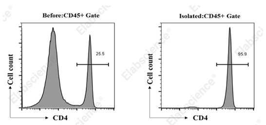
Fig. 3 Detection of CD4+ T cells isolated from mouse tissues. CD4+ T cells were sorted from C57BL/6 mice splenocyte using the EasySort™ Mouse CD4+ T Cell Isolation Kit (MIM002N), then stained with ElabFluor® Violet 450 anti-mouse CD45 antibody (clone 30-F11) and PE anti-mouse CD4 antibody (clone GK1.5). The purities of the initial unseparated population and final enriched CD4+ T cell fraction were 25.5% and 95.9%, respectively. (The data are provided by Elabscience.)
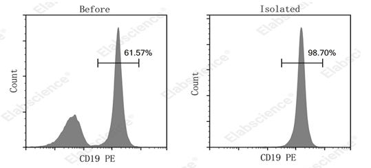
Fig. 4 Detection of B cells isolated from mouse tissues. B cells were sorted from C57BL/6 mice splenocyte using the EasySort™ Mouse B Cell Isolation Kit (MIM004N), then stained with PerCP Anti-Mouse CD45 Antibody[30-F11] (E-AB-F1136F) and PE Anti-Mouse CD19[1D3] (E-AB-F0986D). The purities of the initial unseparated population and final enriched CD45+CD19+ B cell fraction were 61.57% and 98.70%, respectively. (The data are provided by Elabscience.)
Elabscience® Quick Overview of Popular Products:
Table 2. Reagents used for investigating B cell–CD4+ T cell interactions
|
Product Name |
Cat. No. |
|
EasySort™ Mouse B Cell Isolation Kit |
MIM004N |
|
EasySort™ Mouse CD4+T Cell Isolation Kit |
MIM002N |
|
EasySort™ Human CD4+ T Cell Isolation Kit |
MIH002N |
|
EasySort™ Human B Cell Isolation Kit |
MIH004N |
|
PE Anti-Mouse CD19 Antibody[1D3] |
E-AB-F0986D |
|
PE Anti-Human/Monkey CD19 Antibody[CB19] |
E-AB-F1004D |
|
PE Anti-Mouse CD40 Antibody[FGK4.5/FGK45] |
E-AB-F1028D |
|
FITC Anti-Human CD40 Antibody[3A8] |
E-AB-F1037C |
|
Elab Fluor® 647 Anti-Human CD154 (CD40L) Antibody[24-31] |
AN00341M |
|
Mouse IgG(Immunoglobulin G) ELISA Kit |
E-EL-M0692 |
|
Human IgG(Immunoglobulin G) ELISA Kit |
E-EL-H0169 |
|
Porcine IgG(Immunoglobulin G) ELISA Kit |
E-EL-P0004 |
|
QuicKey Pro Mouse IgA(Immunoglobulin A) ELISA Kit |
E-OSEL-M0007 |
|
Rat IgA(Immunoglobulin A) ELISA Kit |
E-EL-R3015 |
|
QuicKey Pro Human IgA(Immunoglobulin A) ELISA Kit |
E-OSEL-H0009 |
|
Human PBMC Separation Solution(P 1.077) |
E-CK-A103 |
|
10×ACK Lysis Buffer |
E-CK-A105 |
|
10× RBC Lysis/Fixation Solution |
E-CK-A106 |
03 Cytokines Secreted by B Cells and Their Immunoregulatory Functions
B cells have long transitioned from being regarded solely as antibody-secreting humoral effector cells to being recognized as multifunctional immune regulators. B cell cytokine production is now established as a central effector mechanism that enables B cells to secrete diverse cytokines to remodel global immune responses. Such cytokine secretion enables B cells to exert both proinflammatory and anti-inflammatory effects, which profoundly shape the onset of autoimmune pathogenesis, sustain immune tolerance, and reshape the tumor microenvironment. The divergent cytokine output of distinct B cell populations recapitulates the functional heterogeneity of B cell subsets; such as regulatory B cells (Bregs) are specialized to produce immunomodulatory mediators, including interleukin-10 (IL-10), transforming growth factor-β (TGF-β), and interleukin-35 (IL-35). These regulatory B cells (Bregs) restrict excessive immune activation, block autoimmune pathology, and maintain immune homeostasis via complex crosstalk with T cells, macrophages and other immune effector populations[12].
Under steady-state physiological conditions, Bregs remain rare, yet their numbers expand markedly during chronic inflammation, infectious challenge, pregnancy, and organ transplantation. Bregs mediate their suppressive function predominantly via secretion of anti-inflammatory cytokines, with IL-10, TGF-β and IL-35 as the most well-characterized mediators. IL-10-competent Bregs represent the most extensively studied Breg subset, which potently limit pathological inflammatory cascades in autoimmune disorders including multiple sclerosis, systemic lupus erythematosus and rheumatoid arthritis. Quantitative detection of these key immune mediators via IL-10 elisa serves as a classic experimental approach to assess Breg anti-inflammatory activity in both preclinical models and clinical sample analysis. These cells suppress the proliferation of proinflammatory Th1 and Th17 cells and drive the differentiation of Foxp3+ regulatory T cells (Tregs), thereby establishing a stable tolerogenic microenvironment[13].
TGF-β-producing B cells constitute another critical arm of B cell-dependent immune regulation, with prominent roles in tumor immunity and tissue repair. Melanoma patients exhibit elevated frequencies of circulating and tumor-resident TGF-β+ Bregs, which expand Foxp3+ Tregs and establish an immune-suppressive tumor niche. Likewise, co-expression of TGF-β and IL-10 acts as an adverse prognostic biomarker in diffuse large B-cell lymphoma, underscoring the translational value of B cell cytokine signatures. TGF-β blocks effector T cell activation and proliferation while instructing naive T cells to differentiate into induced Tregs, consequently blunting anti-tumor immune surveillance and accelerating tumor progression. Given its additional capacity to drive tissue fibrosis and remodeling, TGF-β exemplifies the context-dependent bioactivity of B cell-derived cytokines[14].
As a member of the IL-12 superfamily, IL-35 is another powerful immune-inhibitory cytokine secreted by Bregs, though its downstream signaling cascades remain less thoroughly characterized relative to IL-10 and TGF-β. IL-35-expressing B cells mitigate disease severity in experimental autoimmune encephalomyelitis and other inflammatory disorders by restraining Th17 differentiation and fostering IL-10-secreting Treg populations. The synergistic activity of these three cytokines empowers Bregs to coordinate multiple immune signaling axes and confine inflammatory responses to enable timely resolution. Furthermore, Bregs mediate immune suppression via direct intercellular contact through surface molecules such as PD-L1 and CTLA-4, which further amplify their tolerogenic function[15].
Metabolic reprogramming has emerged as a central switch governing B cell cytokine secretion and functional polarization. Cumulative data demonstrate that proinflammatory B cells generating mediators such as GM-CSF and tumor necrosis factor-alpha (TNF-α) rely heavily on enhanced oxidative phosphorylation (OXPHOS) to satisfy bioenergetic demands. Quantitative measurement of proinflammatory TNF-α in B cell culture supernatants using tnf alpha elisa is widely applied to quantify the proinflammatory potential of effector B cell subsets and evaluate immune activation levels. In contrast, anti-inflammatory IL-10-producing Bregs adopt distinct metabolic programs, indicating that intracellular metabolic status dictates whether B cells commit to a pro- or anti-inflammatory phenotype. Specifically, the differentiation and effector function of IL-10+ Bregs are controlled by mitochondrial electron transport chain activity and balanced intracellular reactive oxygen species (ROS) levels, with the redox regulator thioredoxin serving as an essential stabilizer of Breg identity. This metabolic bifurcation creates novel therapeutic opportunities to modulate B cell function in autoimmunity and cancer through targeted perturbation of core metabolic pathways[16].
Dysregulated B cell cytokine secretion represents a core pathogenic driver in multiple sclerosis (MS). B cells isolated from MS patients display skewed cytokine profiles featuring elevated production of proinflammatory GM-CSF and lymphotoxin-α, which exacerbate neuroinflammation and central demyelination. Conversely, the frequency and functional competence of IL-10-producing Bregs are frequently impaired in MS, leading to lost control over autoreactive T cell clones. The dynamic balance between opposing pro- and anti-inflammatory B cell subsets determines disease progression; therapies designed to restore Breg activity or deplete pathogenic pro-inflammatory B cells have achieved substantial clinical benefits. For instance, the therapeutic efficacy of CD20-targeted B cell depletion in MS confirms the causal contribution of B cells to disease pathology, yet the exact cytokine-dependent regulatory circuits remain under active investigation[17].
Beyond autoimmune pathologies, B cell-derived cytokines exert prominent effects in allergic disorders and respiratory immunity. In childhood allergic asthma, IL-10-secreting Bregs are indispensable for restraining allergen-specific Th2 responses and suppressing IgE synthesis. Defects in Breg generation or IL-10 secretion correlate with aggravated allergic manifestations, implying that boosting Breg activity may constitute a viable therapeutic strategy. Similarly, during respiratory infection and chronic obstructive pulmonary disease (COPD), Bregs restrain pulmonary inflammation via IL-10 and TGF-β release to prevent excessive tissue injury, though overactivated Bregs may simultaneously hinder pathogen clearance[18].
Within the tumor microenvironment, B cell cytokines exert dual opposing effects on anti-tumor immunity. While certain B cell subsets mediate protective anti-tumor responses via antibody production and antigen presentation, Bregs facilitate tumor outgrowth by silencing cytotoxic T cell function. Malignant tissues exploit Breg-derived IL-10 and TGF-β to construct an immune-suppressive niche that enables tumor immune evasion. In melanoma and breast cancer, high frequencies of TGF-β+ and IL-10+ tumor-infiltrating B cells correlate with unfavorable clinical prognosis and resistance to immune checkpoint therapy. Therefore, decoding the unique cytokine signatures of intratumoral B cells is critical to develop interventions that either neutralize Breg-mediated suppression or harness the protective functions of effector B cells for clinical treatment[19].
Bidirectional crosstalk between B cells and other innate immune compartments including macrophages and dendritic cells is also orchestrated by B cell cytokine release. B cells secrete IL-10 and IL-35 to drive macrophage polarization toward the anti-inflammatory M2 phenotype, promoting inflammation resolution and tissue repair. By contrast, under inflammatory conditions such as giant cell arteritis, B cells can skew macrophages toward a proinflammatory phenotype to exacerbate tissue damage. This reciprocal signaling highlights B cells as central coordinators bridging innate and adaptive immune networks[20,21].
In summary, B cells act as abundant cytokine producers that serve as core modulators of immune homeostasis. Via secretion of IL-10, TGF-β and IL-35, Bregs suppress pathological inflammation, enforce peripheral tolerance, and stabilize systemic immune equilibrium. The equilibrium between pro-inflammatory and anti-inflammatory B cell populations is shaped by integrated metabolic, genetic and environmental cues, and its disruption underpins a broad spectrum of human diseases ranging from autoimmunity to malignant tumors. Targeting B cell cytokine regulatory networks provides promising therapeutic strategies to remodel immune responses across diverse disorders, highlighting the urgent need to dissect the molecular programs governing B cell plasticity and subset-specific effector function.
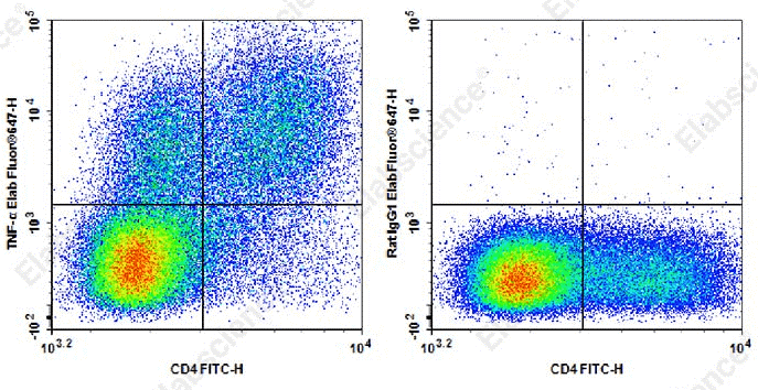
Fig. 5 Detection and analysis of TNF-α in C57BL/6 mouse splenocytes. C57BL/6 mouse splenocytes were stimulated with Cell Stimulation MIX and Protein Transport Inhibitor MIX for 5 h. Cells were stained with FITC anti-mouse CD4 antibody, alongside Elab Fluor® 647 Rat IgG1,κ isotype control (right) or Elab Fluor® 647 anti-mouse TNF-α (XT3.11) (left). Analysis was restricted to lymphocyte-gated cells. (The data are provided by Elabscience.)
Elabscience® Quick Overview of Popular Products:
Table 3. Reagents for research on cytokines Secretion of T/B cell
|
Product Name |
Cat. No. |
|
Elab Fluor® 647 Anti-Mouse TNFα Antibody[XT3.11] |
AN00567M |
|
FITC Anti-Mouse CD4 Antibody[RM4-5] |
E-AB-F1353C |
|
Cell Stimulation and Protein Transport Inhibitor Kit |
E-CK-A091 |
|
Purified Anti-Human TNF-alpha Antibody[Infliximab297.rMAb] |
AN007890P |
|
Purified Anti-Human TNF-α Antibody[MAb11] |
AN008590P |
|
CellaQuant™ Human TNF-α (Tumor Necrosis Factor Alpha) ELISA Kit |
CQH014 |
|
Uncoated Human TNF-α(Tumor Necrosis Factor Alpha) ELISA Kit |
E-UNEL-H0175 |
04 How Regulatory B Cells Suppress Immune Responses
Regulatory B cells (Bregs) constitute a heterogeneous B lymphocyte subset that potently restrains excessive immune activation, sustains peripheral immune tolerance, and limits aberrant inflammatory tissue damage. Bregs primarily execute their immunosuppressive functions through the secretion of anti-inflammatory cytokines, most notably IL-10, TGF-β, and IL-35. These cytokines target multiple immune cell populations, including T cells, macrophages, and dendritic cells, to block proinflammatory signaling cascades and establish a tolerogenic tissue microenvironment. As a gold-standard quantitative approach for evaluating Breg functional activity,IL-10 elisa is widely implemented in immunological research to detect and quantify B cell-derived anti-inflammatory cytokine secretion under both physiological and pathological conditions. IL-10-competent Bregs are critical for curbing pathological inflammatory flares in autoimmune diseases such as multiple sclerosis and systemic lupus erythematosus. By inhibiting the clonal expansion of proinflammatory Th1 and Th17 cells and promoting the differentiation of Foxp3+ regulatory T cells (Tregs), IL-10-producing Bregs effectively mitigate aberrant adaptive immune activation[23,24].
Beyond cytokine-mediated immune regulation, Bregs employ surface molecular ligands to exert contact-dependent immunosuppression. Programmed death-ligand 1 (PD-L1) expressed on Bregs binds to PD-1 on T cells, effectively suppressing T cell activation and proliferation. This contact-dependent inhibitory pathway is highly active in the tumor microenvironment, where Bregs facilitate tumor immune evasion by dampening the cytotoxic function of tumor-infiltrating T cells. Moreover, concurrent expression of TGF-β and PD-L1 on B cells is correlated with poor clinical prognosis in melanoma, highlighting the translational significance of Breg surface-mediated inhibitory mechanisms. Breg-T cell crosstalk is bidirectional: Bregs also modulate the activity of follicular regulatory T cells (Tfr) to stabilize germinal center immune homeostasis, which further fine-tunes B cell differentiation and remodels the overall peripheral B cell repertoire[25].
Intrinsic cellular metabolism acts as a core regulatory switch that dictates B cell immune competence and governs B cell differentiation toward immunosuppressive Breg phenotypes. Accumulating mechanistic evidence indicates that the differentiation and immunosuppressive capacity of IL-10-competent Bregs are tightly controlled by mitochondrial electron transport chain activity and intracellular ROS homeostasis. Thioredoxin, a redox regulatory protein encoded by the TXN gene, is highly expressed in Bregs and is essential for maintaining their stable immunosuppressive phenotype. Unlike pro-inflammatory B cells that rely on enhanced oxidative phosphorylation to sustain effector functions, Bregs undergo unique metabolic reprogramming to support persistent and robust IL-10 production. This distinct metabolic vulnerability provides promising therapeutic targets for modulating Breg activity in autoimmune diseases and malignancies[26].
The stability, survival, and abundance of IL-10+ Bregs are further regulated by cell surface immune checkpoint molecules, with SLAMF5 serving as a key negative modulator. SLAMF5 signaling restricts Breg survival and limits their immunosuppressive potency. Impaired SLAMF1 signaling prolongs Breg viability and enhances IL-10 secretion, which can trigger systemic excessive immunosuppression. This intrinsic negative regulatory circuit constrains Breg activity within a physiological range, preventing immune over-suppression that would compromise host defense against pathogens. The identification of such checkpoint-mediated regulatory mechanisms unveils the sophisticated network that calibrates Breg function and offers novel targets for immune-modulating therapeutic interventions[27].
In tumor microenvironments, Bregs readily acquire potent immunosuppressive phenotypes to drive malignant progression. Melanoma patients exhibit elevated frequencies of circulating and tumor-resident TGF-β+ PD-L1+ Bregs, which promote the expansion of Foxp3+ Tregs and impair anti-tumor immune surveillance. Similarly, dual expression of TGF-β and IL-10 serves as an adverse prognostic biomarker in diffuse large B-cell lymphoma, confirming the pathogenic role of Breg-derived cytokines in tumor progression. These findings demonstrate the dual regulatory roles of B cells in tumor immunity: B cells can mediate protective anti-tumor immune responses via antibody production and antigen presentation, while Bregs facilitate tumor immune escape depending on their unique cytokine profiles and tissue localization[28].
In allergic disorders such as childhood asthma, Bregs confer protective immune regulation by suppressing allergen-specific Th2 immune responses and inhibiting IgE synthesis. IL-10-secreting Bregs are indispensable for maintaining immune tolerance to harmless environmental antigens, and defective Breg function is closely associated with exacerbated allergic symptoms. Quantitative detection via IL-10 elisa reliably verifies impaired breg immunosuppressive function in allergic patients, providing solid experimental evidence for the immunopathological mechanisms of allergic diseases. Collectively, these findings highlight the crucial protective role of Bregs in mitigating hypersensitivity disorders and sustaining respiratory immune homeostasis. Owing to their capacity to bridge and modulate both innate and adaptive immune responses, Bregs function as core orchestrators that maintain systemic immune equilibrium under physiological and pathological conditions[29].
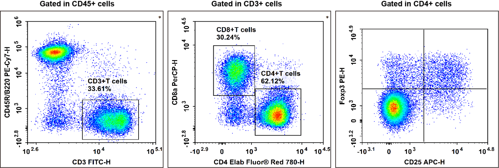
Fig. 6 Detection and analysis of Treg cells in C57BL/6 mouse splenocytes. C57BL/6 mouse splenocytes were surface stained with Elab Fluor® Violet 450 Anti-Mouse CD45, PE/Cyanine7 Anti-Mouse CD45R/B220, FITC Anti-Mouse CD3, Elab Fluor® Red 780 Anti-Mouse CD4, PerCP Anti-Mouse CD8a and APC Anti-Mouse CD25 and then treated with Foxp3/Transcription Factor Staining Kit. Cells were then stained with PE Anti-Mouse Foxp3, followed by analysis via flow cytometry. Regulatory T cells (Treg cells) exhibit the phenotype of CD45+CD45R/B220-CD3+CD4+CD25+Foxp3+. (The data are provided by Elabscience.)
Elabscience® Quick Overview of Popular Products:
Table 4. Reagents for Treg and Breg cell research
|
Product Name |
Cat. No. |
|
Elab Fluor® Violet 450 Anti-Mouse CD45 Antibody[30-F11] |
E-AB-F1136Q |
|
FITC Anti-Mouse CD24 Antibody[M1/69] |
E-AB-F1179C |
|
APC Anti-Mouse CD38 Antibody[NIMR5] |
E-AB-F1193E |
|
Elab Fluor® 647 Anti-Mouse CD27 Antibody[LG.3A10] |
AN00322M |
|
PerCP/Cyanine5.5 Anti-Mouse CD5 Antibody[53-7.3] |
E-AB-F1185J |
|
PE/Cyanine7 Anti-Mouse CD1d Antibody[20H2] |
AN00570H |
|
PE Anti-Mouse IL-10 Antibody[JES5-16E3] |
E-AB-F1197D |
|
PE/Cyanine7 Anti-Mouse CD45R/B220 Antibody[RA3.3A 1/6.1] |
E-AB-F1112H |
|
FITC Anti-Mouse CD3 Antibody[17A2] |
E-AB-F1013C |
|
Elab Fluor® Red 780 Anti-Mouse CD4 Antibody[RM4-5] |
E-AB-F1353S |
|
PerCP Anti-Mouse CD8a Antibody[53-6.7] |
E-AB-F1104F |
|
APC Anti-Mouse CD25 Antibody[PC-61.5.3] |
E-AB-F1102E |
|
PE Anti-Mouse Foxp3 Antibody[3G3] |
E-AB-F1238D |
|
10×ACK Lysis Buffer |
E-CK-A105 |
|
Ready-to-Use Foxp3/Transcription Factor Staining Kit |
E-CK-A108 |
|
Elab Fluor® Violet 450 Anti-Human CD45 Antibody[HI30] |
E-AB-F1137Q |
|
Elab Fluor® Red 780 Anti-Human CD3 Antibody[OKT-3] |
E-AB-F1001S |
|
FITC Anti-Human/Monkey CD4 Antibody[SK3] |
E-AB-F1352C |
|
PerCP/Cyanine5.5 Anti-Human CD8 Antibody[UCHT-4] |
AN00427J |
|
PE Anti-Human CD25 Antibody[BC96] |
E-AB-F1194D |
|
APC Anti-Human CD127/IL-7RA Antibody[A019D5] |
E-AB-F1152E |
|
Human PBMC Separation Solution(P 1.077) |
E-CK-A103 |
|
10×ACK Lysis Buffer |
E-CK-A105 |
|
10× RBC Lysis/Fixation Solution |
E-CK-A106 |
05 Functions of Memory B Cells in Long-Term Immune Protection
Memory B cells (MBCs) constitute the foundational component of long-lived humoral immunity, forming a stable reservoir of antigen-experienced lymphocytes capable of persisting for decades following primary infection or vaccination. Unlike long-lived plasma cells (LLPCs), which constitutively secrete antibodies to confer immediate serological protection, MBCs remain quiescent until reencountering their cognate antigens. Robust memory B cell activation is triggered rapidly upon secondary antigen exposure, enabling MBCs to promptly differentiate into antibody-secreting effector cells or reenter germinal centers (GCs) for additional rounds of affinity maturation. This dual functional endowment equips MBCs to adapt to rapidly mutating pathogens such as viral variants, yielding broadly neutralizing antibodies with heightened antigen-binding specificity and neutralizing potency. The MBC compartment exhibits extensive functional heterogeneity, encompassing distinct subsets defined by unique surface phenotypic markers, antibody isotypes and transcriptional profiles; collectively, these subsets establish robust, flexible immune defense against recurrent pathogen challenge[30].
High-fidelity MBC development primarily occurs within GC reactions, specialized microdomains housed in secondary lymphoid organs where B cells undergo somatic hypermutation and affinity-dependent clonal selection. Inside GCs, B cells compete for limited antigens immobilized on follicular dendritic cells and scarce survival signals secreted by Tfh cells. B cells that efficiently capture and process antigens to present peptide–MHC-II complexes to Tfh cells receive essential costimulatory cues, including CD40 ligand crosslinking and cytokines such as IL-21. These signaling inputs precisely orchestrate B cell differentiation, directing terminal lineage commitment toward either plasma cells or memory B cells. Cell fate determination between the two lineages is tightly controlled by the antagonistic balance of master transcription factors: Bcl-6 maintains the core GC transcriptional program, whereas Blimp-1 drives plasma cell differentiation. Recent mechanistic investigations have identified additional lineage-specifying regulators, including the transcription factor Hhex and corepressor Tle3, both of which are required to support MBC maturation and build a diverse memory B cell repertoire. Moreover, epigenetic remodeling imprints the cellular history of prior antigen stimulation onto MBCs, predetermining their differentiation trajectory, either GC reentry or plasma cell generation, during secondary immune challenge[31,32].
Upon secondary antigen exposure, MBCs mount a rapid recall response that far exceeds the speed and magnitude of the primary response elicited by naive B cells. This accelerated secondary reactivity stems from multiple cell-intrinsic molecular adaptations, including preprimed metabolic readiness and lowered thresholds for antigen-triggered signaling. For instance, the transcription factor ZFP319 modulates mitochondrial homeostasis in MBCs, sustaining potent recall capacity and enabling swift differentiation into antibody-secreting effector cells. Additionally, MBCs can traffic back to germinal centers via CCL21-mediated chemotaxis, supporting iterative affinity maturation and adaptive recognition of antigenic drift. This recycling is particularly vital for sustaining protective immunity against highly mutable pathogens such as influenza A virus and SARS-CoV-2. GC reentry of MBCs also facilitates naive B cell maturation within secondary “recall GCs”, further expanding the breadth of the systemic peripheral antibody repertoire[33].
The MBC pool exhibits pronounced subset heterogeneity, with each subset executing specialized protective functions. Major populations include class-switched (IgG⁺, IgA⁺) and unswitched (IgM⁺) MBCs. Following antigen rechallenge, IgG⁺ MBCs directly differentiate into plasma cells to mediate rapid antibody secretion, while IgM⁺ MBCs readily adopt a GC B cell phenotype during secondary infection to further diversify the antibody repertoire. Accumulating evidence has also characterized tissue-resident MBC subsets localized at mucosal barrier surfaces, including the respiratory and gastrointestinal tracts, that deliver frontline immune protection against invading pathogens. Strategically positioned at primary pathogen entry portals, tissue-resident MBCs mediate swift local immune responses, complementing systemic protection provided by circulating MBCs and LLPCs. The composition of the MBC repertoire is further shaped by the modality of initial antigen exposure; different pathogens drive the expansion of specialized MBC subsets tailored to counter distinct immune threats[34,35].
Long-term MBC survival is governed by multilayered molecular and metabolic regulatory networks. Prosurvival cytokines such as BAFF (B-cell activating factor) and lineage-defining transcription factors including SpiB are indispensable for maintaining stable MBC populations over prolonged timeframes. SpiB regulates the transcription of pan-B cell signature genes and extends MBC lifespan, highlighting its central role in sustaining durable immunological memory. Furthermore, the IgM Fc receptor (FcμR) amplifies recall responses by promoting the generation of mature CD80⁺ PD-L2⁺ MBCs, a population correlated with enhanced secondary immune potency. Metabolic reprogramming is equally critical to MBC effector function: mitochondrial remodeling and oxidative phosphorylation are mandatory to sustain memory B cell activation and the subsequent B cell differentiation into downstream effector subsets. Mitochondrial dysfunction, such as that induced by Tfam deletion, disrupts GC formation and blunts secondary immune responses, demonstrating that intact metabolic homeostasis is an absolute prerequisite for functional B cell memory[36,37].
Immunosenescence during aging impairs MBC function and dampens GC reaction efficiency, leading to weakened protective immunity against natural pathogens and vaccination. This age-related decline arises from multiple cellular defects, including compromised Tfh cell activity and structural deterioration of secondary lymphoid tissue architecture. The senescence-associated effector DOCK11 modulates secondary B cell responses; genetic ablation of DOCK11 abolishes clonal expansion of antigen-specific B cells within GCs. Elucidating these age-dependent immune defects is critical to developing optimized vaccination regimens for elderly populations, who display elevated susceptibility to infectious diseases. Despite age-associated functional impairments, MBCs remain an indispensable pillar of systemic immune defense, and their protective potential can be augmented through vaccination and natural pathogen encounter. The long-term persistence of virus-specific MBCs following SARS-CoV-2 infection and immunization attests to their exceptional longevity and capacity to sustain persistent humoral immunity[38].
The unique biological characteristics of MBCs carry profound translational value, informing rational vaccine design and therapeutic development for autoimmune disorders and malignancies. Vaccines that induce robust GC activity and high-affinity MBC formation confer prolonged cross-protection against divergent viral variants. As a representative example, SARS-CoV-2-targeted mRNA vaccines drive sustained GC reactions and generate a diversified MBC repertoire capable of recognizing multiple viral mutant strains. In autoimmunity, aberrant memory B cell activation drives pathological autoantibody production and chronic inflammatory tissue damage, marking MBCs as promising therapeutic targets. In the context of cancer, MBCs exert dichotomous effects on anti-tumor immunity: they can either initiate protective anti-tumor surveillance or promote tumor immune evasion, depending on their surface phenotype and tissue localization. Dissecting the multifaceted, context-dependent roles of MBCs under physiological and pathological conditions is essential to harness their therapeutic utility and develop novel interventions for infectious diseases and human malignancies[39].
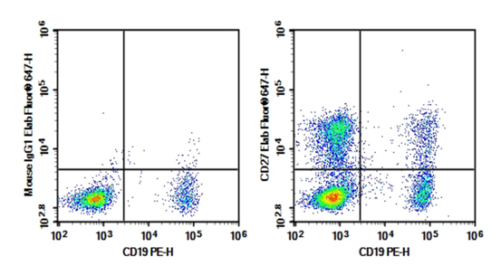
Fig. 7 Detection of memory B cells in human peripheral blood lymphocytes. Human peripheral blood lymphocytes were stained with 0.2 μg Purified Anti-Human CD27 Antibody[O323] (Right) and 0.2 μg Mouse IgG1, κ Isotype Control (Left), followed by Elab Fluor® 647-conjugated Goat Anti-Mouse IgG Secondary Antibody, then anti-Human CD19 PE-conjugated Monoclonal Antibody. The memory B cells exhibit the phenotype of CD19+CD27+IgD+. (The data are provided by Elabscience.)
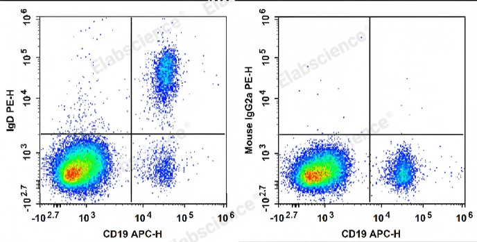
Fig. 8 Detection of memory B cells in human peripheral blood lymphocytes. Human peripheral blood lymphocytes are stained with APC Anti-Human CD19 Antibody and PE Anti-Human IgD Antibody (Left). Lymphocytes are stained with APC Anti-Human CD19 Antibody and PE Mouse IgG2a, κ Isotype Control (Right). The memory B cells exhibit the phenotype of CD19+CD27+IgD+. (The data are provided by Elabscience.)
Elabscience® Quick Overview of Popular Products:
Table 5. Reagents for memory B cell research
|
Product Name |
Cat. No. |
|
Purified Anti-Human CD27 Antibody[O323] |
E-AB-F11400P |
|
Elab Fluor® 647 Anti-Human/Monkey CD27 Antibody[O323] |
E-AB-F1140M |
|
PE Anti-Human/Monkey CD19 Antibody[CB19] |
E-AB-F1004D |
|
APC Anti-Human CD19 Antibody[HI19a] |
E-AB-F1304E |
|
FITC Anti-Human IgD Antibody[IA6-2] |
E-AB-F1171C |
|
PE Anti-Human IgD Antibody[IA6-2] |
E-AB-F1171D |
|
APC Anti-Mouse CD27 Antibody[LG.3A10] |
AN00322E |
|
Elab Fluor® Violet 450 Anti-Mouse IgD Antibody[11-26c.2a] |
E-AB-F1189Q |
|
APC Anti-Mouse IgD Antibody[11-26c.2a] |
E-AB-F1189E |
|
PE Anti-Mouse CD19 Antibody[1D3] |
E-AB-F0986D |
|
APC Anti-Mouse CD24 Antibody[M1/69] |
E-AB-F1179E |
|
PE/Cyanine7 Anti-Human CD38 Antibody[HIT2] |
E-AB-F1058H |
|
Elab Fluor® 700 Anti-Human/Monkey CD95/Fas Antibody[DX2] |
E-AB-F1168M1 |
|
Cell Staining Buffer |
E-CK-A107 |
|
Cell Stimulation and Protein Transport Inhibitor Kit |
E-CK-A091 |
|
Intracellular Fixation/Permeabilization Buffer Kit |
E-CK-A109 |
06 Plasma Cells as Specialized Antibody-Secreting Effector Cells
Plasma cells (PCs) represent the terminal differentiation state of B lymphocytes and serve as the primary effector arm of humoral immunity through sustained, high-volume plasma cell antibody secretion of high-affinity immunoglobulins. Unlike memory B cell (MBC) precursors, which remain quiescent until secondary antigen exposure, PCs are irreversibly committed to robust immunoglobulin synthesis and release, enabling rapid serological defense against invasive pathogens. This extreme functional specialization stems from comprehensive transcriptional reprogramming, metabolic remodeling, and ultrastructural intracellular reorganization, which convert naive or memory B cells into professional antibody manufacturing units. Long-lived plasma cells maintain stable, persistent antibody titers and form a central pillar of enduring immunological memory, with protective humoral responses detectable decades after infection or vaccination[40].
Terminal B cell differentiation toward the plasma cell lineage is orchestrated by a tightly controlled transcriptional cascade. The master lineage-specifying transcription factor Blimp-1, encoded by the PRDM1 gene, actively suppresses the canonical B cell transcriptional program governed by Pax5 and Bcl-6. Blimp-1 induction drives a sweeping transcriptional shift: it silences genes responsible for BCR signaling and antigen presentation while upregulating gene modules supporting enhanced protein synthesis and secretory machinery assembly. Concurrently, X-box binding protein 1 (XBP1) is markedly upregulated to expand the endoplasmic reticulum (ER) and construct the secretory infrastructure required for high-throughput antibody production. Recent mechanistic investigations further reveal that the transcription factor Hhex cooperates with the corepressor Tle3 to balance memory B cell and plasma cell fate decisions, restricting terminal differentiation to germinal center clones with optimized antigen affinity. This multilayered transcriptional circuitry links plasma cell formation directly to germinal center affinity maturation, ultimately elevating the overall quality of systemic antibody repertoires[40,41].
PCs undergo extensive metabolic rewiring to meet their exceptional biosynthetic demands. Elevated oxidative phosphorylation and glycolytic flux supply sufficient ATP to support immunoglobulin folding and secretion. The unfolded protein response (UPR) is constitutively activated in PCs to mitigate ER stress induced by heavy immunoglobulin load and prevent ER-triggered apoptosis. This distinct metabolic signature distinguishes PCs from naive and memory B cells, whose metabolic pathways shift dynamically according to activation status. Bone marrow-resident long-lived PCs depend on niche-derived trophic signals, including IL-6 and TNF-α secreted by stromal cells and eosinophils within the bone marrow microenvironment. Quantification of stromal TNF-α levels that sustain PC survival is commonly performed using tnf alpha elisa, a standard in vitro bioassay for measuring proinflammatory cytokine output within supportive tissue niches. These survival signals preserve the long-lived antibody-secreting compartment that sustains steady-state protection against previously encountered pathogens[40,41].
Plasma cells display substantial heterogeneity in lifespan and tissue residency. Short-lived PCs predominantly emerge in extrafollicular regions during the early immune response and mediate transient, immediate antibody protection. By contrast, long-lived PCs migrate to specialized survival niches, most notably the bone marrow, with secondary reservoirs in the spleen and mucosal barriers, where they can persist for the entirety of the host’s lifespan. PC trafficking to these supportive microenvironments relies on the chemokine receptor CXCR4, which binds stromal CXCL12 expressed within bone marrow niches. Such tissue partitioning sustains persistent circulating high-affinity antibodies and establishes an immediate frontline defense against pathogen re-challenge. PCs carry a unique surface phenotypic signature that simplifies their isolation and functional profiling: they lose surface B cell receptors and MHC-II while abundantly expressing CD138 (syndecan-1). Moreover, bidirectional crosstalk between PCs and regulatory B cell subsets calibrates systemic immune equilibrium. Quantifying anti-inflammatory IL-10 derived from Bregs via IL-10 elisa enables researchers to delineate how IL-10 signaling constrains pathological PC expansion and autoantibody overproduction in inflammatory and autoimmune settings[42,43].
Plasma cell biology holds profound translational significance for both protective vaccination and human immune-mediated disease. In vaccine development, robust long-lived PC formation serves as a reliable biomarker of durable immune protection. For example, SARS-CoV-2 mRNA vaccines elicit sustained germinal center activity that generates high-affinity PCs capable of secreting broadly neutralizing antibodies targeting divergent viral variants. Conversely, dysregulated PC function underpins the pathogenesis of numerous autoimmune disorders such as systemic lupus erythematosus, in which autoantibody-producing PCs drive progressive tissue damage. Malignant transformation of plasma cells additionally causes multiple myeloma, a hematological neoplasm defined by unchecked clonal proliferation of antibody-secreting PCs within the bone marrow. Deciphering the molecular circuits governing PC differentiation, niche retention and secretory effector function is therefore critical for developing novel therapeutics that enhance vaccine potency and treat antibody-driven autoimmune and malignant conditions[44].
In summary, plasma cells are highly specialized terminal effector cells that form the core structural foundation of humoral immunity via robust plasma cell antibody secretion of high-affinity immunoglobulins. Their developmental trajectory is governed by a rigid transcriptional program centered on Blimp-1 and XBP1, accompanied by widespread metabolic and structural adaptations that sustain continuous high-volume antibody production. Colonization of tissue-resident survival niches by long-lived PCs sustains lifelong immunological memory and constant systemic protection against recurrent pathogens. Ongoing research continues to dissect the intricate signaling crosstalk between PCs and their local microenvironment, uncovering new mechanistic insights into humoral response regulation and identifying actionable therapeutic targets for immune disorders. Complementary quantitative immunological tools, including tnf alpha elisa and IL-10 elisa, enable precise measurement of niche and regulatory cytokines, refining our ability to evaluate PC function in preclinical models and clinical specimens alike.
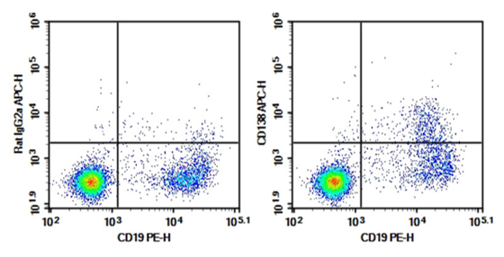
Fig. 9 Detection of plasma cells in C57/BL6 mouse bone marrow. Bone marrow lymphocytes were stained with 0.2 μg Purified Anti-Mouse CD138 Antibody[281-2] (Right) and 0.2 μg Rat IgG2a, κ Isotype Control (Left), followed by APC-conjugated Goat Anti-Rat IgG Secondary Antibody, then anti-Human CD19 PE-conjugated Monoclonal Antibody. (The data are provided by Elabscience.)
Elabscience® Quick Overview of Popular Products:
Table 6. Reagents for plasma cell research
|
Product Name |
Cat. No. |
|
Purified Anti-Mouse CD138 Antibody[281-2] |
E-AB-F1394A |
|
FITC Anti-Human CD138/Syndecan-1 Antibody[B-B4] |
E-AB-F1411C |
|
APC Anti-Human CD138/Syndecan-1 Antibody[DL-101] |
E-AB-F1154E |
|
PE/Cyanine7 Anti-Human CD19 Antibody[HI19a] |
E-AB-F1304H |
|
PerCP/Cyanine5.5 Anti-Human/Monkey CD27 Antibody[O323] |
E-AB-F1140J |
|
Purified Anti-Human CD27 Antibody[O323] |
E-AB-F11400P |
|
PE/Elab Fluor® 594 Anti-Human/Monkey CD19 Antibody[CB19] |
E-AB-F1004P |
|
PE Anti-Mouse CD19 Antibody[1D3] |
E-AB-F0986D |
|
APC Anti-Mouse CD38 Antibody[NIMR5] |
E-AB-F1193E |
|
PE Anti-Mouse CD27 Antibody[LG.3A10] |
AN00322D |
|
Cell Staining Buffer |
E-CK-A107 |
|
Cell Stimulation and Protein Transport Inhibitor Kit |
E-CK-A091 |
|
Intracellular Fixation/Permeabilization Buffer Kit |
E-CK-A109 |
References:
[1] RIPPERGER T J, BHATTACHARYA D. Transcriptional and Metabolic Control of Memory B Cells and Plasma Cells[J]. Annual Review of Immunology, 2021, 39(1): 345-368.
[2] LAM N, LEE Y, FARBER D L. A guide to adaptive immune memory[J]. Nature Reviews Immunology, 2024, 24(11): 810-829.
[3] LUO W, CONTER L, ELSNER R A, et al. IL-21R signal reprogramming cooperates with CD40 and BCR signals to select and differentiate germinal center B cells[J]. Science Immunology, 2023, 8(80).
[4] ROY S, TABIB T, DAS J, et al. NFkB cRel protein amount determines the selection of germinal center B cells 3140[J]. The Journal of Immunology, 2025, 214(Supplement_1).
[5] BELLO A, HIRTH G, VOIGT S, et al. Mechanism and regulation of secondary immunoglobulin diversification[J]. Cell Cycle, 2023, 22(18): 2070-2087.
[6] TURNER J S, O’HALLORAN J A, KALAIDINA E, et al. SARS-CoV-2 mRNA vaccines induce persistent human germinal centre responses[J]. Nature, 2021, 596(7870): 109-113.
[7] WISHNIE A J, CHWAT-EDELSTEIN T, ATTAWAY M, et al. BCR Affinity Influences T-B Interactions and B Cell Development in Secondary Lymphoid Organs[J]. Frontiers in Immunology, 2021, 12.
[8] VICTORA G D, NUSSENZWEIG M C. Germinal Centers[J]. Annual Review of Immunology, 2012, 30(1): 429-457.
[9] BETZLER A C, USHMOROV A, BRUNNER C. The transcriptional program during germinal center reaction - a close view at GC B cells, Tfh cells and Tfr cells[J]. Frontiers in Immunology, 2023, 14.
[10] MERKENSCHLAGER J, FINKIN S, RAMOS V, et al. Dynamic regulation of TFH selection during the germinal centre reaction[J]. Nature, 2021, 591(7850): 458-463.
[11] MUPPIDI J R, KLEIN U. Directing traffic in the germinal center roundabout[J]. Nature Immunology, 2020, 21(6): 599-601.
[12] DE GRUIJTER N M, JEBSON B, ROSSER E C. Cytokine production by human B cells: role in health and autoimmune disease[J]. Clinical and Experimental Immunology, 2022, 210(3): 253-262.
[13] WANG L, FU Y, CHU Y. Regulatory B Cells[M/OL]//Advances in Experimental Medicine and Biology. Springer Singapore, 2020: 87-103.
[14] HARRIS R J, WILLSMORE Z, LADDACH R, et al. Enriched circulating and tumor-resident TGF-β+regulatory B cells in patients with melanoma promote FOXP3+Tregs[J]. OncoImmunology, 2022, 11(1).
[15] MENON M, HUSSELL T, ALI SHUWA H. Regulatory B cells in respiratory health and diseases[J]. Immunological Reviews, 2021, 299(1): 61-73.
[16] LI R, LEI Y, REZK A, et al. Oxidative phosphorylation regulates B cell effector cytokines and promotes inflammation in multiple sclerosis[J]. Science Immunology, 2024, 9(95).
[17] LEE D S W, ROJAS O L, GOMMERMAN J L. B cell depletion therapies in autoimmune disease: advances and mechanistic insights[J]. Nature Reviews Drug Discovery, 2020, 20(3): 179-199.
[18] KLIEM C V, SCHAUB B. The role of regulatory B cells in immune regulation and childhood allergic asthma[J]. Molecular and Cellular Pediatrics, 2024, 11(1).
[19] ZHANG E, DING C, LI S, et al. Roles and mechanisms of tumour-infiltrating B cells in human cancer: a new force in immunotherapy[J]. Biomarker Research, 2023, 11(1).
[20] GRAVER J C, JIEMY W F, ALTULEA D H A, et al. Cytokine producing B-cells and their capability to polarize macrophages in giant cell arteritis[J]. Journal of Autoimmunity, 2023, 140: 103111.
[21] SU Y, LIU S, LONG C, et al. The cross-talk between B cells and macrophages[J]. International Immunopharmacology, 2024, 143: 113463.
[22] RADOMIR L, KRAMER M P, PERPINIAL M, et al. The survival and function of IL-10-producing regulatory B cells are negatively controlled by SLAMF5[J]. Nature Communications, 2021, 12(1).
[23] CATALÁN D, MANSILLA M A, FERRIER A, et al. Immunosuppressive Mechanisms of Regulatory B Cells[J]. Frontiers in Immunology, 2021, 12.
[24] TAN D, YIN W, GUAN F, et al. B cell-T cell interplay in immune regulation: A focus on follicular regulatory T and regulatory B cell functions[J]. Frontiers in Cell and Developmental Biology, 2022, 10.
[25] SCHWARTZ M, ZHANG Y, ROSENBLATT J D. B cell regulation of the anti-tumor response and role in carcinogenesis[J]. Journal for ImmunoTherapy of Cancer, 2016, 4(1).
[26] BRADFORD H F, MCDONNELL T C R, STEWART A, et al. Thioredoxin is a metabolic rheostat controlling regulatory B cells[J]. Nature Immunology, 2024, 25(5): 873-885.
[27] MISHINA T, MIYOSHI H, TAKEUCHI M, et al. Co-expression of regulatory B-cell markers, transforming growth factor β and interleukin-10 as a prognostic factor in diffuse large B-cell lymphoma[J]. Pathology - Research and Practice, 2024, 254: 155117.
[28] FILLATREAU S, GRAY D, ANDERTON S M. Not always the bad guys: B cells as regulators of autoimmune pathology[J]. Nature Reviews Immunology, 2008, 8(5): 391-397.
[29] VAN DER VLUGT L E P M, MLEJNEK E, OZIR‐FAZALALIKHAN A, et al. CD24hiCD27+ B cells from patients with allergic asthma have impaired regulatory activity in response to lipopolysaccharide[J]. Clinical & Experimental Allergy, 2014, 44(4): 517-528.
[30] GLAROS V, FRANCIS N, KRESLAVSKY T. The multilayered identity of B cell memory[J]. Cellular & Molecular Immunology, 2026, 23(2): 150-167.
[31] LUO W, CONTER L, ELSNER R A, et al. IL-21R signal reprogramming cooperates with CD40 and BCR signals to select and differentiate germinal center B cells[J]. Science Immunology, 2023, 8(80).
[32] LAIDLAW B J, DUAN L, XU Y, et al. The transcription factor Hhex cooperates with the corepressor Tle3 to promote memory B cell development[J]. Nature Immunology, 2020, 21(9): 1082-1093.
[33] ZHANG Y, GARCIA-IBANEZ L, ULBRICHT C, et al. Recycling of memory B cells between germinal center and lymph node subcapsular sinus supports affinity maturation to antigenic drift[J]. Nature Communications, 2022, 13(1).
[34] PIETRZAK H M, IOANNIDIS L J, HANSEN D S. IgM+ memory B cells induced in response to Plasmodium berghei adopt a germinal centre B cell phenotype during secondary infection[J]. Parasitology, 2020, 147(9): 994-998.
[35] LEE C M, OH J E. Resident Memory B Cells in Barrier Tissues[J]. Frontiers in Immunology, 2022, 13.
[36] HORIUCHI S, KOIKE T, TAKEBUCHI H, et al. SpiB regulates the expression of B-cell-related genes and increases the longevity of memory B cells[J]. Frontiers in Immunology, 2023, 14.
[37] ZHANG R, SUN J, DONG L, et al. FcμR enhances recall responses by promoting the generation of CD80+PD-L2+ memory B cells[J/OL]. International Immunology, 2026.
[38] SUGIYAMA Y, FUJIWARA M, SAKAMOTO A, et al. The immunosenescence-related factor DOCK11 is involved in secondary immune responses of B cells[J]. Immunity & Ageing, 2022, 19(1).
[39] CIABATTINI A, PASTORE G, LUCCHESI S, et al. Trajectory of Spike-Specific B Cells Elicited by Two Doses of BNT162b2 mRNA Vaccine[J]. Cells, 2023, 12(13): 1706.
[40] RIPPERGER T J, BHATTACHARYA D. Transcriptional and Metabolic Control of Memory B Cells and Plasma Cells[J]. Annual Review of Immunology, 2021, 39(1): 345-368.
[41] LAIDLAW B J, DUAN L, XU Y, et al. The transcription factor Hhex cooperates with the corepressor Tle3 to promote memory B cell development[J]. Nature Immunology, 2020, 21(9): 1082-1093.
[42] CANCRO M P, TOMAYKO M M. Memory B cells and plasma cells: The differentiative continuum of humoral immunity[J]. Immunological Reviews, 2021, 303(1): 72-82.
[43] LAM N, LEE Y, FARBER D L. A guide to adaptive immune memory[J]. Nature Reviews Immunology, 2024, 24(11): 810-829.
[44] INOUE T, KUROSAKI T. Memory B cells[J]. Nature Reviews Immunology, 2023, 24(1): 5-17.

