

Lysosomes are the cell's degradation centers and signaling hubs. Their functional status is closely linked to the onset and progression of many diseases. Efficient, intuitive assessment of lysosomal activity is a key research focus.
Lysosomes contain over 40 hydrolytic enzymes. Among them, proteolytic activity is a core indicator of lysosomal function, directly reflecting the ability of cathepsins and other proteases to degrade substrate proteins. Dysregulation of cathepsin function is a critical step in many diseases.
Therefore, measuring proteolytic activity effectively characterizes overall lysosomal function. Based on this strategy, Elabscience® proudly introduces the Lysosomal Activity Fluorometric Assay Kit (E-BC-F202): a stable, convenient solution for lysosomal activity assessment.
Table of Contents
1. Detection principle of the lysosomal activity assay
2. Key advantages for live-cell lysosomal imaging
3. Experimental validation of lysosomal activity changes
4. Research applications in lysosome biology and disease studies
5. Related products for comprehensive lysosomal function analysis
01 Detection principle of the lysosomal activity assay
This kit uses a red fluorescent dye-labeled BSA probe (Ex/Em: 530 nm/620 nm, bright red fluorescence). The BSA molecule is heavily labeled with red dye at multiple sites, creating strong fluorescence self-quenching. In its intact state, background fluorescence is extremely low. After endocytosis into functional lysosomes, the BSA probe is cleaved by lysosomal proteases, releasing small fragments with fluorophores, thereby relieving quenching and producing bright red fluorescence.
02 Key advantages for live-cell lysosomal imaging
2.1 Ultra-Low Background, Clear Signal
Self-quenching design ensures minimal background — fluorescence only develops upon degradation in lysosomes, providing an excellent signal-to-noise ratio.
2.2 Live-Cell Direct Imaging
Simply add the dye to live cells 1-6 hours before imaging — no complex fixation, staining, or lysis steps.
2.3 Function-Oriented Detection
Unlike conventional lysosomal dyes that rely on low-pH labeling, this probe directly reflects proteolytic activity, giving a more genuine readout of lysosomal function.
2.4 Good Compatibility
Narrow fluorescence emission spectrum allows co-staining with other proteins or dyes of different wavelengths, meeting multiplexing needs.
2.5 Stable and Reliable
Long-lasting, clear imaging with excellent photostability and chemical stability, ideal for long-term confocal microscopy observation.
03 Experimental validation of lysosomal activity changes
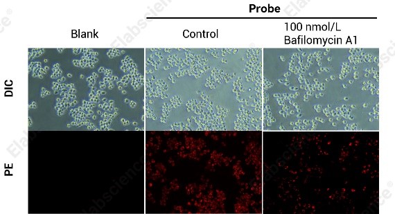
Fig. 1 Representative Results. RAW264.7 cells were seeded at 2×10⁵ cells/mL and incubated overnight at 37°C, 5% CO2. Cells were then treated under different conditions for another overnight before staining. Compared to the Control group, cells treated with 100 nmol/L Bafilomycin A1 for 24 h showed reduced lysosomal activity and weaker red fluorescence after probe staining.
Note: Actual results may vary depending on experimental conditions and instruments. The image is for reference only.
04 Research applications in lysosome biology and disease studies
4.1 Basic lysosomal function assessment
Compare proteolytic activity across cell types or treatment conditions.
4.2 Effects of compounds on lysosomal function
Screen for small molecules that enhance or inhibit lysosomal activity.
4.3 Lysosomal toxicity evaluation
As shown with Bafilomycin A1 (a V-ATPase inhibitor that disrupts acidification and inhibits function), this kit can assess compound-induced lysosomal damage.
4.4 Neurodegenerative diseases
Study lysosomal dysfunction in Alzheimer's, Parkinson's, and other disorders.
4.5 Autophagy research
Evaluate the integrity of the autophagy-lysosome pathway and determine if autophagic flux is blocked.
05 Related products for comprehensive lysosomal function analysis
Table 1. Assay kits for comprehensive lysosomal function analysis
|
Product Name |
Instrument |
Cat. No. |
|
Lysosomal Activity Fluorometric Assay Kit |
Flow Cytometry, Fluorescence Microscope |
E-BC-F202 |
|
Cathepsin B Activity Fluorometric Assay Kit |
Fluorescence Microplate Reader (Ex/Em=355 nm/460 nm) |
E-BC-F059 |
|
Cathepsin D (CatD) Activity Fluorometric Assay Kit |
Fluorescence Microplate Reader (Ex/Em=328 nm/393 nm) |
E-BC-F052 |
With one-step imaging and real functional assessment, Elabscience® Lysosomal Activity Assay Kit simplifies your lysosome research. Follow us for the latest tools and protocols!

