

Immunoprecipitation (IP) technology has a history of more than 60 years since its development. Over this period, it has evolved from concentration determination to the study of unknown protein interactions and has been combined with Western blotting, mass spectrometry, and other techniques. It plays an increasingly important role and has given rise to a wide variety of tools. Nanobody magnetic bead gel, starting from the two sources of antibody structure and solid-phase format, provides a new option with stronger affinity and lower non-specific binding.
Table of Contents
1. The development history and main application directions of immunoprecipitation
2. Key challenges in immunoprecipitation and Co-IP
3. Optimization strategies of nanobody magnetic bead gel for immunoprecipitation technology
01 The development history and main application directions of immunoprecipitation
In the 1960s, Barrett et al. first used[1] "immunoprecipitation" to measure the concentration of gamma globulins in human serum, favoring its greater precision over the electrophoresis methods of the time.
Around the same time, the concept of using antibodies to capture target proteins from solution began to take shape and gradually evolved into a "scaled-down" version of traditional column affinity chromatography: rather than slowly passing samples through a column, capture was performed in a batch-wise manner using agarose resin in a centrifuge tube, making protein purification feasible for small volumes and multiple samples.
Today, immunoprecipitation (IP) and co-immunoprecipitation (Co-IP) have long been established as one of the most fundamental and frequently used staple techniques in life science laboratories.
Their application scenarios and primary research directions have long since expanded far beyond protein purification into multiple cutting-edge frontiers:
① Identifying novel interactions: IP-MS to discover new binding partners and map interaction networks.
② Validating known interactions: Confirming physical interactions between proteins under specific physiological or pathological conditions.
③ Epigenetics and transcriptional regulation: ChIP for dissecting protein-DNA interactions, and RIP for protein-RNA interactions.
④ Post-translational modification (PTM) studies: Enriching modified proteins using specific antibodies against ubiquitination, phosphorylation, and other PTMs.
⑤ Drug discovery and target validation: Such as confirming the formation of the target protein–E3 ligase ternary complex in PROTAC technology.
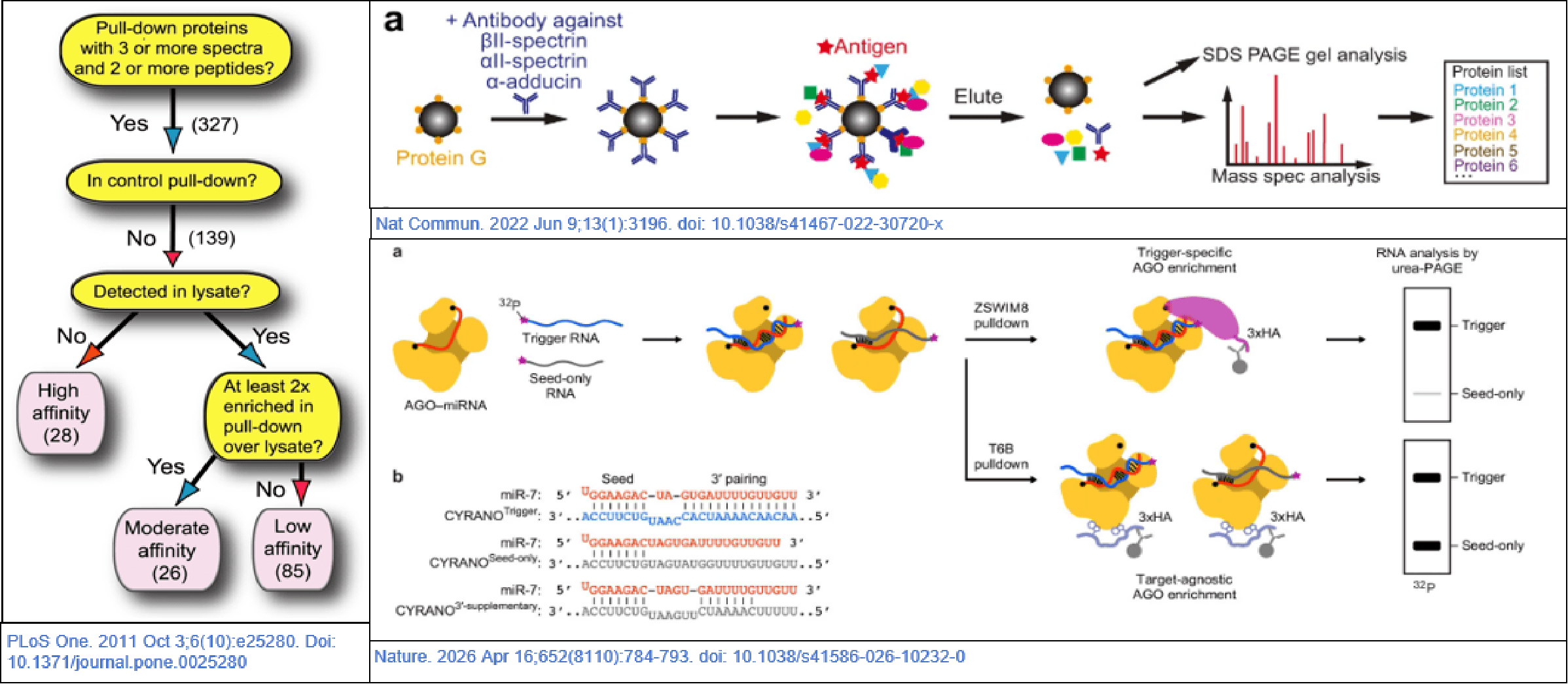
Fig. 1 Applications of IP/Co-IP across various fields[2-4].
02 Key challenges in immunoprecipitation and Co-IP
Contemporary experimental requirements are increasingly precise, which has consequently raised the standards for IP technology. At present, despite the maturity of IP technology, the failure rate of Co-IP in practical applications remains notably high. The difficulties encountered are typically not due to "intricate principles," but rather stem from several recurrent, specific issues.
Heavy and light chain interference. During denaturing elution, conventional IgG antibodies release both their heavy (~55 kDa) and light (~25 kDa) chains. When the IP primary antibody and the WB primary antibody are derived from the same species, the WB secondary antibody will simultaneously recognize the heavy and light chains of the IP primary antibody, as well as the WB primary antibody. Consequently, heavy and light chain bands will appear directly adjacent to the target protein signal. If the target protein happens to fall within this molecular weight range, the masking of its signal is almost inevitable.
High background and excessive non-specific bands. Non-specific adsorption between the antibody Fc region and the surfaces of magnetic beads/agarose will co-precipitate a large amount of unrelated proteins. This results in a multitude of non-specific bands in Western blot (WB) results and the introduction of background proteins in mass spectrometry (MS) analysis.
Insufficient capture efficiency for low-abundance or weak interactions. The limited affinity of conventional antibodies, coupled with the potential dissociation of complexes during overnight incubation, makes signal loss highly likely for low-expression target proteins or weakly interacting proteins.
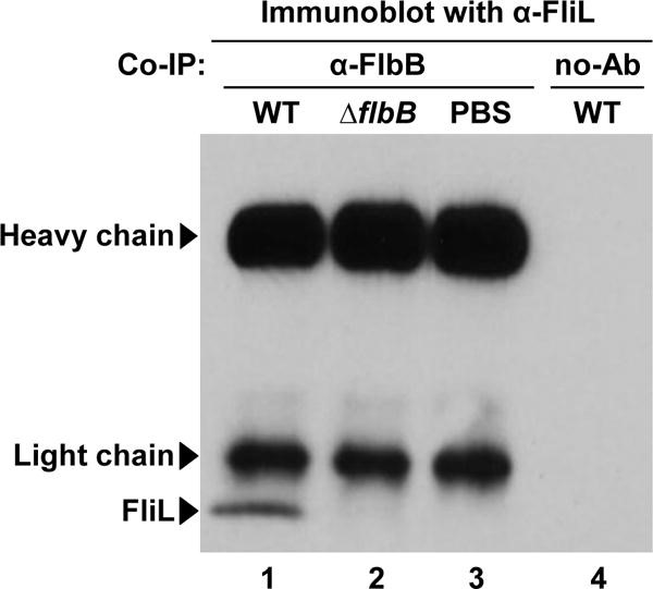
Fig. 2 Co-IP WB results showing heavy and light chain interference[5].
03 Optimization strategies of nanobody magnetic bead gel for immunoprecipitation technology
Regarding heavy and light chain interference, it achieves a "structural elimination." Nanobodies are alpaca-derived single-domain antibodies (VHH) that inherently lack light chains and the Fc region, and possess a lower molecular weight. Moreover, as they are not derived from conventional host species (rabbit, rat, or mouse), elution does not produce the typical 55 kDa and 25 kDa bands associated with conventional IgG. Following IP, samples can be directly subjected to WB, and the target protein signal will appear exactly at its expected position, completely resolving the issue of heavy and light chain interference.
Regarding high background, its single-domain structure similarly reduces the "sites" for non-specific adsorption at the source. The absence of the Fc region, the smaller size, and the limited hydrophobic surface area of nanobodies lead to reduced non-specific binding with magnetic beads. Combined with the washing efficiency of the magnetic bead system, background signals can be further minimized.
Regarding low-abundance and weak interactions, the affinity of nanobodies can reach the picomolar (pM) level, yielding a capture efficiency for tagged or target proteins that far exceeds that of conventional antibodies. The small size and high specific surface area of the magnetic beads further enhance the effective binding area, ensuring that low-abundance complexes are stably captured.
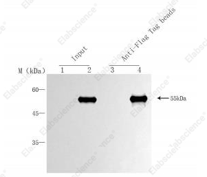
Fig. 3 Representative results of the Anti-Flag Nanobody Immunomagnetic Beads (EA-IP-001MN).
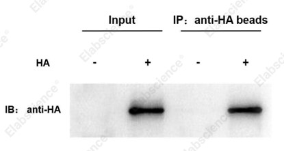
Fig. 4 Representative results of the Anti-HA Nanobody Immunomagnetic Beads (EA-IP-002MN).
Nanobodies offer stronger affinity and reduced non-specific binding. Elabscience® has launched nanobody magnetic bead gels for common tags including GFP, mCherry, MBP, Flag, HA, and V5. These products help Co-IP experiments move from “being feasible” to “being clean, fast, and reliable,” enabling better experimental results.
References:
[1] Barrett B, Wood P A, Volwiler W. Quantitation of gamma globulins in human serum by immunoprecipitation[J]. The Journal of Laboratory and Clinical Medicine, 1960, 55(4): 605-615.
[2] Kozubowski L, Thompson J W, Cardenas M E, et al. Association of calcineurin with the COPI protein Sec28 and the COPII protein Sec13 revealed by quantitative proteomics[J]. PLoS One, 2011, 6(10): e25280.
[3] Zhou R, Han B, Nowak R, et al. Proteomic and functional analyses of the periodic membrane skeleton in neurons[J]. Nature Communications, 2022, 13(1): 3196.
[4] Farnung J, Slobodyanyuk E, Wang P Y, et al. The E3 ubiquitin ligase mechanism specifying targeted microRNA degradation[J]. Nature, 2026, 652(8110): 784-793.
[5] Moon K H, Zhao X, Manne A, et al. Spirochetes flagellar collar protein FlbB has astounding effects in orientation of periplasmic flagella, bacterial shape, motility, and assembly of motors in Borrelia burgdorferi[J]. Molecular Microbiology, 2016, 102(2): 336-348.

