In biological research, we sometimes encounter situations where apoptosis detection is required in cells that carry fluorescent labels, such as EGFP. EGFP (Enhanced Green Fluorescent Protein) is one of the most commonly used cell labeling proteins, emitting green fluorescence when excited by blue light (488 nm laser). The GFP gene is typically introduced into target cells via transfection to label specific cell types, observe protein expression, or monitor gene expression activity.
Annexin V is a widely used method for apoptosis detection. Based on its ability to bind to phosphatidylserine (PS) under specific conditions, it can identify cells that have undergone PS externalization to the outer leaflet of the plasma membrane due to apoptosis. With the additional use of a nucleic acid dye such as PI, it is possible to further distinguish between early apoptotic cells and late apoptotic (including secondary necrotic) cells.
Table of Contents
1. Spectral interference between EGFP and Annexin V-FITC
2. Choosing appropriate Annexin V fluorophores for EGFP-expressing cells
3. Selecting compatible nucleic acid dyes for EGFP-expressing cells
4. Recommended staining protocols based on flow cytometer configuration
01 Spectral interference between EGFP and Annexin V-FITC
How can the Annexin V method be used to detect apoptosis in EGFP-expressing cells? Annexin V is typically detected via flow cytometry using a conjugated fluorophore, the most common being FITC (green fluorescence). However, the excitation and emission spectra of FITC and EGFP are highly overlapping, meaning they are detected in the same channel on a conventional fluorescence flow cytometer (as shown in Fig. 1). Therefore, when cells are labeled with EGFP, it is no longer possible to use FITC-labeled Annexin V for apoptosis detection.
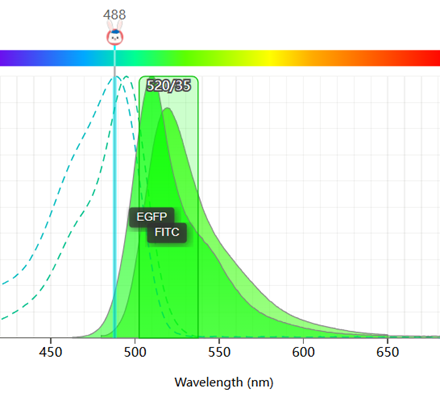
Fig. 1 Spectral overlap between FITC and EGFP.
02 Choosing appropriate Annexin V fluorophores for EGFP-expressing cells
From the above, it can be concluded that when detecting apoptosis in EGFP-expressing cells, the spectral conflict with the fluorophore used to label Annexin V must first be avoided. In this context, using APC-labeled Annexin V is one of the ideal choices. The excitation and emission spectra of APC and EGFP are significantly different, and their detection channels are far apart, resulting in little to no cross-interference (as shown in Fig. 2). However, it should be noted that APC requires excitation using a 638 nm or 640 nm red laser, so it is necessary to confirm that the flow cytometer is equipped with the corresponding laser before use.
%20and%20APC%20(right)_.png)
Fig. 2 Excitation and emission spectra of EGFP (left) and APC (right).
When the flow cytometer is not equipped with an excitation laser suitable for APC, PE can be used as the labeling fluorophore for Annexin V. However, the emission spectrum of EGFP has some spectral spillover into the detection channel of PE (as shown in Fig. 3). Therefore, it is necessary to compare the results from control cells (the same cell type without EGFP) and cells that are not stained with Annexin V (containing only EGFP fluorescence) to perform compensation adjustment, thereby obtaining more accurate results.
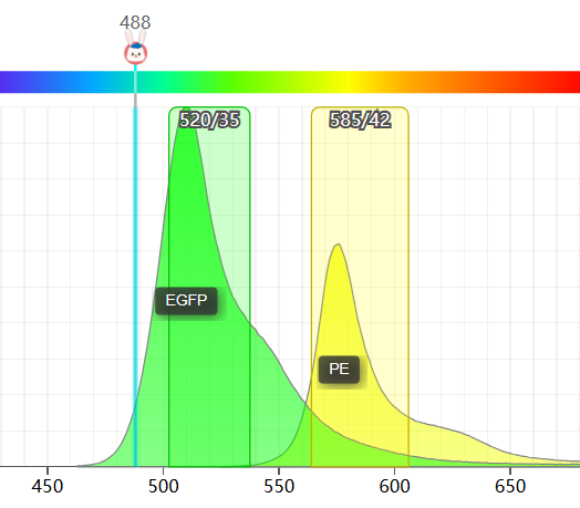
Fig. 3 Illustration of the spectral overlap between EGFP and PE fluorescence emission spectra.
03 Selecting compatible nucleic acid dyes for EGFP-expressing cells
Furthermore, when using nucleic acid dyes to analyze early and late apoptosis, since the emission spectra of EGFP and PI also have significant overlap, it is recommended to use other types of nucleic acid dyes. The ideal choice is to use DAPI as the nucleic acid dye, which ensures no interference with EGFP. However, DAPI requires excitation with a violet laser (405 nm), so instrument compatibility must be confirmed. If the corresponding laser is not available, 7-AAD can be used as the nucleic acid dye. The degree of spectral overlap between 7-AAD and EGFP is smaller than that with PI (as shown in Fig. 4), but single-stained EGFP samples are still required for compensation adjustment.
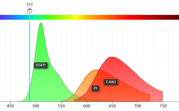
Fig. 4 Illustration of the spectral overlap between EGFP and the fluorescence emission spectra of PI and 7-AAD.
04 Recommended staining protocols based on flow cytometer configuration
For apoptosis detection using Annexin V in EGFP-expressing cells, different experimental protocols can be selected based on the configuration of the flow cytometer.
(1) If the instrument is equipped with 488 nm, 405 nm, and 640 nm lasers, the Annexin V-APC/DAPI Apoptosis Kit (E-CK-A258) can be used for staining. No compensation adjustment is required.
(2) If the instrument is equipped with 488 nm and 640 nm lasers, the Annexin V-APC/7-AAD Apoptosis Kit (E-CK-A218) can be used for staining. In this case, an EGFP single-stained sample is required to adjust compensation for the 7-AAD detection channel.
(3) If the instrument is equipped with only a 488 nm laser, the Annexin V-PE/7-AAD Apoptosis Kit (E-CK-A216) can be used for staining. In this case, an EGFP single-stained sample is mandatory to adjust compensation for both the PE and 7-AAD detection channels.

