Cytotoxicity assessment is a cornerstone of in vitro toxicology, drug development, and biomedical research. Among the most widely employed methods, the MTT (3-(4,5-dimethylthiazol-2-yl)-2,5-diphenyltetrazolium bromide) assay and the lactate dehydrogenase (LDH) release assay represent two mechanistically distinct approaches for evaluating cell viability and cell death. The MTT assay quantifies mitochondrial metabolic activity as a surrogate for viable cell number, while the LDH release assay measures plasma membrane integrity by detecting the leakage of cytoplasmic LDH into the extracellular milieu. This review provides a comprehensive comparative analysis of these two assays, examining their respective principles, methodological considerations, sensitivity profiles, and limitations. We further discuss the utility of a dual assay strategy combining MTT and LDH measurements for robust cytotoxicity validation, the capacity of LDH leakage assays to differentiate between apoptotic and necrotic cell death, and the impact of serum and media composition on LDH background levels. Finally, we contextualize these assays within the broader landscape of cell death detection methodologies, including annexin V binding and TUNEL assays.
Table of Contents
1. MTT assay as a readout of mitochondrial metabolic activity
2. LDH release assay as a marker of membrane integrity damage
3. Comparative sensitivity of MTT vs LDH in cytotoxicity screening
4. Dual assay strategy combining MTT and LDH for cytotoxicity validation
5. LDH leakage vs apoptosis vs necrosis differentiation
6. Serum and media composition effects on LDH background levels
01 MTT assay as a readout of mitochondrial metabolic activity
MTT assay provides a rapid colorimetric endpoint for measuring cellular growth and survival in microtiter plates.Viable cells convert water-soluble yellow MTT into an insoluble purple formazan via reductase activity; after solubilization (with DMSO), formazan absorbance is measured near 570 nm and is proportional to metabolically active cell number under linear ranges[1,2]. Because formazan formation depends on functional mitochondrial dehydrogenases yet also on non-mitochondrial reductase systems, MTT is best defined as a readout of cellular reducing capacity that serves as a practical surrogate for cell viability assay when assay conditions are controlled[1].
Key operational variables include pH sensitivity of the formazan spectrum and the need for high-purity (e.g., Uvasol-grade) DMSO to avoid acidic impurities that skew absorbance[1]. Linearity breaks down at high cell densities owing to substrate depletion and medium acidification; optimal MTT concentration (commonly 0.5–1 mg/mL) and incubation time require empirical validation per cell type[1,2]. Proliferating cells generate more formazan per cell than quiescent cells, so the signal embeds proliferative status and metabolic vigor, not merely head-count.
The 96-well format supports standardization for adherent and suspension cultures, with acceptable between-assay variation and advantages over clonogenic assays in throughput and applicability to non-colony-forming lines[1,2]. Major caveats remain: compounds that intercept electron transport, alter NAD(P) or glucose availability, or chemically react with MTT or formazan can decouple the signal from true cell number, producing false-positive or false-negative interpretations[1,3]. Finally, MTT cannot reliably separate cytostasis from cytotoxicity, a critical limitation when the question is explicitly "killing vs growth inhibition"[3].
02 LDH release assay as a marker of membrane integrity damage
The LDH release assay quantifies cell death via the release of lactate dehydrogenase (LDH), a stable cytoplasmic enzyme constitutively expressed in mammalian cells, into the extracellular milieu following plasma membrane disruption[5,6]. This release occurs during necrosis and late-stage apoptosis with secondary necrosis[5-7]. LDH activity is measured through a coupled reaction where lactate oxidation reduces NAD⁺ to NADH, which subsequently reduces a tetrazolium salt (e.g., INT) to a water-soluble red formazan via an electron coupler (e.g., MPMS); absorbance at 490–492 nm correlates directly with LDH activity and the extent of cell damage[5,6,8].
This assay offers distinct advantages over metabolic methods. By measuring a definitive marker of membrane rupture rather than a surrogate metabolic parameter, LDH provides superior specificity for cell death events compared to assays like MTT, which can be confounded by transient redox changes[7,9]. The water-soluble formazan eliminates the need for a solubilization step, streamlining the workflow[5,6]. Furthermore, its compatibility with 96- and 384-well formats facilitates high-throughput screening[4,5].
Standard protocols require critical controls: spontaneous release (baseline leakage), medium background (serum-derived LDH), and maximum release (total LDH). Cytotoxicity is calculated as: % Cytotoxicity = (Experimental – Spontaneous) / (Maximum – Spontaneous) × 100[5,6]. A major caveat is the potential for direct chemical inactivation of LDH by test compounds. Certain toxicants (e.g., para-aminophenol, menadione) can degrade LDH activity in a concentration-dependent manner, yielding false-negative results despite cell death[4]. While antioxidants like GSH may partially preserve activity, they risk confounding outcomes. Investigators should verify assay compatibility by spiking purified LDH into compound-containing media to confirm recovery[4].
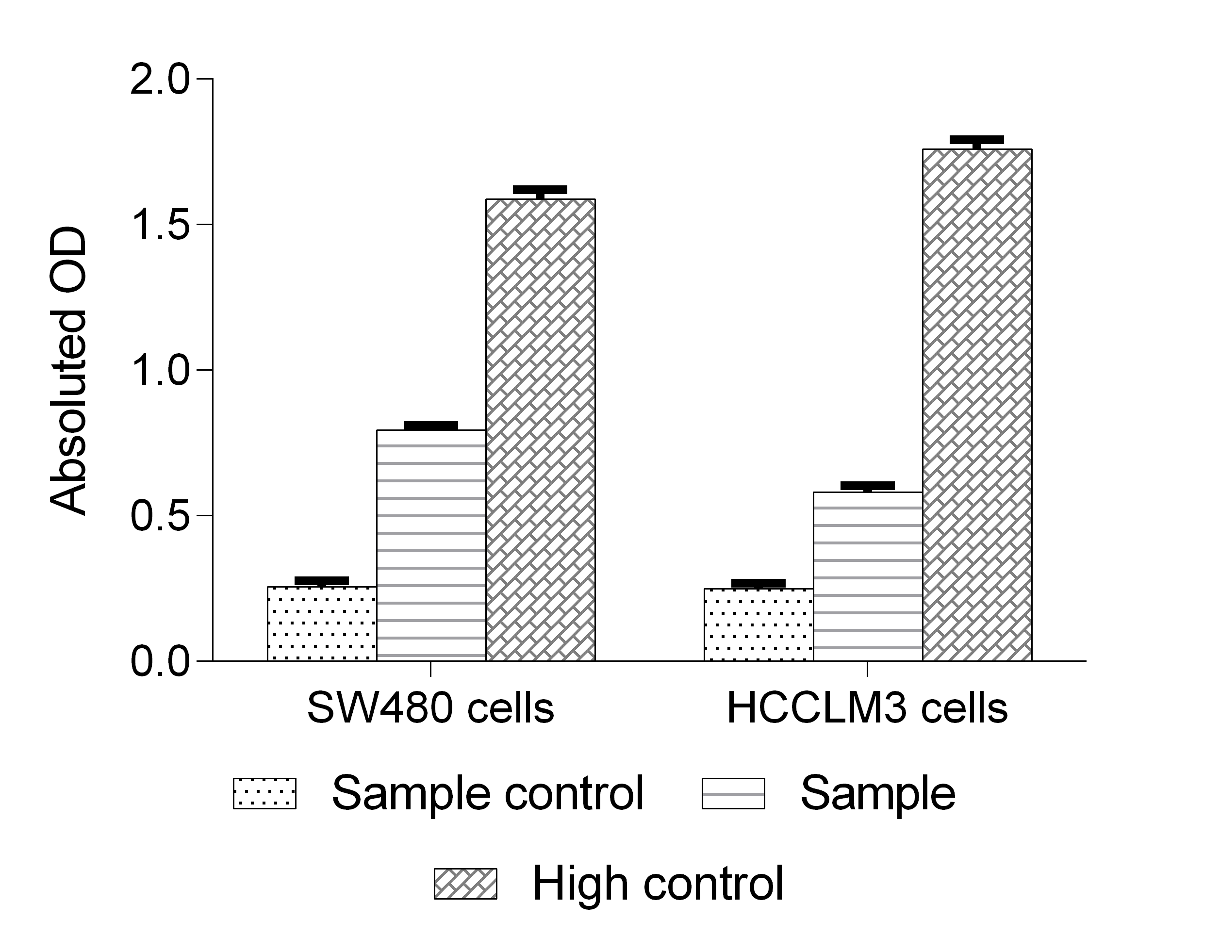
Fig. 1 SW480 and HCCLM3 cells were treated with or without cisplatin for 24 h, and then changes in LDH activity were detected. (The data are provided by Elabscience)
03 Comparative sensitivity of MTT vs LDH in cytotoxicity screening
Comparative analyses reveal fundamental differences between MTT and LDH assays regarding cellular endpoints and cytotoxicity screening sensitivity. MTT activity decreased at H₂O₂ concentrations well below those required to induce trypan blue uptake or LDH release in HEK 293T and Raw264.7 cells, indicating MTT detects early metabolic perturbations preceding irreversible membrane damage[7]. Conversely, LDH assays exhibited threshold behavior, requiring substantial membrane disruption before signal increases. Cell-type dependency is proven critical; CHO cells with elevated GSH and NADPH levels maintained unchanged MTT reduction across all H₂O₂ concentrations despite significant cell death, rendering MTT unreliable in cells with robust antioxidant defenses[7].
The MTT assay cannot distinguish cytotoxicity from growth inhibition. Chan et al. showed MTS (water-soluble MTT analog) produced identical dose-response curves for bortezomib (inducing cell death) and PLX4720 (causing cytostatic arrest) in A375 melanoma cells, despite morphological confirmation of differential killing[3]. In contrast, properly configured LDH assays successfully differentiated these mechanisms[3].
Chan et al. addressed a critical LDH limitation regarding growth-arresting compounds[3]. Standard protocols using vehicle-control lysis for maximum release calculations underestimate cytotoxicity when treatments inhibit proliferation, as control wells accumulate more cells and total LDH. Implementing condition-specific controls (lysing replicate wells for each drug concentration) corrected this bias, accurately quantifying cell death (36% at 10 µM PLX4720) versus standard protocol underestimation (<10%)[3].
In summary, MTT assays offer superior sensitivity for early metabolic stress but lack cell death specificity, while LDH provides definitive cell death markers at the expense of early detection sensitivity[7,9]. Assay selection should align with research objectives: MTT for metabolic stress and growth inhibition, LDH for quantifying actual cell death events.
04 Dual assay strategy combining MTT and LDH for cytotoxicity validation
Given the complementary nature of MTT (metabolic activity) and LDH (membrane integrity) assays, dual-assay strategies are widely recommended for rigorous cytotoxicity assessment[7,9]. Combining these orthogonal readouts captures both early metabolic dysfunction and late-stage cell death, providing a holistic view of cellular health.
Evidence supports this approach, as the assays can diverge under specific conditions. Decreased MTT without increased LDH release, indicating metabolic compromise without cell death[7]. Conversely, compound-mediated LDH inactivation may cause false negatives, detectable via reduced MTT viability[4]. Concordance between assays strengthens cytotoxicity conclusions, while discordance warrants mechanistic investigation.
A cost-effective dual-MTT method using a single well has been developed: supernatant is collected for an LDH assay (using an MTT-based substrate) before MTT is added directly to the remaining medium[8]. This approach reduces costs by approximately 20-fold compared with commercial kits while maintaining comparable sensitivity, as validated in astrocytes exposed to tert-butyl hydroperoxide and neurons exposed to glutamate[8].
The necessity of multi-assay redundancy in nicotinamide N-methyltransferase (NNMT) neuroprotection studies has been demonstrated[9]. In NNMT-expressing SH-SY5Y cells, toxins reduced ATP but also decreased LDH release; reliance on a single assay would have yielded erroneous conclusions about NNMT’s effects[9]. This underscores the risk of single-assay data and advocates for the use of two or more mechanistically distinct assays.
Assay selection should consider: (1) principles (metabolic vs. membrane integrity); (2) compound interference; (3) expected cell death mechanism (apoptosis vs. necrosis); (4) experimental time course relative to endpoint kinetics[7,9]. While MTT is typically endpoint-based, the LDH assay allows real-time kinetic monitoring via serial supernatant collection[5,8].
05 LDH leakage vs apoptosis vs necrosis differentiation
A critical consideration in any cytotoxicity assay is selecting a cell death assay panel to distinguish apoptosis from necrosis. While the LDH release assay (often employed as a functional necrosis assay) detects late-stage membrane disruption, it cannot differentiate them alone[5-7], necessitating complementary methods such as an apoptosis assay.
During apoptosis, phosphatidylserine (PS) translocates to the outer membrane leaflet[10,12]. Annexin V detects this early event[10,12,13]. Combined with PI or 7-AAD, annexin V staining discriminates four populations: viable (annexin V⁻/PI⁻), early apoptotic (annexin V⁺/PI⁻), late apoptotic/secondary necrotic (annexin V⁺/PI⁺), and primary necrotic (annexin V⁻/PI⁺) cells[10,13]. This provides greater insight than LDH release or MTT assays alone.
Specific apoptosis assay techniques, including annexin V and TUNEL assay kit protocols, provide sensitive detection[10,13]. The TUNEL method labels DNA breaks to detect fragmentation. Steensma et al. found annexin V detected apoptosis earlier than TUNEL, reflecting PS externalization preceding DNA damage[10].
LDH release kinetics differ by death mode. Apoptotic cells retain membrane integrity initially, releasing LDH only during secondary necrosis[5-7]. Early LDH assays may underestimate cytotoxicity, while late measurements cannot distinguish apoptotic from primary necrotic LDH release[3,5,10].
Real-time cell death assay formats enable kinetic monitoring. A bioluminescent annexin V assay using NanoBiT technology has been developed for continuous measurement of PS exposure[12]. Incorporating a cell-impermeant dye allows real-time discrimination between apoptosis (lag between PS exposure and permeability) and primary necrosis (concurrent increases)[12], offering resolution beyond endpoint LDH or MTT assays[12].
A tiered approach is recommended: initial LDH assay (or MTT and LDH dual strategy) for overall cytotoxicity, followed by annexin V/PI flow cytometry for apoptotic vs. necrotic contributions, and TUNEL assay kit or caspase-3/7 measurements for confirmation[10,13]. The TUNEL assay kit applies to both flow cytometry and microscopy[13].
%20or%20without%20(Right)%205%20%CE%BCM%20Camptothecin%20for%204%20h_.png)
Fig. 2 Jurkat cells were cultured with (Left) or without (Right) 5 μM Camptothecin for 4 h. Annexin V-FITC single-positive cells were early apoptotic cells, Annexin V-FITC and PI double-positive cells were necrotic or late apoptotic cells, and PI single-positive cells were naked nuclei. (The data are provided by Elabscience)
06 Serum and media composition effects on LDH background levels
Serum-derived LDH significantly impacts background absorbance in the LDH release assay, a critical factor often overlooked. Fetal bovine serum (FBS), standard in mammalian cell culture media, contains substantial endogenous LDH that elevates baseline absorbance and compresses the assay’s dynamic range[9,11].
FBS increases LDH assay absorbance dose-dependently at 5%, 10%, and 15% (v/v)[9]. At 15% FBS, background absorbance reached ~0.46 units, reducing the relative dynamic range (rDR) to 46.3% versus serum-free conditions[9]. This attenuation risks false-negative results for biologically relevant LDH release, especially with mildly cytotoxic compounds[9].
Mitigation strategies exist. Reducing FBS concentration during assays enhances dynamic range; lowering FBS from 10% to 1% increased Triton X-100-induced LDH detection by >300% in NIH-3T3 cells. However, serum reduction may compromise viability or drug response in cell lines requiring high serum[9].
Heat inactivation decreases FBS LDH content by ~50% via enzyme denaturation. Substituting heat-inactivated FBS (HI-FBS) raised rDR from 36.8% to 70.5%[9]. Phenol red, a pH indicator, contributes minimally to background absorbance. Using phenol red-free medium during assays is recommended, as it lacks essential biological functions and does not affect viability[9].
Because chemically defined, serum-free media lack serum, they eliminate intrinsic LDH activity and thus entirely remove serum background[9]. Supplements like B-27 provide nutrients or growth factors without FBS-associated LDH contamination[9]. Methodologically, all LDH assays require medium-only controls (no cells) to establish baseline absorbance from serum or media components, which should be subtracted from experimental readings[5,6,9]. When comparing conditions, ensure identical medium composition (especially serum content) to prevent confounding background LDH differences[9].
Quick Overview of Popular Products:
Table 1. Reagents for cell viability assessment and cell death
|
Cat. No. |
Product Name |
|
E-CK-A341 |
MTT Cell Proliferation and Cytotoxicity Assay Kit |
|
E-CK-A211 |
Annexin V-FITC/PI Apoptosis Kit |
|
E-CK-A320 |
One-step TUNEL In Situ Apoptosis Kit (Green, FITC) |
|
E-CK-A331 |
TUNEL In Situ Apoptosis Kit (HRP-DAB Method) |
|
E-CK-A383 |
Caspase 3/7 Activity Assay Kit(Colorimetric Method) |
|
E-CK-A483 |
Caspase 3/7 Activity Detection Substrate for Flow Cytometry |
|
E-CK-A481 |
Caspase 1 Activity Detection Substrate for Flow Cytometry |
|
E-BC-F200 |
Cell Viability Chemiluminescence Assay Kit |
|
E-BC-K771-M |
Lactate Dehydrogenase (LDH) Cytotoxicity Colorimetric Assay Kit |
|
E-CK-A362 |
Enhanced Cell Counting Kit 8 (WST-8/CCK8) |
References:
[1] van Meerloo J., Kaspers G.J.L., Cloos J. The MTT Tetrazolium Salt Assay Scrutinized: How to Use this Assay Reliably to Measure Metabolic Activity of Cell Cultures in vitro for the Assessment of Growth Characteristics, IC50-Values and Cell Survival. Clinical Chemistry and Laboratory Medicine, 1995. 33(11):813-818.
[2] Kumar P., Nagarajan A., Uchil P.D. Analysis of Cell Viability by the MTT Assay. Cold Spring Harbor Protocols, 2018. 2018(6):pdb.prot095505.
[3] Chan F.K.M., Moriwaki K., De Rosa M.J. A Simple Protocol for Using a LDH-Based Cytotoxicity Assay to Assess the Effects of Death and Growth Inhibition at the Same Time. PLoS ONE, 2011. 6(11):e26908.
[4] Kendig D.M., Tarloff J.B. Inactivation of Lactate Dehydrogenase by Several Chemicals: Implications for in vitro Toxicology Studies. Toxicology in Vitro, 2007. 21(1):125-132.
[5] Kaja S., Payne A.J., Naumchuk Y., Koulen P. Quantification of Lactate Dehydrogenase for Cell Viability Testing Using Cell Lines and Primary Cultured Astrocytes. Current Protocols in Toxicology, 2017. 72(1):2.26.1-2.26.10.
[6] Kumar P., Nagarajan A., Uchil P.D. Analysis of Cell Viability by the Lactate Dehydrogenase Assay. Cold Spring Harbor Protocols, 2018. 2018(6):pdb.prot095497.
[7] Han J., Kim S., Kim D., et al. Discriminative Cytotoxicity Assessment Based on Various Cellular Damages. Toxicology Letters, 2009. 184(1):13-21.
[8] Steensma D.P., Timm M., Witzig T.E. Comparison of the TUNEL, Lamin B and Annexin V Methods for the Detection of Apoptosis by Flow Cytometry. Acta Histochemica, 2002. 104(4):365-373.
[9] Parsons R.B., Brown J.O., Milani M., et al. The Effect of Foetal Bovine Serum Supplementation upon the Lactate Dehydrogenase Cytotoxicity Assay: Important Considerations for in vitro Toxicity Analysis. Toxicology in Vitro, 2015. 34:163-173.
[10] Sgonc R., Wick G. Apoptosis Detection: an Overview. Experimental Gerontology, 1998. 33(6):525-533.
[11] Duellman S.J., Zhou W., Meisenheimer P., et al. A Real-Time, Bioluminescent Annexin V Assay for the Assessment of Apoptosis. Apoptosis, 2018. 23(5-6):319-332.
[12] Ciccolini F., Harsch M., Rothen-Rutishauser B., et al. Impact of Serum in Cell Culture Media on in vitro Lactate Dehydrogenase (LDH) Release Determination. Journal of Cellular Biotechnology, 2017. 3(1):9-16.
[13] Shekhawat A.S., Patil V.R., Pawar S.V., et al. Cytotoxicity Assay. In: Springer Protocols Handbooks, 2023.

