Cell cycle refers to the whole process of continuously dividing cells from the end of one mitosis to the end of the next mitosis. The study of cell cycle is of great significance for understanding the growth and development of organisms and controlling the growth of tumors. For example, in the treatment of cancer, G0 phase cells are not sensitive to chemotherapy, and cancer cells are difficult to be effectively killed, which often becomes the source of cancer recurrence in the future. By inducing G0 phase cancer cells to enter the cell cycle and improving chemotherapy sensitivity, cancer cells can be effectively killed.
Cell cycle detection is mainly based on the ability of intracellular DNA to bind to certain fluorescent dyes (such as propidium iodide, PI). Due to the different content of DNA in each phase of the cell, the amount of fluorescent dye bound to it is also different. The fluorescence intensity detected by flow cytometry can reflect the change of cell cycle.
This method is easy to operate and only requires a simple fixed staining to detect, but the results are still not ideal. This article will take you to look at the common problems and solutions in cell cycle detection:
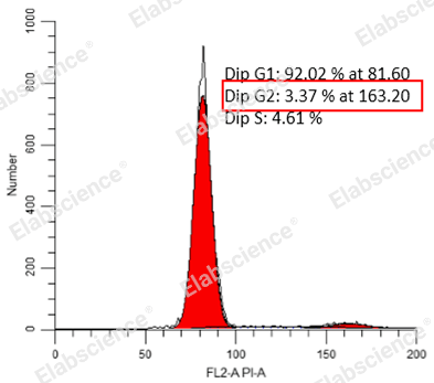
01 CV value is too large
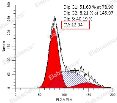
In cell cycle detection, CV Refers to the Coefficient of Variance (CV) of G0/G1 phase peak, which is an important indicator to evaluate the cycle result. The smaller the value, the better the result. The abnormal causes and corresponding solutions of excessive CV values are as follows:
|
Abnormal causes |
Solutions |
|
Interference with RNA |
Add RNase for incubation |
|
Sample loading is too fast resulting in reduced resolution |
Reduce the loading speed |
|
Instrument abnormality |
Perform instrument quality control/calibration |
|
Unreasonable data analysis |
Adjust the position of each phase and G2/G1 ratio |
|
Drug handling |
CV rises after medicated treatment, this is a normal phenomenon |
02 G2/M phase is absent
When the cycle detection is When the cycle detection is performed, it is found that the G2/M phase is missing, first of all, we must make sure that the cells we detect are able to divide and proliferate. For example, peripheral blood lymphocytes will not proliferate under normal circumstances and are in G0 phase.
In addition, if the cell culture conditions are not right or the nutrition is insufficient, resulting in slow cell growth, it will also lead to a large number of cells entering the G0 phase. At this time, it is necessary to optimize the culture conditions in time, otherwise, the cell state will become worse after a long time, until cell death.
Many cells grow at too high density, which will cause contact inhibition, which will also lead to G2/M phase loss. When cells are in contact inhibition, they can no longer proliferate, therefore, if the experimental treatment group is to promote cell growth, or cause G2/M phase arrest, it may be completely undetectable, which seriously affects the judgment of the experimental results. Therefore, when conducting cell cycle experiments, we must ensure that there is still a reasonable growth space for cells when detecting.
03 The normal somatic cell cycle test showed that the proportion of 8 or 4 ploidy was too high
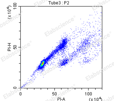
In most cases, normal mammalian somatic cells are diploid or tetraploid, and the proportion of G0/G1 phase is much higher than that of G2/M phase. When the normal somatic cell cycle detection shows 8-ploid, or the proportion of tetraploid is too high, most of them are caused by agglutinate. Therefore, PI-A/PI-H should be used to exclude agglutinate in the cell cycle experiment to get the correct results.
Product recommendation: Cell Cycle Assay Kit (DNA content detection)
For more questions related to cell cycle detection, please consult online customer service on the right side of the page ~
Elabscience® Flow Cytometry Antibodies Features (←Click to view)
Multi-color optional: up to 13 fluorescent markers, suitable for more experimental protocols
Cost-Effective: Small package,only need $15-60
Adequate inventory: conventional antibody, spot stock, delivery within 24 hours
High-quality: derived from classical cloning, the third generation of non-destructive labeling process, grouping is obvious

