Cell Function
Procedure and precautions of cell cycle detection
Source: Elabscience®Published: Jan 16,2024
When doing cell cycle detection experiments, it is necessary to explore the conditions for a long time to get stable and reliable results, which involves more factors. Elabscience introduce the principle, process and precautions of cell cycle detection experiments in detail to facilitate our customers to obtain stable and reliable ideal data.
Cell cycle refers to the activity process of cells from the end of one division to the end of the next division. The division process is divided into two stages: Cell division interphase and division phase. As shown below:
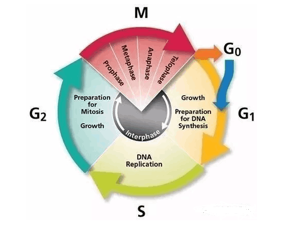
G0 phase: This phase of the cell is called dormant cells. The cell temporarily out of the division cycle and do not carry out DNA replication and division. However, these cells can be induced to restart DNA synthesis and cell division under certain conditions.
G1 phase: This phase main synthesizes RNA, ribosomes, proteins, lipids and carbohydrates.
S-phase: This phase mainly synthesizes DNA and assembles histones.
The enzymes required for DNA replication are synthesized during this period.
G2 stage: Late phase of DNA synthesis. This phase main synthesis of ATP, RNA, proteins, including microfilament tubulin.
M stage: At this time RNA synthesis is stopped, protein synthesis is reduced, and chromosomes are highly spiralized.
Principles of cell cycle detection
PI method is a classic periodic detection method. PI is Propidium iodide (Propidium), a double-stranded DNA fluorescent dye. The combination of propidium iodide and double-stranded DNA can produce fluorescence, and the fluorescence intensity is proportional to the amount of double-stranded DNA. After the DNA in the cells is stained with propyl iodide, the DNA content of the cells can be determined by Flow Cytometry, and then according to the distribution of DNA content, the cell cycle and apoptosis can be analyzed.
Normally, the G0/ G1 phase of normal cells has the DNA content of diploid cells (2N), G2/ M phase has the DNA content of tetraploid cells (4N), and the DNA content of S phase is between 2N and 4N. After the cell is fixed with ice ethanol, PI can bind to the DNA of the cell, and its fluorescence intensity directly reflects the DNA content in the cell.
Therefore, the cell cycle can be divided into G1/G0 phase, S phase and G2/M phase through the detection of intracellular DNA content, and the percentage of cells in each phase can also be obtained.
Cell cycle detection operation procedure
Collect cells: Collect cells 5~20×105 in a centrifuge tube. Centrifuge small cells (such as lymphocytes) at 400 g or centrifuge large cells (such as tumor cells) at 300 g for 5~10 min respectively, then discard the culture medium
Washing: Add 1 mL cold PBS (pre-cooling at 4ºC in advance) and wash once, then centrifuge and discard the supernatant.
Pre-fixation treatment: Use the residual solution at the bottom of the tube, flick the bottom of the tube, and re-suspend the precipitation into cell suspension to avoid cell clumping.
Cell fixation: Add cell solution to approximate 1 mL of anhydrous ethanol (pre-cooled at -20ºC), and gently blow and mix. Fix the cells at -20ºC for 1 h or overnight.
Washing: Add 1 mL cold PBS (pre-cooling at 4ºC in advance) and wash once, centrifuge 300 g for 10min, discard the supernatant.
RNA enzyme digestion: Centrifugate at 300 g for 5 min, discard the supernatant, then add 100 μL of RNase A to fully suspension cells. Put the solution into 37°C water bath for 30 min.
PI staining: Add 400 μL PI solution to cell solution and thoroughly mix, and incubated at 4°C for 30 min in dark.
Detection: Detect red fluorescence at the excitation wavelength of 488 nm by Flow Cytometry. Obtain Cells at low speed, and DNA content is analyzed by analysis software.
Precautions for cell cycle detection
1. Pay attention to the sterile environment of cultured cells.
2. Dyeing solution and other substances are certain toxic, pay attention to protection.
3. In this experiment, the amount of cells needed to be collected by detection is large, collect cells as many as possible.
4. This experiment have many cell centrifugation and resuspension procedures. When doing these operations, pay attention to slow movements to avoid excessive damage to cells.
5. When fixed cells, pre-cooled PBS must be re-suspended into anhydrous ethanol, and the order of the two cannot be reversed.
6. When cultured cells, it is appropriate to treat them in logarithmic phase to avoid experimental errors caused by insufficient number of cells collected due to few cells or excessive number of cells that start to die in large numbers.
7. Although the fixed cells can be stored for a long time before detection, in order to avoid the influence of uncontrollable factors, it is best to detect as early as possible.
Recommended -- Elabscience Cell Cycle Detection Kit (DNA content detection)
Cat. No.: E-CK-A351
Experimental result
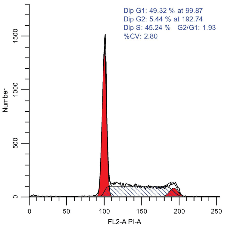
In addition to cell cycle detection kits, Elabscience has also developed a variety of cell related detection kits, covering apoptosis, cell proliferation and toxicity detection, etc., which can meet your multi-dimensional experimental research, if you have any questions, You can also click on the online consultation on the right, and our technicians will provide you with answers.

