Mitochondria are crucial hubs determining cellular fate, playing key regulatory roles in cellular physiological changes, signaling pathways, and metabolism. They are essential for regulating cell proliferation, differentiation, survival, and maintaining homeostasis. Mitochondria are involved in the development and progression of numerous diseases, including cancer, cardiovascular diseases, neurodegenerative disorders, diabetes, and immune-related conditions. The link between mitochondria and various diseases stems from their role as vital organelles for energy production; when their function declines, so does cellular activity. Research in this area has exploded over the past decade, making mitochondria a frequent focus of high-impact publications.
Mitochondria participate in and regulate processes such as growth, development, metabolism, aging, disease, and death, and are involved in metabolic processes like intracellular Ca2+ homeostasis, reactive oxygen species (ROS) production, and cytochrome C release. Detecting mitochondrial function often reflects the state of the organism, commonly through the following indicators.
Table of Contents
1. Mitochondrial ATP
2. Cell Mitochondrial Complex & Tricarboxylic Acid (TCA) Cycle
3. Reactive Oxygen Species (ROS)
4. Mitochondrial Membrane Potential (MMP, Δψm)
01 Mitochondrial ATP
Mitochondria are the primary sites where cellular substances are metabolized and converted into ATP. ATP is widely regarded as the most important energy metabolite in organisms, powering life activities and participating in various biological processes. Intracellular ATP levels are susceptible to the cellular environment, such as toxic stress or hypoxia[1]. Methods for ATP detection typically include chemiluminescence, enzyme-coupled assays, and high-performance liquid chromatography (HPLC).
02 Cell Mitochondrial Complex & Tricarboxylic Acid (TCA) Cycle
The Cell Mitochondrial Complexes (I-V) are the engines of oxidative phosphorylation. Assessing their individual and collective function is critical for understanding energetic capacity and identifying defects. Similarly, measuring key enzymes and metabolites of the TCA cycle provides insights into the crucial substrate supply for these processes.
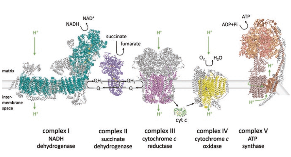
Fig. 1 The Cell Mitochondrial Complexes work together to create the proton gradient essential for ATP production[2].
03 Reactive Oxygen Species (ROS)
Under normal conditions, cells possess an antioxidant system comprising reducing substances and antioxidant enzymes, which helps reduce intracellular ROS levels. Reactive Oxygen Species (ROS) are a class of chemically reactive molecules or ions with high oxidative activity, primarily including superoxide anion, hydrogen peroxide (H2O2), and hydroxyl radicals (HO·). When organism function is impaired, the balance between the intracellular antioxidant system and ROS is disrupted, leading to increased oxygen free radicals. This can further damage mitochondrial DNA, proteins, lipid membranes, etc., resulting in functional changes such as reduced ATP synthesis and altered membrane potential[3]. Therefore, measuring ROS levels can indicate whether mitochondrial function is normal.
04 Mitochondrial Membrane Potential (MMP, Δψm)
Due to the unique ion permeability of the inner mitochondrial membrane, metabolically generated H+ creates a negative potential difference across it, typically ranging from -180 mV to -200 mV. This is known as the Mitochondrial Membrane Potential (MMP, Δψm). Changes in MMP, even minor ones, can significantly impact mitochondrial function. Abnormal MMP changes can further lead to mitochondrial diseases[4]. Monitoring MMP is a common method for assessing mitochondrial functional status.
Elabscience® Quick Overview of Popular Products
Table 1. Assay Kits for Mitochondrial Functional Research
|
Detection Focus |
Target |
Cat. No. |
|
Mitochondrial ATP |
ATP |
E-BC-F002 |
|
E-BC-F300 |
||
|
Cell Mitochondrial Complex |
Cell Mitochondrial Complex I |
E-BC-K834-M |
|
Cell Mitochondrial Complex II |
E-BC-K835-M |
|
|
Cell Mitochondrial Complex Ⅲ |
E-BC-K836-M |
|
|
Cell Mitochondrial Complex IV |
E-BC-K837-M |
|
|
Cell Mitochondrial Complex Ⅴ |
E-BC-K838-M |
|
|
TCA Cycle |
α-Ketoglutarate Dehydrogenase (α-KGDH) |
E-BC-K083-M |
|
α-Ketoglutarate (α-KG) |
E-BC-F047 |
|
|
Citrate Synthase (CS) |
E-BC-K178-M |
|
|
NAD+/NADH |
E-BC-K804-M |
|
|
Status Assessment |
Mitochondrial Membrane Potential (JC-1) |
E-CK-A301 |
|
Mitochondrial Permeability Transition Pore (mPTP) |
E-BC-F064 |
|
|
Mitochondrial Stress |
E-BC-F078 |
|
|
Mitochondrial Superoxide |
E-BC-F008 |
|
|
Reactive Oxygen Species (ROS) |
E-BC-K138-F |
|
|
E-BC-F005 |
References:
[1] Suxing, Jin, Yigang, et al. Impact of Mitochondrion-Targeting Group on the Reactivity and Cytostatic Pathway of Platinum(IV) Complexes. Inorganic Chemistry, 2018.
[2] Sousa J S, D’Imprima E, Vonck J. Mitochondrial respiratory chain complexes. Membrane Protein Complexes: Structure and Function, 2018.
[3] Yang F, Liao J, Pei R, et al. Autophagy attenuates copper-induced mitochondrial dysfunction by regulating oxidative stress in chicken hepatocytes. Chemosphere, 2018.
[4] Chen L B. Mitochondrial membrane potential in living cells. Annual Review of Cell Biology, 1988.

