

Tumor Necrosis Factor (TNF) is a master pro-inflammatory cytokine that orchestrates a complex and often paradoxical spectrum of cellular responses, ranging from essential host defense mechanisms to the pathogenesis of chronic inflammatory disorders. Its biological activity is mediated via two distinct cell surface receptors, TNFR1 and TNFR2, which initiate divergent signaling cascades that ultimately dictate a cell’s fate, specifically, whether it undergoes survival, inflammation, apoptosis, or necroptosis. Understanding these signaling pathways, the methodologies employed to detect and quantify TNF as well as its biological activity, and their extensive research applications is fundamental to modern immunology, oncology, and drug discovery and development.
This article reviews the molecular identity of TNF as a pro-inflammatory cytokine family, provides a comparative analysis of tumor necrosis factor-alpha (TNF-α) and tumor necrosis factor-beta (TNF-β), including their cellular origins and functional roles in immunity, explores TNF-mediated amplification of the cytokine cascade in systemic inflammation, describes the ELISA-based quantification of TNF-α in serum and cell culture supernatants, discusses the simultaneous measurement of TNF-α, TNF-β, and IL-6 using multiplex cytokine panels, and addresses the emerging functional reassessment of TNF-β in the contexts of immune homeostasis and disease.
Table of Contents
1. Molecular identity of TNF as a pro-inflammatory cytokines family
2. Comparative analysis of TNF-α and TNF-β: cellular sources and functional roles in immunity
3. TNF-mediated cytokine cascade amplification in systemic inflammation
4. ELISA-based quantification of TNF-α in serum and cell culture supernatants
5. Multiplex cytokine panels for simultaneous measurement of TNF-α, TNF-β, and IL-6
6. Emerging functional reassessment of TNF-β in immune homeostasis and disease contexts
01 Molecular identity of TNF as a pro-inflammatory cytokines family
TNF does not exist as a single independent molecule; instead, it belongs to a structurally homologous and functionally heterogeneous cytokine superfamily termed the TNF superfamily (TNFSF). In humans, the TNFSF comprises at least 19 ligands (e.g., TNF-α, TNF-β/LT-α, FasL, TRAIL, CD40L, RANKL and BAFF) and 29 receptors (e.g., TNFR1, TNFR2, Fas, DR4/5 and BCMA)[1,2]. The majority of these ligands and receptors are transmembrane proteins. Certain ligands such as TNF-α can be proteolytically cleaved into soluble isoforms, whereas other members including FasL and CD40L exert their biological functions predominantly as membrane-bound molecules[1]. At the molecular level, the TNF superfamily constitutes a class of potent pro-inflammatory cytokines. Almost all TNFSF members trigger the expression of inflammatory genes via activation of the NF-κB signaling pathway and MAPK signaling cascades[1,3]. In addition to mediating inflammatory responses, the TNF signaling pathway and TNFSF also participates extensively in the regulation of apoptosis, necroptosis, cell proliferation, cell differentiation, immune homeostasis and tissue morphogenesis[3,4,5]. Accordingly, the conceptual definition of TNF in academic research specifically refers to a multicomponent ligand-receptor system typified by TNF-α and TNF-β, rather than an individual molecule. This superfamily exerts pivotal biological functions in the pathogenesis of cancer, autoimmune diseases, neurological disorders, cardiovascular disorders and metabolic diseases[1,2].
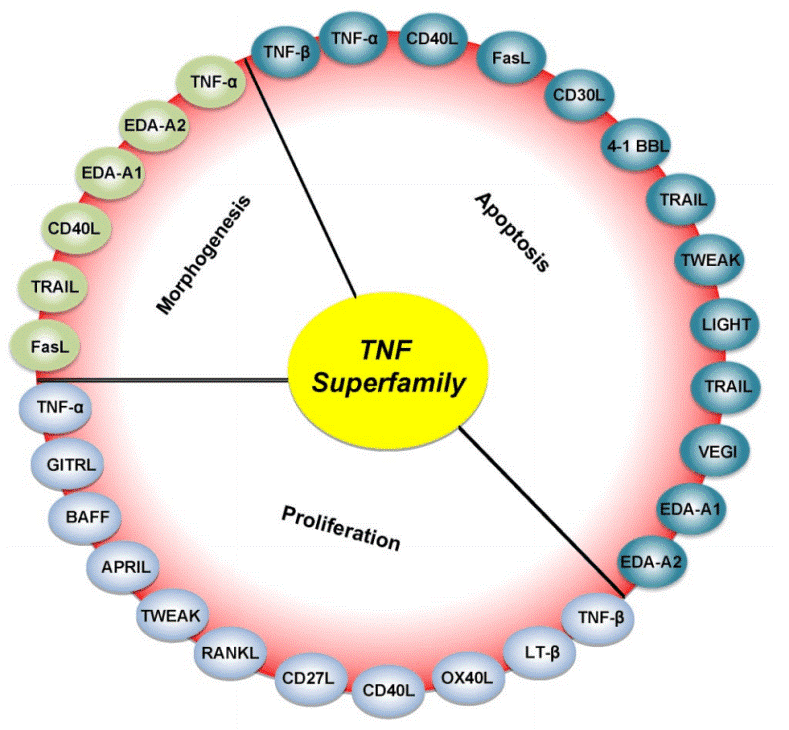
Fig. 1 Roles of various members of the TNF superfamily in inflammation, cellular proliferation, apoptosis, and morphogenesis. All members of the TNF superfamily exhibit pro-inflammatory activity, in part through activation of the transcription factor NF-κB (full red circle); OX40L, CD40L, CD27L, APRIL, and BAFF exhibit proliferative activity in part through activation of various mitogen-activated kinases (sky blue); TNF-α, TNF-β, FasL, and TRAIL control apoptosis (bluish-green); and EDA-A1, EDA-A2, TNF-α, FasL, and TRAIL regulate morphogenesis (green)[1].
02 Comparative analysis of TNF-α and TNF-β: cellular sources and functional roles in immunity
TNF-α primarily acts to amplify inflammatory cascades, whereas TNF-β is mainly responsible for fine-tuning adaptive immune responses. These two cytokines function synergistically in immune regulation rather than exhibiting simple functional redundancy. TNF-α and TNF-β (also referred to as lymphotoxin) differ substantially in their cellular origins and immunological functions, indicative of their distinct roles in innate and adaptive immunity. TNF-α is secreted by both innate immune cells (e.g., macrophages and NK cells) and adaptive immune cells (including T and B lymphocytes). Its expression can be rapidly triggered by pathogen-associated molecular patterns such as lipopolysaccharide (LPS). Characterized by a high transcription rate and a short mRNA half-life of approximately 30 min, TNF-α is well positioned to rapidly amplify inflammatory reactions[6,8].In comparison, TNF-β is predominantly generated by adaptive immune cells, especially activated T and B lymphocytes. Under physiological conditions, its basal transcription remains at a very low level; upon stimulation, however, the mRNA of TNF-β displays a much longer half-life (approximately 5.5 h), implying its crucial involvement in immune homeostasis and lymphocyte-mediated effector functions[6,7]. Functionally, TNF-α broadly activates the NF-κB signaling and apoptotic pathways, thereby modulating inflammation, cell survival and tumor progression, which endows it with the characteristic of a double-edged sword. Researchers commonly adopt TNF alpha elisa and TNF beta elisa to detect their expression levels. Although TNF-β shares the same receptors (TNFR1 and TNFR2) with TNF-α, it is capable of forming membrane-bound heterotrimeric complexes and participates in the development of secondary lymphoid organs, maintenance of T cell homeostasis, and initiation of cell apoptosis[6,7]. Moreover, TNF-β is indispensable for B cell maturation and antibody production during adaptive immune responses. By contrast, TNF-α is essential for host defense against bacterial pathogens and the mediation of endotoxic shock.
03 TNF-mediated cytokine cascade amplification in systemic inflammation
TNF serves as a central "apex cascade initiator" in systemic inflammatory responses. It not only directly induces the expression of inflammatory genes but also indirectly amplifies inflammatory responses by triggering cell death[9].
In chronic inflammatory disorders such as rheumatoid arthritis, TNF occupies a pivotal position at the apex of a complex cytokine network. It stimulates a variety of cell types, including synovial fibroblasts and macrophages, to secrete a broad spectrum of secondary pro-inflammatory cytokines and chemokines, such as interleukin-1 (IL-1), interleukin-6 (IL-6), interleukin-8 (IL-8), monocyte chemoattractant protein-1 (MCP-1), and interferon-inducible protein-10 (IP-10), thereby rapidly expanding the magnitude and diversity of inflammatory signaling[10].
Furthermore, the amplifying effect of TNF is also manifested in its regulation of cell death pathways. Upon TNF binding to TNFR1, in addition to activating the classical NF-κB and MAPK signaling pathways, under specific conditions (e.g., when cell death checkpoints are impaired), TNF can switch to inducing receptor-interacting serine/threonine-protein kinase 1 (RIPK1) kinase activity-dependent apoptosis, necroptosis, or pyroptosis. These lytic forms of cell death release substantial amounts of damage-associated molecular patterns (DAMPs), which further activate adjacent cells and immune cells, forming a self-sustaining inflammatory amplification loop[9].This cell death-driven inflammation is particularly prominent when the epithelial barrier is disrupted by microorganisms or when genetic defects lead to the inactivation of cell death checkpoints. For example, in mouse models, TNF-induced RIPK1 kinase activity-dependent cell death directly results in lethal endotoxic shock; in SHARPIN-deficient mice, TNF-mediated keratinocyte death drives severe cutaneous inflammation[9].More importantly, TNF can also reshape the cellular chromatin landscape through epigenetic mechanisms, inducing a "short-term transcriptional memory" that primes cells to mount faster and more robust inflammatory responses upon subsequent stimulation. This "priming" or "training" effect transforms inflammation from a localized defense mechanism into a persistent, dysregulated pathological state[10].Collectively, the amplifying effect of TNF operates through multi-dimensional crosstalk at the transcriptional, cell death, and epigenetic levels, which collectively drives the transition from local inflammation to a systemic inflammatory storm.
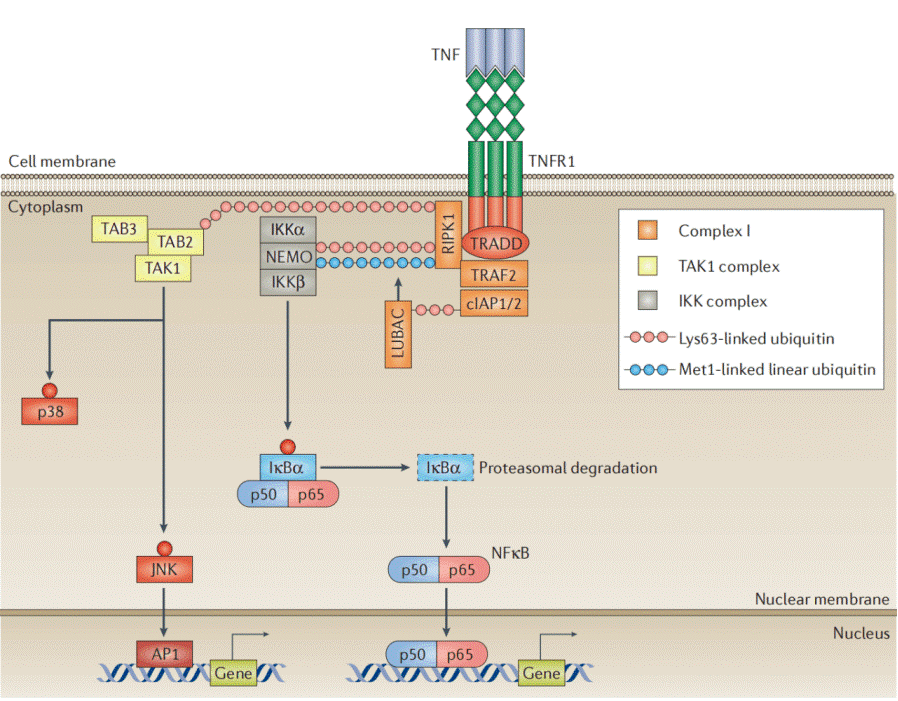
Fig. 2 A model of TNFR–complex I signalling. The binding of homotrimeric TNF to homotrimeric TNF receptors (TNFRs) induces the formation of complex I, comprising TNFR1-associated death domain protein (TRADD), receptor-interacting serine/threonine-protein kinase 1 (RIPK1), TNFR-associated factor 2 (TRAF2), cellular inhibitor of apoptosis protein 1 (cIAP1) or cIAP2, and linear ubiquitin chain assembly complex (LUBAC)[10].
04 ELISA-based quantification of TNF-α in serum and cell culture supernatants
ELISA assay represents a classic technique for the detection of TNF-α and is widely recognized as the gold standard for cytokine quantification. In practical experiments, the TNF-α elisa kit and commercial TNF alpha elisa kit are the most commonly used laboratory tools. In basic research, ELISA is routinely employed to assess TNF-α secretion in cell culture supernatants. For instance, following LPS stimulation of THP-1 cells, the collected culture supernatants can be subjected to sandwich ELISA to sensitively quantify TNF-α concentrations[11]. ELISA is also suitable for testing clinical serum samples; nevertheless, commercially available ELISA kits often exhibit distinct discrepancies in analytical performance, including accuracy, linearity and sensitivity. To guarantee experimental reliability, validation of the selected kit is strongly recommended. Specifically, international reference standards such as the NIBSC standard can be utilized to evaluate kit recovery and precision[12]. Additionally, pre-analytical sample stability is a critical consideration. Serum samples remain stable at 4°C for up to one week, while TNF-α levels in EDTA plasma tend to elevate under identical storage conditions. Accordingly, storage at -20°C or lower is optimal for sample preservation. Of note, serum samples from patients treated with anti-TNF-α monoclonal antibodies (e.g., infliximab) are prone to false-negative ELISA results, as therapeutic antibodies may interfere with targeted epitope recognition.
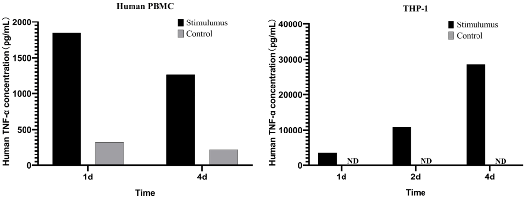
Fig. 3 Detection of TNF-α levels in cell culture supernatants. Human peripheral blood mononuclear cells (PBMCs) were stimulated with 10 μg/mL phytohemagglutinin (PHA) for 1, 2 and 4 days, followed by detection of related contents in cell supernatants; untreated cells were set as the control group. THP-1 cells were treated with 100 ng/mL PMA combined with 500 ng/mL LPS for 1, 2 and 4 days. The corresponding indicators in cell supernatants were then determined, with untreated cells serving as controls. (All experimental data were provided by Elabscience®)
05 Multiplex cytokine panels for simultaneous measurement of TNF-α, TNF-β, and IL-6
Research has demonstrated that TNF-α, TNF-β, and interleukin-6 (IL-6) are key components of the complex cytokine network, playing pivotal roles in immune-mediated disorders such as rheumatoid arthritis (RA). A study by Skrzypkowska et al. revealed that even in RA patients with low disease activity, serum IL-6 levels exhibited an upward trend compared to those in the osteoarthritis (OA) control group, whereas TNF-α levels were comparable between the two groups[13]. To simultaneously and accurately capture the dynamic changes of these TNF family members and related cytokines, highly sensitive multiplex cytokine assay and multiplex elisa technologies are indispensable. For instance, microfluidic nanoplasmonic digital immunoassays have enabled the parallel detection of up to six cytokines, including TNF-α and IL-6, with limits of detection as low as 0.46-1.36 pg/mL and dynamic ranges spanning 1-10,000 pg/mL[14]. Therefore, the simultaneous quantification of TNF-α, TNF-β (also referred to as lymphotoxin-alpha, LT-α), and IL-6 using a multiplex panel (the 6-plex nanoplasmonic chip) not only overcomes the limitations of single-plex assays but also more accurately reflects their regulatory network in diseases such as RA, thereby providing critical data support for mechanistic research and precision diagnostics.
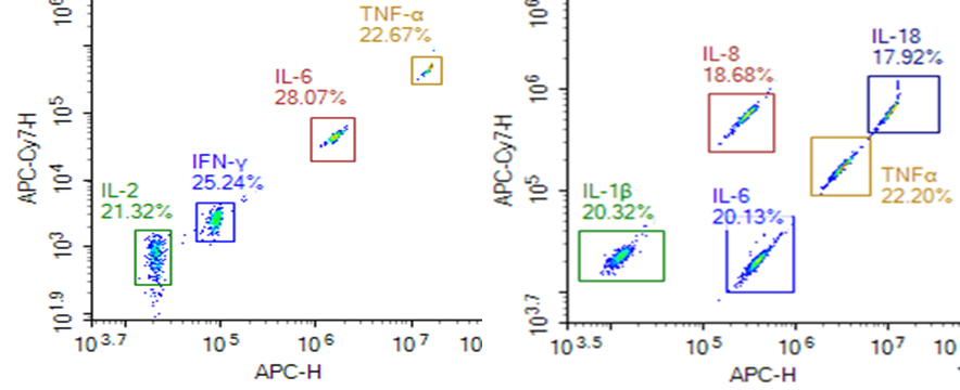
Fig. 4 The detection results of multiple cytokines. Various detection panels are available, enabling simultaneous quantification of 3 to 15 cytokines in a single assay based on experimental requirements. (All experimental data were provided by Elabscience®)
06 Emerging functional reassessment of TNF-β in immune homeostasis and disease contexts
The role of TNF-β, also referred to as lymphotoxin-alpha (LTα), has undergone a progressive redefinition, shifting from its traditional classification as a "helper" cytotoxic factor to a critical immune regulator[15]. Early research posited that TNF-β was functionally redundant to TNF-α; however, subsequent studies utilizing mice deficient in TNF-β or lymphotoxin beta receptor (LTβR) have uncovered its non-redundant roles in secondary lymphoid organ development, intestinal IgA production, CD4+ T cell differentiation, and innate antiviral immunity[16]. More importantly, recent discoveries have demonstrated that the TNF-β signaling pathway not only sustains immune homeostasis but also modulates the composition of commensal microbiota, thereby exerting an influence on metabolic diseases such as obesity[16]. Furthermore, under pathological conditions including autoimmune pancreatitis, TNF-β facilitates disease progression by inducing the formation of tertiary lymphoid tissues[17]. Collectively, these findings reposition TNF-β from a mere "helper factor" to a multifunctional core molecule that bridges immune development, homeostasis, microbiota interaction, and metabolic regulation. This redefinition provides a theoretical basis for future research directions in autoimmune diseases, metabolic disorders, and infectious immunity.
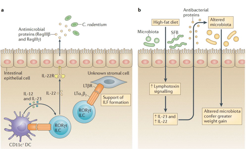
Fig. 5 LT regulates responses to microorganisms at mucosal surfaces. a) During Citrobacter rodentium infection, the lymphotoxin (LT) pathway is part of an innate feedback loop in which retinoic acid receptor related orphan receptor γt (RORγt)+ innate lymphoid cells (ILCs) induce interleukin 12 (IL 12) and IL 23 production by local dendritic cells (DCs) via the LT pathway. In response to IL 23, these ILCs produce IL 22, which promotes gut epithelial cells to produce antimicrobial peptides, such as RegIIIβ and RegIIIγ. These antimicrobial peptides have direct killing functions on C. rodentium and members of the commensal microbiota. LT dependent interactions between ILCs and stromal cells can also support the formation of isolated lymphoid follicles (ILFs) during infections. b) In response to a high fat diet, LT signaling is increased within the colon (V.U. and Y. X.F., unpublished observations) and this increases IL 23 and IL 22 production96. This then induces the production of antibacterial proteins, which are essential for the clearance of some microorganisms (such as segmented filamentous bacteria (SFB)) in response to high fat diet administration and which contribute to the alteration of the microbiota, which in turn confers greater weight gain96. IL 22R, interleukin 22 receptor; LTβR, LTβ receptor[16].
In summary, TNF-α and TNF-β represent pivotal pro-inflammatory cytokines with distinct functional profiles in immune regulation. TNF-α predominantly originates from innate immune cells and serves as a rapid initiator of inflammation, triggering NF-κB activation, amplifying the cytokine cascade, and promoting systemic inflammatory responses. In contrast, TNF-β is mainly secreted by adaptive lymphocytes and exerts non-redundant functions in lymphoid organ development, immune homeostasis maintenance, and disease pathogenesis. Enzyme-linked immunosorbent assay (ELISA) remains the gold-standard technique for individual quantification of TNF-α, whereas multiplex assay panels enable simultaneous detection of TNF-α, TNF-β, and IL-6, allowing more comprehensive characterization of the inflammatory regulatory network. TNF signaling drives inflammatory processes via transcriptional regulation, cell death modulation, and epigenetic modification mechanisms; its functional dysregulation is closely implicated in the pathogenesis of autoimmune disorders and chronic inflammatory diseases. An in-depth understanding of the biological functions of the TNF family will facilitate the development of novel targeted immunotherapeutic strategies.
Elabscience® Quick Overview of Popular Products:
Table 1. Research Tools for cytokines
|
Cat. No. |
Product Name |
|
MIH002N |
EasySortTM Human CD4+ T Cell Isolation Kit |
|
MIM005N |
EasySort™ Mouse NK Cell Isolation Kit |
|
E-EL-H0088 |
Human IL-1α(Interleukin 1 Alpha) ELISA Kit |
|
CQH015 |
CellaQuant™ Human IL-1β (Interleukin 1 Beta) ELISA Kit |
|
E-HSEL-H0003 |
High Sensitivity Human IL-6 (Interleukin 6) ELISA Kit |
|
CQH004 |
CellaQuant™ Human IL-8 (Interleukin 8) ELISA Kit |
|
CQH019 |
CellaQuant™ Human IL-12 (Interleukin 12) ELISA Kit |
|
E-EL-H0109 |
Human TNF-α(Tumor Necrosis Factor Alpha) ELISA Kit |
|
E-EL-H2306 |
Human TNF-β(Tumor Necrosis Factor Beta) ELISA Kit |
|
MPA019 |
Aptplex™ Human Cytokine 6-Plex Panel |
|
MPD012 |
Aptplex™ Human Cytokine 13-Plex Panel (C) |
References:
[1] Aggarwal B B, Gupta S C, Kim J H. Historical perspectives on tumor necrosis factor and its superfamily: 25 years later, a golden journey[J]. Blood, The Journal of the American Society of Hematology, 2012, 119(3): 651-665.
[2] Xu J. The role of tumor necrosis factor receptor superfamily in cancer: insights into oncogenesis, progression, and therapeutic strategies[J]. NPJ Precision Oncology, 2025, 9(1): 275.
[3] Chu W M. Tumor necrosis factor[J]. Cancer letters, 2013, 328(2): 222-225.
[4] Balkwill F. Tumor necrosis factor or tumor promoting factor?[J]. Cytokine & growth factor reviews, 2002, 13(2): 135-141.
[5] Goetz F W, Planas J V, MacKenzie S. Tumor necrosis factors[J]. Developmental & Comparative Immunology, 2004, 28(5): 487-497.
[6] Aggarwal B B. Signalling pathways of the TNF superfamily: a double-edged sword[J]. Nature reviews immunology, 2003, 3(9): 745-756.
[7] Li K, Qiu H, Yan J, et al. The involvement of TNF-α and TNF-β as proinflammatory cytokines in lymphocyte-mediated adaptive immunity of Nile tilapia by initiating apoptosis[J]. Developmental & Comparative Immunology, 2021, 115: 103884.
[8] Ruddle N H. Tumor necrosis factor (TNF-α) and lymphotoxin (TNF-β)[J]. Current opinion in immunology, 1992, 4(3): 327-332.
[9] Van Loo G, Bertrand M J M. Death by TNF: a road to inflammation[J]. Nature Reviews Immunology, 2023, 23(5): 289-303.
[10] Kalliolias G D, Ivashkiv L B. TNF biology, pathogenic mechanisms and emerging therapeutic strategies[J]. Nature reviews rheumatology, 2016, 12(1): 49-62.
[11] Leister K P, Huang R, Goodwin B L, et al. Two high throughput screen assays for measurement of TNF-α in THP-1 cells[J]. Current chemical genomics, 2011, 5: 21.
[12] Valaperti A, Li Z, Vonow-Eisenring M, et al. Diagnostic methods for the measurement of human TNF-alpha in clinical laboratory[J]. Journal of pharmaceutical and biomedical analysis, 2020, 179: 113010.
[13] Skrzypkowska M, Stasiak M, Sakowska J, et al. Cytokines and chemokines multiplex analysis in patients with low disease activity rheumatoid arthritis[J]. Rheumatology International, 2022, 42(4): 609-619.
[14] Icoz K, Tas Z, Azizieh F. Transforming cytokine diagnostics: AI, multiplexing, and point-of-care biosensing technologies[J]. Microchimica Acta, 2025, 192(11): 784.
[15] McDevitt H, Munson S, Ettinger R, et al. Multiple roles for tumor necrosis factor-α and lymphotoxin α/β in immunity and autoimmunity[J]. Arthritis Research & Therapy, 2002, 4(Suppl 3): S141.
[16] Upadhyay V, Fu Y X. Lymphotoxin signalling in immune homeostasis and the control of microorganisms[J]. Nature Reviews Immunology, 2013, 13(4): 270-279.
[17] Seleznik G M, Reding T, Romrig F, et al. Lymphotoxin β receptor signaling promotes development of autoimmune pancreatitis[J]. Gastroenterology, 2012, 143(5): 1361-1374.

