Introduction:
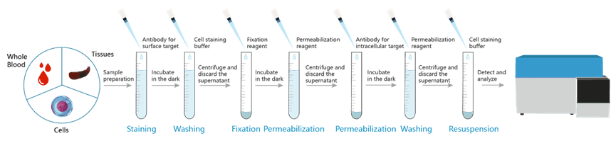
A modification of the basic immunofluorescent staining and flow cytometric analysis can be used for simultaneous analysis of surface molecules and intracellular antigens at the single-cell level by flow cytometry. Typically, cells are fixed with formaldehyde to stabilize the cell membrane, then permeabilized with detergent or ethanol to allow antibodies against intracellular antigens, to access to stain intracellularly.
Protocol-- intracellular (cytoplasmic) proteins
1. Prepare cells
More detail you can view Sample Preparation for Flow Cytometry
(1). Collect cells, filtere through a 200-mesh sieve and collect the filtrate.Centrifuge at 300 × g for 5 min, and discard the supernatant.
(2). Add Cell Staining Buffer [E-CK-A107] (or PBS with 1% BSA) to resuspend the sample.
2. Cell Counting
After counting the suspension with a hemocytometer or other instruments, adjust the cell concentration to about 1 × 10⁷/mL.
3. Set Sample and Control
|
Groups
|
Tubes
|
|
Controls
|
Blank
|
|
Single staining control
|
|
|
Isotype control
|
|
|
FMO
|
|
|
Biological control
|
|
|
Sample
|
Experimental sample
|
4. Block Fc Receptor
Block Fc receptors may reduce nonspecific immunofluorescent staining.
For human cells, EasyStain™ Human Fc Receptor Blocking Solution [E-CK-A171] can be used as an FcR blocking reagent. Add 5 μL of EasyStain™ Human Fc Receptor Blocking Solution, mix well, and incubate at room temperature for 10 min.
For mouse cells, Purified Anti-Mouse CD16/CD32 Antibody specific for FcγR III/II can be used to block nonspecific staining of antibodies, and reduces the background fluorescence of negative cells to the level of unlabeled cells. Add 1 μg of Purified Anti-Mouse CD16/32 Antibody [E-AB-F0997A] and incubate at room temperature for 10 min.
For rat cells, excessive purified Ig from the same source and subtype as fluorescent antibodies or serum from the same source can be directly used for blocking, or commercial FcR blocking agents can be used.
5. Cell Surface Staining
(1). Add 5 μL corresponding antibody to each sample tube except blank.
(2). Incubate at 4°C for 30 min in the dark.
6. Fixation and Permeabilization
If the markers include those in the nucleus(e.g. Foxp3, STAT3), please refer to the Cells Intranuclear Targets Staining for Flow Cytometry.
(1). Dilute Fixation and Permeabilization Solution [E-CK-A109] according to the manual.
(2). Add 2 mL cell staining buffer [E-CK-A107] (or PBS with 1% BSA) to each tube, centrifuge at 300 × g for 5 min, and discard the supernatant.
(3). Add 200 μL cell staining buffer [E-CK-A107] (or PBS with 1% BSA) to resuspend the sample.Intranuclear
(4). Add 200 μL 1× Fixation Buffer to each tube, mix gently.
(5). Incubate at RT for 30-60 min in the dark.
(6). Add 1 mL 1× Permeabilization Working Solution to each tube, centrifuge at 600 × g for 5 min, and discard the supernatant.
7. Cell Intracellular Staining
(1). Add 100 μL 1× Permeabilization Working Solution to each tube, resuspend the sample.
(2). Add 5 μL corresponding antibody to the tube required.
(3). Incubate at RT for 30 min in the dark.
(4). Add 2 mL cell staining buffer [E-CK-A107] (or PBS with 1% BSA) to each tube, centrifuge at 600 × g for 5 min, and discard the supernatant.
8. Detection
(1). Add 200 μL cell staining buffer [E-CK-A107] (or PBS with 1% BSA) to resuspend the sample.
(2). Adjust instrument parameters, detection.

