

10 minutes on the cytometer, yet 5 hours of sample preparation and analysis, and all you get are aggregates and debris?” Many researchers invest heavily in panel design and compensation, only to be tripped up by the most unassuming step: sample preservation. In this article, we won’t dive into complex panel design. Instead, we’ll focus on one thing: how to keep your samples in optimal condition until just before acquisition. We’ll cover four key aspects: pre-staining, post-staining, overnight storage, and pause points for intracellular staining.
Table of Contents
1. Why do flow cytometry results look poor?
2. How to store samples before staining (unstained)?
3. How to stabilize fluorescence signals in stained samples?
4. Flow cytometry intracellular staining: how to pause?
5. Special reminders for tissue-derived samples (spleen, tumor, etc.)
6. How should control samples be stored?
01 Why do flow cytometry results look poor?
Once a single-cell suspension leaves the body, it faces three "killers":
(1) Cell death: Necrotic cells release DNA, causing aggregates, and also bind antibodies non-specifically, increasing background signal.
(2) Antigenic modulation: Certain surface molecules (e.g., chemokine receptor CCR7) are highly sensitive to temperature and enzymatic treatment. These molecules are rapidly internalized or downregulated ex vivo, leading to distorted results.
(3) Fluorescence quenching: Tandem dyes (e.g., PE-Cy7, APC-Cy7) are sensitive to light and fixatives.
Core principle: After preparation, stain as soon as possible; after staining, acquire as soon as possible. This is the optimal strategy. When conditions are limited, clearly understand "when you can wait and when you cannot."
02 How to store samples before staining (unstained)?
2.1 For unstained whole blood, samples should be processed as soon as possible
(1) Whole blood preserves the most natural survival microenvironment for cells. Short-term storage at room temperature (18–25 °C) has minimal impact on cell viability. It is recommended to complete staining and analysis within 6 hours after collection. If processing within 6 hours is not possible, it is recommended to complete testing within 24 hours after collection. At this time point, the expression stability of activation markers (e.g., CD25, HLA-DR) is still acceptable and individual variability is minimal, but absolute cell counts may no longer be reliable due to cell death and selective loss of myeloid populations. Samples stored at room temperature for 24–48 hours can still be analyzed, but reliability decreases significantly, and selective loss of myeloid cells becomes evident.
(2) Next, the choice of anticoagulant:
EDTA anticoagulant is the most commonly used option for flow cytometric immunophenotyping. According to published data, EDTA-anticoagulated whole blood can be stored stably at room temperature for approximately 12 to 24 hours. Beyond 24 hours, selective loss of myeloid cells (such as neutrophils, eosinophils, and CD16+ monocytes) occurs, and the fluorescence intensity of multiple surface markers, including CD27 and TCRγδ, declines significantly. The same literature also points out that EDTA interferes with enzymatic reactions by chelating (or removing) calcium ions, so caution is needed when functional assays are involved.
Sodium heparin performs better in maintaining cell viability and surface marker stability. Published data confirm that at room temperature, cell population distribution and marker stability are significantly superior to EDTA, and whole blood can be stored stably for up to 48 hours. Because heparin does not chelate calcium ions, it is more suitable for scenarios requiring combined detection of cell function, sample transport, or prolonged storage in multicenter studies.
Sodium citrate is more commonly used for coagulation function testing.
Recommendations: Unstained whole blood should be processed as much as possible within 6 hours. If the expected transport time is 24 to 48 hours, use sodium heparin as the anticoagulant and transport at room temperature. For unstained EDTA whole blood, prolonged transport at 4 °C is not recommended. However, if the experimental endpoint specifically focuses on monocytes and cold transport is unavoidable, in-house validation is required.
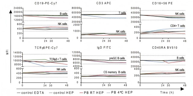
Fig. 1 Longitudinal analysis of blood samples collected into Na-Heparin tubes. Peripheral blood samples were collected into Na-Heparin tubes, stained and acquired directly or stored at 4 °C for delayed acquisition. Additional samples were collected into Na-Heparin tubes and stored either at RT or at 4 °C for delayed staining. All results were compared to freshly stained K3EDTA samples of the same donors, n ≥ 3. Mean fluorescence intensity (MFI) of selected cell surface markers on positive and negative reference populations.
2.2 Storage of Single-Cell Suspensions Prepared from Tissues
Tissue samples are more delicate than whole blood because the preparation process itself (enzymatic digestion, mechanical dissociation) imposes stress on cells.
(1) Immediate processing is strongly recommended: After tissue dissociation, cells are in a stressed state and should be stained and acquired as soon as possible (within 2 hours).
(2) Short-term (within 24 hours) storage protocol (if immediate staining is not possible):
Storage buffer: Always use a protein-supplemented buffer (staining buffer or complete culture medium). Never use plain PBS.
Conditions: Store at 4 °C protected from light. Before acquisition the next day, always perform a viable cell count or add a viability dye at the time of acquisition.
(3) Long-term storage (months to years): cryopreservation in liquid nitrogen
If long-term storage is needed, the only reliable method is controlled-rate freezing and storage in liquid nitrogen.
Use a cryopreservation medium consisting of 90% FBS and 10% DMSO (or a commercial cryopreservation medium), cool gradually using a controlled-rate freezing container, keep at -80 °C overnight, and then transfer to liquid nitrogen for storage.
Results should be interpreted with caution. Published data confirm that after long-term cryopreservation of human peripheral blood or PBMC samples, many target antigens undergo changes.
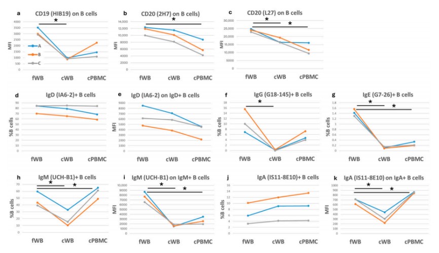
Fig. 2 Comparison of B cells in fresh and corresponding cryopreserved samples. Each graph shows either the frequency of B cells positive for a specific antigen or the corresponding mean fluorescence intensity (MFI) on the y-axis. The three colored lines (blue, orange, and grey) correspond to three blood samples, each of which was assessed under three different conditions: fresh (fWB), after blood cryopreservation (cWB), and after PBMC isolation and cryopreservation (cPBMC). These conditions are indicated on the x-axis.
03 How to stabilize fluorescence signals in stained samples?
After staining is completed, antibodies have already bound to their targets. At this point, the core task is to prevent fluorescence quenching and antibody dissociation. Processing strategies differ among sample types, as described below.
3.1 Stained Whole Blood Samples
After red blood cell lysis and staining are completed, centrifuge and wash once to remove free antibodies and residual lysis buffer. Then resuspend the cell pellet in staining buffer and store at 4 °C protected from light.
It is recommended to complete acquisition within 6 hours to obtain optimal signal intensity and cell condition. If this is not possible, detection should be completed within 24 hours. Otherwise, signal decay and reduced cell viability will significantly compromise data quality.
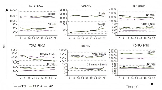
Fig. 3 Longitudinal analysis of stained samples stored without or with additional fixative. Peripheral blood samples were collected into K3EDTA tubes stained and stored at 4 °C for delayed acquisition. Additional stained samples were treated with 1% paraformaldehyde (PFA) or Fix&Perm Solution A (F&P) directly after staining and prior to storage at 4 °C, n ≥ 3. Mean fluorescence intensity (MFI) of selected cell surface markers on positive and negative reference populations.
3.2 Stained Single-Cell Suspensions
The storage strategy for single-cell suspensions (e.g., tissue-derived or cultured cells) should be selected flexibly according to the timing of acquisition:
a. Acquisition within 4 hours
After staining, wash once to remove excess antibodies, resuspend in staining buffer, and store at 4 °C protected from light. Within this window, fluorescence signals are stable and cell viability is good. This is the ideal arrangement.
b. Acquisition within 24 hours
If detection cannot be completed within 4 hours, choose one of the following two options:
Unfixed option (suitable for most routine assays): After staining and washing, resuspend cells directly in staining buffer, store at 4 °C protected from light, and acquire within 24 hours. This option is simple, but note that the viability of certain fragile cell populations (e.g., granulocytes) may decline over time. It is recommended to confirm cell status with a viability dye before acquisition.
Fixed option: After staining and washing, fix immediately with a final concentration of 1% to 2% paraformaldehyde (PFA) at 4 °C, protected from light, for 15 to 20 minutes. After fixation, wash twice with staining buffer to thoroughly remove residual fixative, and finally resuspend in fresh staining buffer. Store at 4 °C protected from light. Fixation can effectively stabilize antigen-antibody complexes and delay signal decay, making it especially suitable for scenarios requiring overnight or next-day detection.
Note: Tandem dyes such as PE-Cy7, APC-Cy7, and PerCP-Cy5.5 are sensitive to fixatives. After fixation, fluorescence signals will decay to varying degrees, and batch-to-batch variation is considerable.
Long-term storage after staining is not recommended.
04 Flow cytometry intracellular staining: how to pause?
4.1 After Fixation and Before Permeabilization and Intracellular Staining
This is a relatively safe and recommended overnight window within the entire intracellular staining workflow. The procedure is as follows: after surface staining and fixation are completed, wash cells thoroughly twice with staining buffer to completely remove residual fixative. Finally, resuspend cells in staining buffer and store at 4 °C protected from light. Under these conditions, cell morphology and antigenic epitopes are stably cross-linked, allowing safe overnight storage (18 to 24 hours). Permeabilization and intracellular staining can be performed the next day with minimal impact on most markers.
Important note: The above is a general recommendation. Fixation / permeabilization kits from different manufacturers may have specific requirements for the storage buffer after fixation and before permeabilization. For example, some transcription factor staining kits require cells to be kept in permeabilization buffer. Before formal experiments, always read the official protocol of the kit carefully and follow its recommendations strictly. If the protocol does not specify, staining buffer can be used as the preferred resuspension buffer for storage, or contact the manufacturer's technical support for confirmation.
4.2 After All Intracellular Staining Is Completed
Data acquisition on the same day is recommended, and prolonged storage is not advisable. If it is absolutely necessary to postpone acquisition to the next day, stained cells may be kept in 1x permeabilization buffer at 4 °C, protected from light overnight.
However, special attention should be paid: overnight storage may lead to fluorescence signal decay (especially for tandem dyes such as PE-Cy7 and APC-Cy7), and background may increase due to enzymes released by dead cells or free antibodies.
Therefore, this approach should only be used as an emergency backup measure and is not recommended for routine use. If it is adopted, it is essential to ensure that experimental and control groups are processed under exactly the same conditions, and signal changes should be carefully considered when interpreting results.
05 Special reminders for tissue-derived samples (spleen, tumor, etc.)
(1) Thoroughly wash away residual collagenase.
(2) Tissue samples are highly prone to aggregation. It is recommended to add DNase I to the buffer, with the final concentration adjusted according to the degree of sample aggregation.
(3) It is recommended to proceed to the staining procedure within 2 hours.
(4) Myeloid cells in tissues (macrophages, monocytes, granulocytes) express high levels of Fc receptors. Treat with an Fc receptor blocking reagent for 10 minutes before staining to avoid non-specific binding or increased background.
06 How should control samples be stored?
For gating controls (FMO control, isotype control, single-stain controls): the principle of strictly matched handling: same conditions, same timing, same treatment. If the experimental group is fixed, the FMO must also be fixed; if the experimental group is stored overnight, the FMO must also be stored overnight. Otherwise, the gating position will shift, leading to invalid gating.
Finally, the reliability of flow cytometry experiments depends heavily on sample processing and preservation. If your sample preservation is not done properly, how elegant your panel design is or how expensive your antibodies are, you will not be able to recover the true biological differences buried under technical noise.
Best recommendation: use fresh samples, process immediately after collection, and acquire as soon as possible.
References:
[1] Diks AM, et al. Impact of blood storage and sample handling on quality of high dimensional flow cytometric data in multicenter clinical research. J Immunol Methods. 2019; doi:10.1016/j.jim.2019.06.007.
[2] Davis C, et al. Stability of immunophenotypic markers in fixed peripheral blood for extended analysis using flow cytometry. J Immunol Methods. 2011;363(2):158-65. doi:10.1016/j.jim.2010.09.029.
[3] Gorog DA, Becker RC. Point-of-care platelet function tests: relevance to arterial thrombosis and opportunities for improvement. J Thromb Thrombolysis. 2021;51(1):1-11. doi:10.1007/s11239-020-02170-z.
[4] Reichard A, et al. Best Practices for Preparing a Single Cell Suspension from Solid Tissues for Flow Cytometry. Cytometry A. 2019;95(2):219-226. doi:10.1002/cyto.a.23690.
[5] Serra V, et al. FlowCLOc, a New Tool for Selecting the Most Appropriate Antibodies in Flow Cytometry. Int J Mol Sci. 2026;27(4):1664. doi:10.3390/ijms27041664.
[6] Sedek L, et al. Impact of Pre-Analytical and Analytical Variables Associated with Sample Preparation on Flow Cytometric Stainings Obtained with EuroFlow Panels. Cancers. 2022;14(3):473. doi:10.3390/cancers14030473.
[7] Cossarizza A, et al. Guidelines for the use of flow cytometry and cell sorting in immunological studies (third edition). Eur J Immunol. 2021;51(12):2708-3145. doi:10.1002/eji.202170126.

