

The development of flow cytometry is inextricably linked to technological innovations in fluorescent dyes. From early organic small-molecule dyes to the increasingly diverse categories of tandem dyes and polymer dyes, the continuous refinement of fluorescent reagent systems has driven the realization of multiparameter flow cytometric analysis. This article systematically elaborates on the performance characteristics, representative dyes, and practical applications of the four core types of fluorescent dyes in flow cytometry: small-molecule dyes, macromolecular dyes (phycobiliproteins), tandem dyes, and polymer dyes. In addition, drawing on an example of 20-color spectral flow cytometric analysis of mouse splenic immune cells, it analyzes the core principles of dye selection and practical considerations in spectral multicolor flow cytometry experiments.
Table of Contents
1. Organic small-molecule fluorescent dyes
2. Macromolecular fluorescent dyes and phycobiliproteins
3. Tandem fluorescent dyes and Förster resonance energy transfer
4. Polymer-based fluorescent dyes for multicolor flow cytometry
01 Organic small-molecule fluorescent dyes
Organic small-molecule dyes are the longest-established and most widely used class of fluorescent compounds in flow cytometry. These dyes are characterized by a small molecular weight (typically on the order of several hundred Da), a relatively small Stokes shift (approximately 50–100 nm), a well-defined structure, good photostability, and ease of conjugation to antibodies. Common small-molecule dyes used in flow cytometry applications are summarized in Table 1.
Table 1. Summary and Comparison of Performance Characteristics of Organic Small-Molecule Dyes
|
Dye Name |
Excitation(nm) |
Emission (nm) |
Common Detection Channel |
Molecular Weight (Da) |
Brightness |
Key Features |
|
FITC |
488~495 |
518~530 |
530/30(Green channel) |
~389 |
Moderate |
The most classic green fluorescent dye, widely used; the human eye is highly sensitive to yellow-green light; sensitive to pH; moderate photostability. |
|
Elab Fluor® 488 |
488~495 |
516~519 |
530/30(Green channel) |
~643 |
Moderate |
An excellent alternative to FITC, with better photostability, suitable for experiments requiring imaging analysis. |
|
Elab Fluor® 647 |
647~655 |
665~680 |
660/20 nm (Red channel) |
~1300 |
Bright |
Excellent photostability, pH-insensitive (stable across pH 4–10), compatible with the 633 nm laser, and a workhorse dye in the red channel for multicolor experiments. |
|
Elab Fluor® 700 |
675~702 |
710~740 |
725/40 nm (Near-infrared channel) |
1200~1500 |
Bright |
Emission in the near-infrared region, where biological autofluorescence is extremely low; minimal spectral overlap with other common dyes; enhanced tissue penetration; suitable for multicolor flow cytometry and in vivo imaging. |
|
Pacific Blue |
401~410 |
452~455 |
450/50 (Violet-blue channel) |
242~406 |
Moderate |
Violet (405 nm)-excited blue fluorescent dye; small molecular weight, suitable for multicolor panel design. |
|
Texas Red |
586~596 |
605~615 |
610/20(Red channel) |
625~1109 |
Moderate |
Red fluorescent dye, commonly used as the acceptor molecule for tandem dyes |
|
Cy5 |
648~650 |
662~670 |
670/30(Far-red channel) |
657~792 |
Moderate |
Far-red fluorescent dye, commonly used as the acceptor for tandem dyes; effectively avoids sample autofluorescence background |
|
Elab Fluor® Red 780 |
620~660 |
760~800 |
780/60(Near-infrared channel) |
~930 |
Dim |
Strong photostability and high resistance to photobleaching; supports long-term fluorescence imaging. |
|
Elab Fluor® Violet 450 |
395~415 |
430~470 |
450/50(Violet channel) |
841 |
Dim |
A workhorse dye for the classic violet laser channel; minimal spectral overlap with other violet laser dyes; excellent stability; suitable for labeling medium- to high-abundance antigens. |
|
Elab Fluor® Violet 500 |
395~415 |
480~520 |
525/50(Violet channel) |
672 |
Dark |
Green fluorescent dye for the violet laser channel; spectrally separable from FITC under the 488 nm laser, greatly reducing compensation burden in multicolor panels. |
|
Elab Fluor® Violet 540 |
395~415 |
520~560 |
545/40(Violet channel) |
983 |
Dim |
Fills the gap in the yellow-green region of the violet laser channel; almost no spectral overlap with the PE channel; adds an independent detection dimension in multicolor experiments |
|
Elab Fluor® Violet 610 |
395~415 |
590~630 |
610/20(Violet channel) |
1293 |
Dim |
Emission in the orange-red region; minimal interference from biological autofluorescence; suitable for high-resolution detection of low-abundance antigens; meets the need for channel expansion in multicolor panels. |
02 Macromolecular fluorescent dyes and phycobiliproteins
Macromolecular fluorescent dyes mainly refer to high-molecular-weight protein-based fluorescent dyes extracted from natural organisms, and they represent a classic class of flow cytometry labeling dyes. The classic macromolecular fluorescent dyes are primarily phycobiliproteins such as PE (phycoerythrin), APC (allophycocyanin), and PerCP (peridinin-chlorophyll-protein). These dyes have high molar extinction coefficients and high fluorescence quantum yields, and their brightness is far greater than that of ordinary small-molecule dyes, making them suitable for labeling low-abundance antigens. However, they are also susceptible to photobleaching. The characteristics of the major macromolecular fluorescent dyes are summarized in Table 2.
Table 2. Summary and Comparison of Performance Characteristics of Macromolecular Dyes
|
Dye Name |
Excitation (nm) |
Emission (nm) |
Common Detection Channe |
Molecular Weight (kDa) |
Brightness |
Key Features |
|
PE(Phycoerythrin) |
488/565 |
575~578 |
585/42 (Orange-yellow channel) |
~240 |
Bright |
One of the brightest dyes in flow cytometry, with a quantum yield >90%; suitable for low-abundance antigen detection; susceptible to photobleaching |
|
APC(Allophycocyanin) |
633~652 |
660~662 |
660/20 (Far-red channel) |
~104 |
Bright |
Far-red emission, avoids sample autofluorescence background; good stability; suitable for multichannel experimental design |
|
PerCP |
488 |
~675 |
670/30(Red channel) |
~35.5 |
Dim |
Relatively low quantum yield and weak signal, suitable only for high-abundance antigen detection; extremely light-sensitive |
03 Tandem fluorescent dyes and Förster resonance energy transfer
Tandem dyes are a special class of fluorescent molecules designed based on the principle of fluorescence resonance energy transfer (FRET, i.e., Förster resonance energy transfer). Their basic structure consists of a donor fluorophore (such as a phycobiliprotein or polymer dye) chemically conjugated to an acceptor small-molecule organic dye (such as Cy3, Cy5, or Cy7). When the donor is excited by a laser, its emission light excites the acceptor through the FRET effect, ultimately emitting fluorescence at the acceptor's characteristic wavelength. Major examples include PE-Cy5, PE-Cy7, APC-Cy7, and PerCP-Cy5.5. Their core advantage is a greatly extended Stokes shift (150–300 nm), enabling "single-laser excitation with multi-wavelength emission." Their fluorescence brightness is markedly greater than that of single-molecule dyes, and they expand the range of fluorescent dyes excitable by a single laser. They can be used for multiparameter flow cytometric analysis and are especially suitable for experimental designs requiring more detection parameters with a limited number of lasers. However, they also have drawbacks that need to be addressed, such as sensitivity to light, requiring minimization of light exposure during experiments, and potential differences in protein-to-dye ratios between different batches and suppliers. The main characteristics and applications of tandem dyes are summarized in Table 3.
Table 3. Summary and Comparison of Performance Characteristics of Tandem Dyes
|
Dye Name |
Excitation(nm) |
Emission (nm) |
Common Detection Channe |
Molecular Weight (kDa) |
Brightness |
Key Features |
|
PE-Cy7 |
488/565 |
~780 |
780/60(Near-infrared channel) |
~240(PE-based) |
Very bright |
Extremely large Stokes shift (~215 nm), enabling far-red detection; a top choice for multicolor flow cytometry; light-sensitive |
|
APC-Cy7 |
633~650 |
~780 |
780/60(Near-infrared channel) |
~104(APC-based) |
Moderate brightness |
Red-light excitation with near-infrared emission, further expanding detection parameters; potential batch-to-batch variation in energy transfer efficiency |
|
PerCP-Cy5.5 |
482~489 |
676~695 |
695/40(Far-red channel) |
~35 |
Bright |
Brighter than PerCP and with better photostability; excited by the 488 nm laser with far-red emission |
|
PE/Cy5 |
488/561 |
660~680 |
670/30 or 695/40(Far-red channel) |
~240(PE-based) |
Bright |
A classic PE tandem dye with a large Stokes shift and good spectral separation from FITC and PE channels; enables multicolor detection with a single 488 nm laser; suitable for labeling medium- to high-abundance antigen |
|
PE/Cy5.5 |
488/561 |
680~710 |
690/50 or 700/50(Near-infrared channel) |
~240(PE-based) |
Bright |
Emission in the near-infrared I region, with minimal interference from biological autofluorescence; can be paired with PE/Cy5 and PE/Cy7 to expand the detection channels of the 488 nm laser; suitable for multicolor flow cytometry panel design |
|
PE/Elab Fluor® 594 |
488/561 |
600~630 |
610/20 or 620/30(Orange channel) |
~240(PE-based) |
Very bright |
Emission in the orange-red region, with low spectral overlap with PE and PE/Cy5; good photostability and resistance to photobleaching; suitable for immunofluorescence and multicolor flow cytometry experiments |
04 Polymer-based fluorescent dyes for multicolor flow cytometry
Polymer dyes are a rapidly emerging class of novel fluorescent dyes developed in recent years, composed of polymer chains capable of harvesting light signals. By adjusting the length of the polymer chains and the attached molecular subunits, the absorption and emission spectra of the dyes can be "customized." Major examples include the Elab Bright™ Violet series, such as Elab Bright™ Violet 421 and Elab Bright™ Violet 510. Their core advantages include excellent photostability and high brightness, enabling better discrimination between weakly positive and negative cell populations, far surpassing traditional dyes. Their quantum efficiency is comparable to that of phycobiliproteins, and they absorb only specific wavelengths of light, avoiding the compensation complexity caused by multi-laser excitation of quantum dots in multiparameter experiments. They can be used for multiparameter flow cytometric analysis, especially for multicolor panel design with violet and ultraviolet laser excitation. Polymer dyes have largely replaced quantum dots in multiparameter staining applications. Commonly used polymer dyes are summarized in Table 4.
Table 4. Summary and Comparison of Performance Characteristics of Polymer Dyes
|
Dye Name |
Benchmark Dye |
Excitation (nm) |
Emission (nm) |
Common Detection Channe |
Molecular Weight (kDa) |
Brightness |
Key Features |
|
Elab Bright™ Violet 421 |
BV421 |
405~407 |
421~423 |
450/50 (Violet channel) |
~70 |
Very bright |
A violet-excited, high-brightness dye; one of the brightest fluorescent dyes currently available; excellent photostability; a non-tandem polymer |
|
Elab Bright™ Violet 510 |
BV510 |
395~415 |
490 ~530 |
525/50 (Violet channel) |
~77 |
Moderate brightness |
Far more efficiently excited under the 405 nm violet laser than FITC, with extremely low compensation interference with far-red dyes such as APC and PE-Cy7; a commonly used dye in multicolor flow cytometric analysis |
|
Elab Bright™ Violet 650 |
BV650 |
395~415 |
630~670 |
660/20 (Violet channel) |
~63 |
Very bright |
Emission in the far-red region, with extremely low spectral overlap with the APC channel; enables independent detection of far-red signals under the violet laser, further expanding the channel ceiling of multicolor experiments |
The above provides an introduction to the four core types of fluorescent dyes in flow cytometry. It should be noted that in multicolor flow cytometry applications, dye selection must follow three core principles: Adopt a strong-weak pairing strategy: use weak fluorescent dyes for highly expressed targets and strong fluorescent dyes for low-abundance or rare targets, to prevent signal spillover from masking weak signals; Follow a spectral optimization approach: prioritize dye combinations excited by different lasers to reduce spectral overlap and alleviate compensation burden; Accommodate scenario-specific requirements: for intracellular and nuclear fixation/permeabilization experiments, prioritize small-molecule and macromolecular dyes, and use conventional tandem dyes with caution. At the same time, be aware of pitfalls: tandem dyes must be stored protected from light at 2–8°C throughout, must never be frozen, and repeated freeze-thaw cycles can cause dye disaggregation and loss of efficacy.
As shown in Figure 1, when selecting fluorescent dyes for spectral flow cytometry, it is important to ensure that the spectral similarity between fluorescent dyes is ≤0.98, which yields good unmixing analysis results.
Fig. 1 20-color full-spectrum flow cytometric analysis of mouse splenic immune cells. A spectral flow cytometer (Agilent Novocyte Opteon VBRY) was used to detect and analyze mouse splenic immune cell subsets.
Table 5. Product application list for 20-color spectral flow cytometric analysis of mouse splenic immune cells
|
Marker |
Fluorescent Dye |
Antibody |
Cat. No. |
|
CD45 |
Elab Fluor® Red 780 |
Elab Fluor® Red 780 Anti-Mouse CD45 Antibody[30-F11] |
E-AB-F1136S |
|
CD3 |
Elab Fluor® Violet 500 |
Elab Fluor® Violet 500 Anti-Mouse CD3 Antibody[17A2] |
E-AB-F1013R |
|
CD4 |
PerCP |
PerCP Anti-Mouse CD4 Antibody[GK1.5] |
E-AB-F1097F |
|
CD8 |
Elab Fluor® Violet 540 |
Elab Fluor® Violet 540 Anti-Mouse CD8a Antibody[53-6.7] |
E-AB-F1104T3 |
|
CD25 |
PE/Elab Fluor® 594 |
PE/Elab Fluor® 594 Anti-Mouse CD25 Antibody[PC-61.5.3] |
E-AB-F1102P |
|
NK1.1 |
Elab Fluor® 700 |
Elab Fluor® 700 Anti-Mouse CD161/NK1.1 Antibody[PK136] |
E-AB-F0987M1 |
|
CD44 |
Elab Bright™ Violet 650 |
Elab Bright™ Violet 650 Anti-Human/Mouse CD44 Antibody[IM7] |
E-AB-F1100U |
|
CD62L |
APC |
APC Anti-Mouse CD62L Antibody[MEL-14] |
E-AB-F1011E |
|
CD127 |
Elab Bright™ Violet 421 |
Elab Bright™ Violet 421 Anti-Mouse CD127/IL-7RA Antibody[A7R34] |
E-AB-F1023Q2 |
|
B220 |
Elab Fluor® Violet 450 |
Elab Fluor® Violet 450 Anti-Mouse CD45R/B220 Antibody[RA3.3A 1/6.1] |
E-AB-F1112Q |
|
CD19 |
PE/Cyanine7 |
PE/Cyanine7 Anti-Mouse CD19 Antibody[1D3] |
E-AB-F0986H |
|
CD5 |
PE |
PE Anti-Mouse CD5 Antibody[53-7.3] |
E-AB-F1185D |
|
CD27 |
FITC |
FITC Anti-Mouse CD27 Antibody [LG.3A10] |
AN00322C |
|
CD11b |
Elab Bright™ Violet 510 |
Elab Bright™ Violet 510 Anti-Mouse/Human/Monkey CD11b Antibody[M1/70] |
E-AB-F1081R1 |
|
CD11c |
Elab Fluor® Violet 610 |
Elab Fluor® Violet 610 Anti-Mouse CD11c Antibody[N418] |
E-AB-F0991T |
|
MHC-II |
Elab Fluor® 488 |
Elab Fluor® 488 Anti-Mouse MHC II (I-A/I-E) Antibody[M5/114] |
E-AB-F0990L |
|
F4/80 |
PerCP/Cyanine5.5 |
PerCP/Cyanine5.5 Anti-Mouse F4/80 Antibody[CI:A3-1] |
E-AB-F0995J |
|
Ly6G |
Elab Fluor® 647 |
Elab Fluor® 647 Anti-Mouse Ly6G Antibody[1A8] |
E-AB-F1108M |
|
Ly6C |
PE/Cyanine5.5 |
PE/Cyanine5.5 Anti-Mouse Ly6C Antibody[Monts 1] |
E-AB-F1121I |
|
Viability |
APC/Cy7 |
STYX™ Near-IR Fixable Viability Kit |
E-CK-A168 |
Elabscience® has its own independent fluorescent dye brand, the Elab Fluor® series, whose spectra cover the ultraviolet to infrared range, with a total of 21 dyes, including multiple fluorescent groups such as Elab Fluor® Violet 450. These are compatible with multi-laser flow cytometers equipped with 405 nm, 488 nm, and 633 nm lasers, among others. The iteratively upgraded third-generation non-destructive optimized labeling process maximally preserves the natural structure and biological function of antibodies, ensuring that labeled antibodies can still efficiently and specifically recognize target antigens, providing strong support for 10–21 color high-parameter spectral flow cytometry detection.
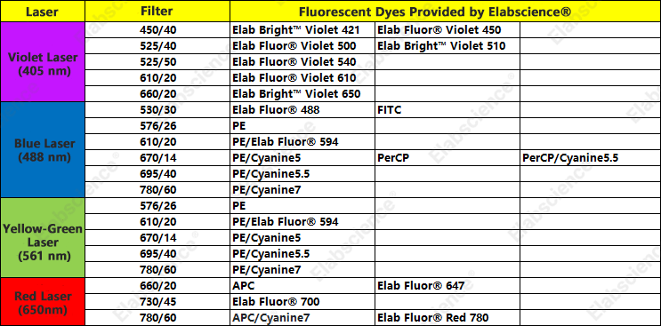
The fluorescence dye brightness and filter comparison information now available from Elabscience® is shown in Table 6 and Table 7:
Table 6. Summary of Brightness Comparison of Currently Available Fluorescent Dyes from Elabscience®
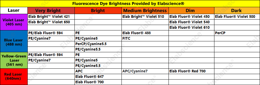
Table 7. Comparison of Filter Distribution of Currently Available Fluorescent Dyes from Elabscience®
New Product Preview: Elab Fluor® 659 — A Strong Preferred Alternative to PerCP
3-4× brightness improvement, combined with excellent photostability and highly matched spectral characteristics. It can serve not only as an ideal replacement for PerCP dyes in conventional flow cytometry, but also open up a new independent detection channel on spectral flow cytometry platforms.
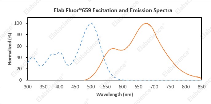
Fig. 2 Spectral scan of Elab Fluor® 659.
Table 8. Spectral information of Elab Fluor® 659
|
Parameter |
Measured Value |
|
Maximum Absorption Wavelength (Exmax) |
500 nm |
|
Maximum Emission Wavelength (Emmax) |
672 nm |
|
Stokes Shift |
172 nm |
|
Recommended Laser |
488 nm |
|
Recommended Detection Filter |
667/30 nm (PerCP) |
The spectral similarity between Elab Fluor® 659 and PerCP is below 0.9, allowing it to share the PerCP channel in spectral flow cytometry detection.
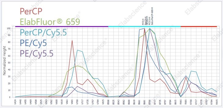
Fig. 3 Comparison of the unmixed spectral profiles of five dyes.
Elabscience® flow cytometry antibodies are dedicated to providing a superior experience for your flow cytometric analysis experiments. Elabscience® products cover human/non-human primate/rat/mouse targets, among others, with a rich selection of markers. Elabscience® also offer free flow cytometry panel design and flow cytometry data analysis services, as well as complete flow cytometry solutions! If you have any questions related to flow cytometry detection, please feel free to consult Elabscience® technical team at any time!

