

Megakaryocytes (MKs) are among the rarest cells in the bone marrow, yet they release roughly one hundred billion platelets every day. Identifying them reliably, and judging how mature they are, underpins research on thrombopoiesis, platelet biogenesis, inherited bleeding disorders and the immune functions of the megakaryocytic lineage.
● The integrin αIIbβ3 (CD41/CD61, GPIIb/IIIa) is expressed from the earliest committed MK progenitor and persists on platelets. It is the practical anchor of megakaryocyte identification.
● CD42b (GPIbα), the von Willebrand factor-binding subunit of the GPIb-IX-V complex, appears later than CD41 and CD61 and is the standard discriminator between immature (CD41a+CD42b−) and mature (CD41a+CD42b+) megakaryocytes.
● Maturation should be read on two axes at once: surface phenotype (CD41/CD61 density, CD42a and CD42b gain, CD34 and CD45 loss) and DNA content, measured with DNA-binding dyes or imaging flow cytometry.
● Megakaryocytes are immune effector cells. Toll-like receptors, FcγRIIA (CD32a), CD40L (CD154), MHC class I and II and CD80/CD86 allow them to sense pathogens, present antigen and shape inflammation.
● Activation and inflammation are reported by CD62P (P-selectin), CD40L, the PAC-1 neoepitope and loss of CD42b through shedding. These readouts must be separated from lineage identity.
This article summarises the biology behind these megakaryocyte markers, the combinations that work in practice, and the artefacts that most often distort megakaryocyte phenotyping.
Table of Contents
1. Megakaryocyte development and maturation
2. Key markers for identifying megakaryocytic lineage cells
3. How do CD41 and CD61 identify megakaryocytic lineage cells?
4. CD42b as a marker of megakaryocyte maturation and platelet identity
5. Megakaryocyte–immune cell interactions and inflammatory markers
6. Practical guidance for panel design and data interpretation of megakaryocytes and platelets
7. Related resources
01 Megakaryocyte development and maturation
Megakaryopoiesis converts a small number of haematopoietic stem cells into extremely large, polyploid cells that package their cytoplasm into platelets. It is best understood as two intertwined programmes running in parallel: nuclear polyploidisation and cytoplasmic maturation[1].
1.1 Two routes into the megakaryocyte lineage
The classical model places megakaryocytes downstream of the megakaryocyte-erythroid progenitor (MEP), the bipotent progenitor that also gives rise to the erythroid lineage. Commitment is followed by a sequence of colony-forming intermediates, the burst-forming unit-megakaryocyte (BFU-Mk) and the colony-forming unit-megakaryocyte (CFU-Mk)[1,2].
A second, stem-cell-adjacent route has been described in which committed megakaryocyte progenitors arise before and independently of the MEP split. In mouse bone marrow, GPIbα (CD42b) is detectable on a small fraction of common myeloid progenitors (megakaryocytes in bone marrow) that also express CD9, CD41 and SLAMF1[1,2].
These cells differentiate almost exclusively into megakaryocytes in culture and generate platelets after transplantation. The two routes differ in their dependence on thrombopoietin signalling, which matters when interpreting c-Mpl (CD110) staining in progenitor populations[1,2].
1.2 Endomitosis and polyploidisation
Megakaryocytes abandon the normal cell cycle. They enter mitosis, duplicate their DNA, and then fail to complete cytokinesis, a specialised process called endomitosis. The result is a single cell carrying 4N to 128N DNA, most commonly 16N or 32N in humans[3].
Ploidy is the most direct measure of nuclear maturation. It is quantified with DNA-binding fluorescent dyes such as propidium iodide and reported as the distribution of cells across the 2N, 4N, 8N, 16N and higher classes[3].
Endomitosis depends on a defined transcriptional programme. GATA1, FOG1, NF-E2, RUNX1, FLI1, TAL1 and MYB act in a stage-specific manner, and loss of these factors produces small, low-ploidy megakaryocytes[4].
1.3 Cytoplasmic maturation: granules, membranes and proplatelets
Cytoplasmic maturation is marked by a dramatic increase in cell size, from roughly 10 µm in the megakaryoblast to as much as 50 to 100 µm in the mature cell. During this phase the cells assemble their secretory machinery[5].
α-granules store von Willebrand factor, fibrinogen, PF4/CXCL4, RANTES/CCL5 and CD40L, while dense granules store ADP, ATP and serotonin. Both granule types are inherited by the platelets that are eventually released[5,6].
The demarcation membrane system (DMS) is an extensive network of membrane invaginations that acts as a surface-area reservoir. It supplies the membrane needed when the megakaryocyte extends proplatelet branches and finally sheds platelets[7].
Proplatelet formation is a microtubule-driven process that occurs asynchronously in culture. Because it is coupled to a compartmentalised form of apoptosis, the viability of fully mature megakaryocyte cultures is inherently low.
1.4 The thrombopoietin axis
Thrombopoietin binding to its receptor c-Mpl (CD110) is the master signal for megakaryocyte survival, proliferation and ploidy increase. Mice lacking c-Mpl have severely reduced megakaryocyte and platelet numbers, although some thrombopoiesis persists[8].
Inflammatory cytokines including IL-6, IL-1 and TNF-α raise thrombopoietin levels and shift megakaryopoiesis towards platelet release. This is one mechanism behind reactive thrombocytosis in infection and inflammation[7,8].
Table 1. Summarises the developmental stages of the lineage and the surface phenotype that is used to distinguish them
|
Stage |
Defining biology |
Representative surface phenotype |
|
HSC / multipotent progenitor |
Self-renewal with multilineage output |
CD34+ CD38− CD45RA− CD90+ CD110+ |
|
CMP to MEP |
Myeloid restriction; CD38 is progressively lost |
CD34+ CD38low/− CD45RA− CD123+ |
|
BFU-Mk / CFU-Mk |
Megakaryocyte-committed colony-forming progenitors |
CD34+ CD41a+ CD61+ CD42b− |
|
Megakaryoblast |
Earliest morphologically recognisable MK; 2N to 4N |
CD41a+ CD61+ CD42b−, CD34 weakly positive |
|
Promegakaryocyte |
Granule synthesis begins; DMS forms; 4N to 16N |
CD41a++ CD61++ CD42b+ |
|
Mature megakaryocyte |
Polyploid (16N to 64N); proplatelet formation |
CD41a++ CD61++ CD42a+ CD42b+ CD45− |
|
Proplatelet / platelet |
Anucleate fragments released into sinusoids |
CD41a+ CD61+ CD42a+ CD42b+ CD62P variable |
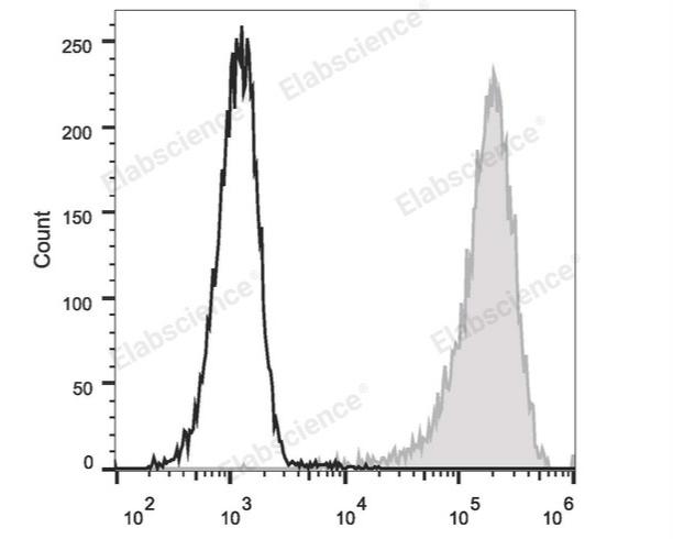
Fig. 1 Flow cytometric analysis of CD34 expression in Molt-4 cells. Human T lymphoblastic leukemia cell line Molt-4 cells were stained with PE Anti-Human CD34 Antibody (filled gray histogram) or Mouse IgG1 Isotype Control PE (empty black histogram). (The data are provided by Elabscience®)
Interpretation: CD34, also known as gp105-120, is a type I monomeric sialomucin-like glycophosphoprotein with an approximate molecular weight of 105-120 kD. Selectively expressed on the majority of hematopoietic stem/progenitor cells, bone marrow stromal cells, capillary endothelial cells, embryonic fibroblasts, and some nervous tissue, CD34 is a commonly used marker to identify human hematopoietic stem/progenitor cells. According to the differential sensitivity to enzymatic cleavage, four groups of epitopes of CD34 have been described. CD34 mediates cell adhesion and lymphocytes homing through binding to L-selectin and E-selectin ligands.
Assay Used for This Experiment:
PE Anti-Human CD34 Antibody[581]
Cat. No.: E-AB-F1143D
Sample Type: Molt-4 cells
Detection Instrument: Flow Cytometer
Fluorophores: PE
Explore the PE Anti-Human CD34 Antibody[581] Used in 15 Publications →
02 Key markers for identifying megakaryocytic lineage cells
Identifying cells of the megakaryocytic lineage (from early progenitors to mature megakaryocytes and platelets) relies on a combination of surface antigens, intracellular proteins, and functional properties. A workable megakaryocyte panel answers three separate questions: is this cell of the megakaryocytic lineage, how mature is it, and what is it currently doing? The markers below are the reagents that answer those questions most reliably[9,10].
Table 2. Lineage, maturation and activation markers used with megakaryocytes and platelets
|
Marker (gene) |
Expression profile |
Principal function |
Role in a panel |
|
|
CD41 / ITGA2B (GPIIb, integrin αIIb) |
From MK commitment onwards; density rises with maturation; retained on platelets |
αIIb subunit of integrin αIIbβ3 |
Anchor lineage marker; CD41+CD34+ identifies early MK progenitors |
|
|
CD61 / ITGB3 (GPIIIa, integrin β3) |
Parallels CD41 in the MK lineage; also pairs with CD51 |
β3 subunit of αIIbβ3 and αvβ3 |
Confirms αIIbβ3 when paired with CD41 |
|
|
CD41/CD61 activation epitope |
Exposed only after conformational change |
Fibrinogen binding; platelet aggregation |
Reports activation, not lineage (clone PAC-1) |
|
|
CD42a / GP9 (GPIX) |
Late MKs and platelets |
GPIb-IX-V complex component |
Refines maturation towards a platelet-like phenotype |
|
|
CD42b / GP1BA (GPIbα, glycocalicin) |
Mature MKs and platelets; appears after CD41 and CD61 |
von Willebrand factor receptor; adhesion under shear |
Maturation discriminator: CD41a+CD42b− versus CD41a+CD42b+ |
|
|
CD42c / GP1BB and CD42d / GP5 |
Same complex as CD42b |
GPIb-IX-V assembly and signalling |
platelet identification and complex integrity |
|
|
GPVI / GP6 |
MKs and platelets |
Collagen receptor; platelet activation |
Functional platelet phenotype |
|
|
CD9 |
MKs and platelets; also B cells and eosinophils |
Tetraspanin; supports αIIbβ3 signalling and proplatelet formation |
Auxiliary marker, especially useful in mouse |
|
|
CD36 / GPIV |
MKs, platelets, erythroid cells, endothelium, monocytes |
Thrombospondin and oxidised LDL receptor |
Additional characterisation; not lineage-specific |
|
|
CD110 / MPL (c-Mpl) |
MK progenitors and MKs |
Thrombopoietin receptor |
Identifies thrombopoietin-responsive progenitors |
|
|
CD34 |
Primitive HSPC; lost near MK commitment |
Progenitor and adhesion molecule |
Negative on mature MK; defines early MK progenitors with CD41 |
|
|
CD45 / PTPRC (LCA) |
Low or absent on mature MKs and platelets |
Protein tyrosine phosphatase |
Exclusion marker for leukocyte contamination |
|
|
CD62P / SELP (P-selectin) |
α-granule membrane; surface after activation |
PSGL-1 ligand; platelet-leukocyte adhesion |
Activation and granule-release readout |
|
|
CD154 / CD40L |
Stored in granules; surface after activation |
CD40 signalling |
Inflammation and immune-crosstalk marker |
|
|
CD32a / FCGR2A (FcγRIIA) |
MKs and platelets |
Immune-complex binding |
Immune-interaction marker |
|
|
CD47 |
MKs and platelets |
Survival signal through SIRPα |
Phagocytic clearance studies |
|
Only a few of these megakaryocyte markers are genuinely specific. CD41 and CD61 together provide the strongest practical evidence of megakaryocytic identity, because the αIIbβ3 heterodimer is assembled almost exclusively in this lineage[9,10].
CD42b adds stage information rather than identity. It is expressed on mature megakaryocytes and platelets but not on early megakaryocyte progenitors, and CD42a behaves in a similar way. The two are frequently used side by side[9,10].
Markers such as CD36, CD9 and CD61 are shared with other cell types. They are useful as supporting evidence, but they should never be the sole basis for calling a cell a megakaryocyte[9,10].
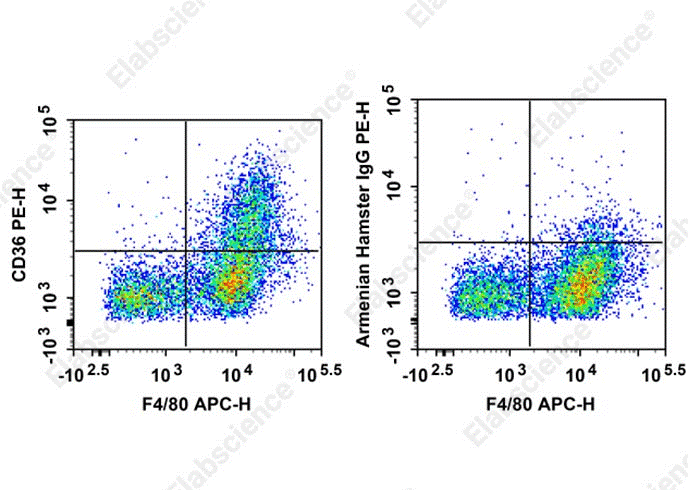
Fig. 2 Flow cytometric analysis of CD36 expression in mouse abdominal macrophages. C57BL/6 murine abdominal macrophages elicited by starch broth were stained with PE Anti-Mouse CD36 Antibody[HM36] (filled gray histogram) or PE Armenian Hamster IgG Isotype Control (empty black histogram). (The data are provided by Elabscience®)
Interpretation: CD36 is a 85 kD glycoprotein, also known as FAT, gpIIIb, or gpIV. It is a member of the class B scavenger receptor family, expressed on platelets, monocytes, macrophages, megakaryocytes, microvasculature, dendritic cells and mammary endothelial cells. The primary ligands for CD36 have been reported to be oxidized low density lipoprotein, anionic phospholipids, and collagens I, IV, and V. CD36 acts as a scavenger receptor thus promoting the removal of apoptotic neutrophils and other apoptotic bodies, as well as clearance of defective erythrocytes.
Assay Used for This Experiment:
PE Anti-Mouse CD36 Antibody[HM36]
Cat. No.: E-AB-F1291D
Sample Type: mouse abdominal macrophages
Detection Instrument: Flow Cytometer
Fluorophores: PE
Explore more applications of CD36 antibodies with various fluorescent labels →
03 How do CD41 and CD61 identify megakaryocytic lineage cells?
Megakaryocytic lineage cells can be reliably distinguished from other hematopoietic populations by detecting surface integrin proteins using anti CD41 antibody and anti CD61 antibody. CD41 (integrin αIIb) non-covalently associates with CD61 (integrin β3) to form the αIIbβ3 complex, which is one of the earliest expressed markers committed to the megakaryocyte lineage. At the stage of hematopoietic progenitor differentiation, the co-expression of CD41 and CD61 can mark cells that have begun megakaryocytic specification, even before full morphological maturation[10,11].
While anti CD41 antibody and anti CD61 antibody are excellent for lineage commitment screening, they are not exclusive to mature megakaryocytes. For further evaluation of terminal differentiation status, anti CD42b antibody is commonly incorporated into the staining panel. CD42b appears later during development and serves as one of the key megakaryocyte maturation markers[12].
In practical flow cytometry assays, a typical panel combines anti CD41 antibody and anti CD61 antibody to gate megakaryocyte lineage cells, then uses anti CD42b antibody to subdivide immature progenitors and mature megakaryocytes. Cells positive for CD41 and CD61 but negative or low for CD42b represent early megakaryocytic precursors; upregulation of CD42b indicates progression toward full maturation. This marker combination allows researchers to trace the whole developmental continuum from lineage commitment to platelet formation[9,11].
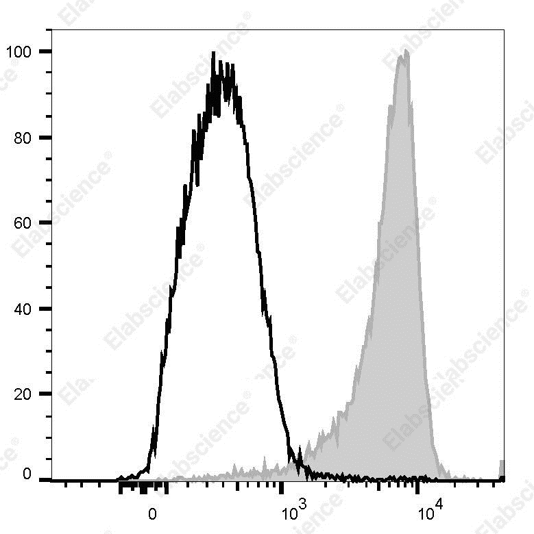
Fig. 3 Flow cytometric analysis of CD41 expression in mouse platelets. Mouse platelets were stained with FITC-conjugated anti-mouse CD41 antibody (filled gray histogram) or FITC rat IgG1, κ isotype control (open black histogram). (The data are provided by Elabscience®)
Interpretation: CD41, also known as integrin α2b and GPIIb, is a transmembrane glycoprotein that is expressed by platelets and megakaryocytes. It was reported that CD41 is also expressed on hematopoietic progenitors. CD41 associates with CD61 (integrin β3) to form complexes that interact with fibrinogen, fibronectin, von Willebrand factor, and thrombin. CD41 is required for platelet adhesion and aggregation. Defect of CD41 leads to disorders of coagulation.
Assay Used for This Experiment:
FITC Anti-Mouse CD41 Antibody[MWReg30]
Cat. No.: E-AB-F1183C
Sample Type: mouse platelets
Detection Instrument: Flow Cytometer
Fluorophores: FITC
Explore the FITC Anti-Mouse CD41 Antibody[MWReg30] Used in 3 Publications →
Fig. 4 Flow cytometric analysis of CD61 expression in mouse bone marrow cells. C57BL/6 murine bone marrow cells were stained with PE Anti-Mouse/Rat CD61 Antibody[2C9.G2] (filled gray histogram)or PE Armenian Hamster IgG Isotype Control[PIP]. (The data are provided by Elabscience®)
Interpretation: CD61 is a 110 kD integrin β chain also known as β3 integrin or gpIIIa. It associates with the integrin αv chain (CD51) to form the vitronectin receptor. In addition, CD61 can associate with the integrin αIIb chain (CD41) to form the gpIIb/IIIa complex. CD61 is expressed on platelets, megakaryocytes, endothelium, smooth muscle, a subset of B cells, myeloid cells, osteoclasts, and mast cells. CD61, in conjunction with CD41 or CD51, mediates adhesion to fibronectin, fibrinogen, vitronectin, thrombospondin, and von Willebrand factor. Leukocyte-endothelial adhesion is mediated by the binding of αv/β3 integrin or vitronectin receptor to CD31 (PECAM-1).
Assay Used for This Experiment:
PE Anti-Mouse/Rat CD61 Antibody[2C9.G2]
Cat. No.: AN00947D
Sample Type: mouse bone marrow cells
Detection Instrument: Flow Cytometer
Fluorophores: PE
Explore more applications of CD61 antibodies with various fluorescent labels →
04 CD42b as a marker of megakaryocyte maturation and platelet identity
CD42b turns a lineage gate into a maturation readout. Where CD41 and CD61 indicate that a cell is megakaryocytic, CD42b indicates how far along the maturation path it has travelled[13].
4.1 GPIb-IX-V: a four-subunit adhesion complex
CD42b is glycoprotein Ibα (GPIbα), encoded by GP1BA. It is disulfide-linked to GPIbβ (CD42c) and associates non-covalently with GPIX (CD42a) and GPV (CD42d) to form the GPIb-IX-V complex[13].
GPIbα binds von Willebrand factor and is essential for the initial capture of platelets at sites of vascular injury under high shear. It also binds thrombin, factor XI and factor XII, linking primary adhesion to coagulation.
Loss of the complex causes Bernard-Soulier syndrome, a bleeding disorder characterised by macrothrombocytopenia and absent ristocetin-induced platelet agglutination. CD42b staining is therefore informative in inherited platelet disorders as well as in basic studies[13].
4.2 CD42b as a maturation switch
CD42b expression begins later than CD41 and CD61 and is confined to mature megakaryocytes and platelets. The combination CD41a+/CD42b− therefore marks immature megakaryocytes, while CD41a+/CD42b+ marks mature cells[14].
The same logic holds in pharmacological studies. Immunomodulatory drugs such as lenalidomide and pomalidomide block endomitosis and maturation, expanding the immature CD41a+/CD42b− pool, a shift that is readily detected by flow cytometry[15].
Table 3. Summary of immune phenotype markers for megakaryocytes and platelets at different stages
|
Immunophenotype |
Suggested stage |
Interpretation |
|
CD34+ CD41a− CD42b− |
MEP / pre-commitment progenitor |
Erythroid potential retained; low ploidy |
|
CD34+ CD41a+ CD42b− |
Early megakaryocyte progenitor |
Earliest gain of CD41; still cycling |
|
CD34− CD41a++ CD61++ CD42b− |
Immature megakaryocyte |
Expanded whenever maturation is blocked |
|
CD34− CD41a++ CD61++ CD42b+ |
Mature megakaryocyte |
High ploidy; active granule synthesis |
|
CD41a++ CD61++ CD42a+ CD42b+ CD45−, high FSC |
Platelet-producing MK / proplatelet |
Preferred population for platelet-release assays |
|
CD41a+ CD61+ CD42a+ CD42b+ CD45−, low FSC |
Platelet or platelet-like particle |
Identity requires scatter or imaging calibration |
In mouse bone marrow, GPIbα is detectable on a rare progenitor population that expresses CD9, SLAMF1 and CD41 and is restricted to megakaryocytic differentiation (megakaryocytes in bone marrow). CD42b is thus informative beyond human cells[15].
4.3 CD42b as a platelet identification marker
Platelets carry CD41a, CD61, CD42a and CD42b. A CD42b-positive population that is also CD41a+CD61+ and CD45− is therefore identified as platelet or platelet-like, which is why CD42b is standard in ex vivo platelet-production workflows[13-15].
In quality control of stem cell-derived platelets, CD41a and CD61 assign lineage while CD42a and CD42b refine the product towards a platelet-like phenotype. Exclusion markers, including CD34, CD45, CD14, CD235a with CD71, and SSEA4, control residual progenitors and contaminating lineages.
4.4 When CD42b is lost
CD42b is not a fixed label. During megakaryocyte apoptosis the ectodomain is cleaved by ADAM17, releasing soluble glycocalicin and generating CD42b-negative mature megakaryocytes.
The same shedding occurs during platelet storage and in thrombocytopenic states. High-granularity CD42b-negative megakaryocytes in a culture should be read as damaged rather than immature cells[13].
This distinction has practical consequences. Protecting mature megakaryocytes from apoptosis preserves CD42b and enriches the subpopulation that actually releases functional platelets in vivo.
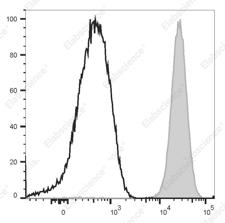
Fig. 5 Flow cytometric analysis of CD42b expression in human peripheral blood platelets cells. Normal human peripheral blood platelets cells were stained with FITC Anti-Human CD42b Antibody[HIP1] (filled gray histogram) or FITC Mouse IgG1, κ Isotype Control (empty black histogram). Cells in the platelets gate were used for analysis. (The data are provided by Elabscience®)
Interpretation: CD42b is a 145 kD glycoprotein known as gpIbα. It is covalently bonded to CD42c to form GPIb. CD42b is expressed on platelets and megakaryocytes. CD42b/c heterodimer forms a complex with CD42a and d and acts as the receptor for von Willebrand factor and thrombin.
Assay Used for This Experiment:
FITC Anti-Human CD42b Antibody[HIP1]
Cat. No.: E-AB-F1327C
Sample Type: Human peripheral blood platelets cells
Detection Instrument: Flow Cytometer
Fluorophores: FITC
Explore more applications of CD42b antibodies with various fluorescent labels →
05 Megakaryocyte–immune cell interactions and inflammatory markers
Megakaryocytes are increasingly recognised as immune cells. They express pattern-recognition receptors, present antigen, engulf leukocytes and release a broad repertoire of inflammatory mediators.
5.1 Immune receptors on megakaryocytes
Bone marrow megakaryocytes express TLR1, TLR2, TLR4, TLR6, TLR7 and TLR8, allowing direct recognition of bacterial and viral structures. TLR2 and TLR4 activation accelerates maturation and proplatelet formation, whereas TLR7 activation impairs megakaryopoiesis[17].
They also carry FcγRIIA (CD32a), which binds immune complexes, together with IL-6R, IFNAR1 and IFNAR2, C-type lectin receptors, CD40, CD80 and CD86. These receptors place megakaryocytes within both innate and cytokine-driven circuits[17,18].
MHC class I is expressed by bone marrow megakaryocytes and is transferred to platelets during proplatelet formation. Megakaryocytes in lung and spleen additionally express MHC class II and CCR7, giving them an antigen-presenting and migratory phenotype[18].
5.2 Emperipolesis and direct cell–cell contact
Neutrophils, monocytes, lymphocytes and erythroid cells pass through the megakaryocyte cytoplasm in a process called emperipolesis, with neutrophils the most common cargo.
Emperipolesis allows bidirectional membrane transfer between leukocytes and the demarcation membrane system. It accelerates platelet production and generates hybrid platelets carrying neutrophil membrane proteins.
CD40L (CD154) provides a second contact route. Megakaryocytes expressing CD40L interact with CD40 on B cells and macrophages, and platelets derived from immune-skewed megakaryocytes can drive neutrophil extracellular trap formation[19].
5.3 Inflammatory mediators and emergency thrombopoiesis
Megakaryocytes and platelets store and release PF4/CXCL4, RANTES/CCL5, CXCL12, PPBP and TGF-β, together with IL-6, IL-1β, TNF-α and IFN-β produced under inflammatory stimulation[16].
Infection triggers emergency megakaryopoiesis. A CXCR4-high megakaryocyte subset appears and supports bacterial phagocytosis by macrophages and neutrophils through TNF-α and IL-6 secretion[16].
Activated megakaryocytes can also release extracellular chromatin webs analogous to neutrophil extracellular traps. In sepsis these structures contribute to microvascular thrombosis and disseminated intravascular coagulation[20].
Table 4. Summary of megakaryocyte–immune cell interactions and inflammatory markers
|
Marker |
Molecule |
Role in megakaryocyte immunity |
|
TLR1, TLR2, TLR4, TLR6, TLR7, TLR8 |
Toll-like receptors |
Pathogen sensing; TLR2 and TLR4 accelerate maturation, TLR7 impairs it |
|
CD32a |
FcγRIIA |
Immune-complex binding and pathogen clearance |
|
CD154 |
CD40L |
Interaction with B cells and macrophages; promotes NETosis |
|
CD40 |
CD40 |
Reciprocal signalling with CD40L-expressing cells |
|
HLA-ABC |
MHC class I |
Cross-presentation of antigen to CD8+ T cells |
|
HLA-DR |
MHC class II |
Antigen presentation by lung and spleen megakaryocytes |
|
CD80, CD86 |
Costimulatory ligands |
T-cell costimulation |
|
CD126 |
IL-6 receptor |
Inflammatory cytokine signalling |
|
IFNAR1, IFNAR2 |
Type I interferon receptor |
Antiviral responses; dendritic-cell-driven megakaryopoiesis |
|
CD62P |
P-selectin |
Platelet-neutrophil and platelet-monocyte binding |
|
CXCR4 |
CXCR4 |
Migration of emergency megakaryocytes to lung and spleen |
|
CD274 |
PD-L1 |
Immunoregulation of T-cell responses |
|
CD47 |
CD47 |
Survival signal; limits phagocytic clearance |
|
CD54, CD11a |
ICAM-1, LFA-1 |
Adhesion to leukocytes and endothelium |
5.4 Markers that report the inflammatory state
Activation is read at the surface. CD62P (P-selectin) moves from the α-granule membrane to the plasma membrane when the cell is activated, making it the standard granule-release marker[21].
CD40L upregulation and binding of the activation-dependent PAC-1 epitope report a second, integrin-based activation axis. Annexin V or phosphatidylserine staining adds the procoagulant surface that supports thrombin generation[21].
Loss of CD42b through ADAM17-mediated shedding is a further inflammatory signature. Read together with CD62P and annexin V, it separates activated platelets from dying megakaryocytes.
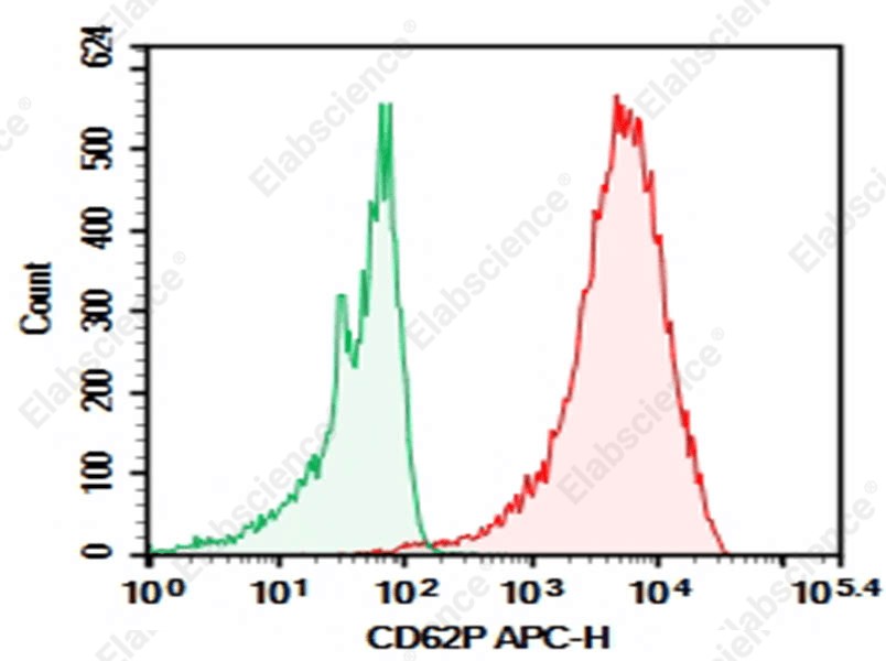
Fig. 6 Flow cytometric analysis of CD62P expression in human peripheral blood platelets cells. Normal human peripheral blood platelets cells were stained with 0.2 μg Purified Anti-Human CD62P Antibody[HI62P] (Right, red) and 0.2 μg Mouse IgG1, κ Isotype Control (Left, green), followed by APC-conjugated Goat Anti-Mouse IgG Secondary Antibody. (The data are provided by Elabscience®)
Interpretation: CD62P, also termed P-selectin, is a signature adhesion molecule rapidly exposed upon activation of platelets and vascular endothelial cells. Under resting conditions, it is primarily stored within platelet α-granules and endothelial Weibel–Palade bodies. Within minutes of stimulation, CD62P translocates to the cell membrane surface and mediates platelet adhesion to neutrophils and monocytes. As a critical mediator linking thrombosis and local inflammatory responses, its soluble plasma fragment sCD62P directly reflects the in vivo activation status of platelets and endothelial cells. CD62P serves as a widely used sensitive biomarker for clinical evaluation of pre-thrombotic states, monitoring antiplatelet drug efficacy, and aiding assessment of vascular injury severity in disorders including atherosclerosis and severe infections.
Assay Used for This Experiment:
Purified Anti-Human CD62P Antibody[HI62P]
Cat. No.: AN008350P
Sample Type: Human peripheral blood platelets cells
Detection Instrument: Flow Cytometer
Fluorophores: APC
Explore more applications of CD62P antibodies with various fluorescent labels →
06 Practical guidance for panel design and data interpretation of megakaryocytes and platelets
This practical guidance systematically covers panel design, pre-analytical control, gating strategy and data interpretation for flow cytometric analysis of megakaryocytes (MKs) and platelets, addressing the unique technical challenges of these two closely linked cell populations.
6.1 Panel Design Principles
The panels below adapt these markers to specific experimental questions. All of them assume a viability dye, a singlet gate and, for bone marrow or culture samples, scatter-based selection of the large-cell population[20-23].
Table 5. Criteria for marker selection in flow cytometric analysis of megakaryocytes and platelets
|
Objective |
Core markers |
Comment |
|
Lineage identification |
CD41a, CD61, CD45, viability dye |
Use CD41a+CD61+CD45− as the backbone; add CD34 for progenitors |
|
Maturation staging |
CD41a, CD61, CD42a, CD42b, CD34 |
Read CD42b gain alongside CD34 loss |
|
Ploidy assessment |
CD41a, CD42b, propidium iodide |
Report the 2N to 128N distribution; requires permeabilisation |
|
Platelet-like particle QC |
CD41a, CD61, CD42a, CD42b, CD34, CD45, CD14, CD235a with CD71 |
Exclusion markers control residual progenitors and contaminants |
|
Activation state |
CD62P, PAC-1, annexin V, CD42b |
Staining conditions matter; avoid EDTA when using PAC-1 |
|
Immune interaction |
CD32a, CD154, HLA-ABC, CD62P, TLR2 and TLR4 |
Add CD45 and CD66b to identify bound leukocytes |
|
Mouse megakaryocytes |
CD41, CD61, GPIbα (CD42b), CD9, CD150 |
Mouse GPIbα also marks a stem-cell-adjacent MK progenitor |
Four artefacts account for most erroneous megakaryocyte data. Platelets adhering to leukocytes create false CD41a+CD61+ events; very large megakaryocytes are lost from the scatter gate or discarded as doublets; CD42b sheds during apoptosis and storage; and activation during blood collection raises CD62P before the experiment begins. Prompt fixation, appropriate anticoagulant choice and imaging flow cytometry each address part of the problem.
6.2 Pre-Analytical Critical Controls
(1) Anticoagulant selection: For platelet activation and function assays, select the anticoagulant according to the assay requirements. Sodium citrate is commonly preferred, while EDTA should be avoided for PAC-1 binding assays because it disrupts αIIbβ3 integrin function. The suitability of heparin should be validated for the specific assay.
(2) Sample processing window: Complete staining and acquisition within 4 hours after venipuncture, ideally within 30 minutes; discard the first 2 mL of drawn blood to avoid in-vitro activation caused by needle trauma .
(3) Prevent artificial activation: Avoid mechanical vibration, high temperature and repeated freeze-thaw of samples; for fixed samples, confirm that the fixative does not destroy the epitopes of target markers such as CD42b and CD62P.
6.3 Standardized Gating Strategy
(1) Platelet gating workflow: Set FSC and SSC to logarithmic scale, first gate CD41+ events to exclude red blood cells, cell debris and noise, then further separate single platelets from platelet-leukocyte aggregates and platelet microparticles. Use FMO (Fluorescence Minus One) controls to define the positive/negative boundary of activation markers, rather than relying solely on isotype controls .
(2) Megakaryocyte gating workflow: For conventional flow cytometry: First gate CD41+ events, then exclude CD45+ leukocytes and dump channel positive cells, and use FSC-A vs SSC-A to identify large MK events and exclude cell clumps.
(3) For imaging flow cytometry: Restrict DNA signal measurement strictly within the CD41+ fluorescence mask, use the ratio of halo mask to CD41 mask to exclude platelets attached to other cells, and use machine learning LDA classifier to distinguish true single MKs from doublets, which can significantly improve the accuracy of ploidy analysis.
6.4 Quality Assurance Requirements
● Run normal donor reference samples in parallel for each batch of experiments, and use the MFI ratio relative to the reference to report results, rather than absolute fluorescence intensity values.
● For rare MK populations, acquire at least 50,000 CD41+ events to ensure statistical reliability of ploidy distribution and subset frequency.
● All gating boundaries must be verified with FMO controls, and the analysis logic must be reproducible across different operators and instruments.
Taken together, the essential discipline is to keep the three questions separate: lineage identity from CD41 and CD61, maturation from CD42b and ploidy, and functional state from CD62P, CD40L and the activation-dependent integrin epitope.
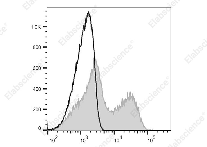
Fig. 7 Flow cytometric analysis of CD40L expression in human peripheral blood mononuclear cells. Normal human peripheral blood mononuclear cells were stained with Elab Fluor® 647 Anti-human CD154 Antibody[24-31] (filled gray histogram) or Elab Fluor® 647 Mouse IgG1, κ Isotype Control (empty black histogram). (The data are provided by Elabscience®)
Interpretation: CD154 (CD40 ligand) is also known as CD40L, gp39, TRAP and T-BAM. CD40 ligand is a 32-39 kD type II transmembrane glycoprotein. It is a member of the TNF superfamily and is expressed on activated T cells. It has been reported to be important for B cell costimulation following binding of its receptor, CD40. Additionally, binding of CD40L to CD40 on B cells promotes the secretion of immunoglobulin and Ig isotype switching. CD40L is also involved in the regulation of cytokine production by T cells.
Assay Used for This Experiment:
Elab Fluor® 647 Anti-Human CD154/CD40L Antibody[24-31]
Cat. No.: AN00341M
Sample Type: human peripheral blood mononuclear cells
Detection Instrument: Flow Cytometer
Fluorophores: Elab Fluor® 647
Explore more applications of CD40L antibodies with various fluorescent labels →
07 Related resources
● Explore common immune cell populations and learn about the markers used for immunophenotyping..
Read Phenotyping of Common Immune Cells →
● Simplify flow cytometry panel design with expert support tailored to your research needs.
Explore Flow Cytometry Panel Design Services →
References:
[1] Deutsch V R , Tomer A .Megakaryocyte development and platelet production[J].British Journal of Haematology, 2006, 134(5).
[2] Shoeb M R , Schinnerl D , Shaw L E ,et al.A stem cell differentiation model reveals two alternative fates in CBFA2T3::GLIS2-driven acute megakaryoblastic leukemia initiation[J].Communications Biology, 2025.
[3] Zhang Y , Wang Z , Ravid K .The Cell Cycle in Polyploid Megakaryocytes Is Associated with Reduced Activity of Cyclin B1-dependent Cdc2 Kinase[J].Journal of Biological Chemistry, 1996, 271(8):4266.
[4] Yokomizo T , Takahashi S , Mochizuki N ,et al.Characterization of GATA-1(+) hemangioblastic cells in the mouse embryo.[J].Embo Journal, 2007, 26(1):184.
[5] Guerrero J A , Bennett C , Weyden L V D ,et al.Gray platelet syndrome: proinflammatory megakaryocytes and α-granule loss cause myelofibrosis and confer metastasis resistance in mice.[J].Blood, 2014, 124(24).
[6] Deutsch V R , Tomer A .Megakaryocyte development and platelet production[J].British Journal of Haematology, 2006, 134(5).
[7] Chang Y , Bluteau D , Debili N ,et al.From hematopoietic stem cells to platelets[J].Journal of Thrombosis & Haemostasis, 2010, 5(s1):318-327.
[8] Novershtern N , Subramanian A , Lawton L N ,et al.Densely interconnected transcriptional circuits control cell states in human hematopoiesis.[J].Cell, 2011, 144(2):296-309.
[9] Miyawaki K , Iwasaki H , Jiromaru T ,et al.Identification of unipotent megakaryocyte progenitors in human hematopoiesis[J].Blood, 2017:blood-2016-09-741611.
[10] Novershtern N , Subramanian A , Lawton L N ,et al.Densely interconnected transcriptional circuits control cell states in human hematopoiesis.[J].Cell, 2011, 144(2):296-309.
[11] Wang H , He J , Xu C ,et al.Decoding Human Megakaryocyte Development[J].Cell Stem Cell, 2020.DOI:10.1016/j.stem.2020.11.006.
[12] Matsumura-Takeda K , Sogo S , Isakari Y ,et al.CD41+/CD45+ Cells Without Acetylcholinesterase Activity Are Immature and a Major Megakaryocytic Population in Murine Bone Marrow[J].STEM CELLS, 2007.
[13] Nishikii H , Kurita N , Chiba S .The Road Map for Megakaryopoietic Lineage from Hematopoietic Stem/Progenitor Cells[J].Stem Cells Transl Med, 2017, 6(8):1661-1665.
[14] Moran B , Shattil S .Megakaryocytes derived from human embryonic stem cells: a genetically tractable system to study megakaryocytopoiesis and integrin function[J].Journal of Thrombosis & Haemostasis, 2010, 4(2):436-442.
[15] Velzen J F V , Gorkom L V , Pop G A M ,et al.Multicolor flow cytometry for evaluation of platelet surface antigens and activation markers[J].Thrombosis Research, 2012, 130(1):92-98.
[16] Rehman J .Decision letter: CXCR4high megakaryocytes regulate host-defense immunity against bacterial pathogens[J]. 2022.
[17] Hua T , Yao F , Wang H ,et al.Megakaryocyte in sepsis: the trinity of coagulation, inflammation and immunity[J].Critical Care, 2024, 28(1):1-17.
[18] D'Atri L P , Etulain J , Rivadeneyra L ,et al.Expression and functionality of Toll‐like receptor 3 in the megakaryocytic lineage[J].Journal of Thrombosis and Haemostasis, 2015, 13(5):839-850.
[19] Marcoux G ,Audrée.Laroche, Romero J E ,et al.Role of platelets and megakaryocytes in adaptive immunity[J].Platelets, 2021, 32(3):340-351.
[20] Velzen J F V , Gorkom L V , Pop G A M ,et al.Multicolor flow cytometry for evaluation of platelet surface antigens and activation markers[J].Thrombosis Research, 2012, 130(1):92-98.
[21] Bendas G , Gobec M , Schlesinger M .Modulating Immune Responses: The Double-Edged Sword of Platelet CD40L[J].Seminars in Thrombosis and Hemostasis[2026-10-19].
[22] Shattil S J , Hoxie J A , Cunningham M J ,et al.Changes in the platelet membrane glycoprotein IIb.IIIa complex during platelet activation.[J].Journal of Biological Chemistry, 1985, 260(20):11107.
[23] Detection of Activated Feline Platelets in Platelet-rich Plasma by Use of Fluorescein-labeled Antibodies and Flow Cytometry[J].Veterinary Pathology, 1994, 31(5):553.

