

In tumor animal experiments, the three most frustrating things are: cells are implanted but no tumors grow; tumors are harvested but single cells cannot be isolated; and when running flow cytometry, there are too many nontarget cells and the background explodes. Many experiments fail not because the study design is flawed, but because details in animal modeling and sample preprocessing go wrong.
Today, Elabscience® has compiled a complete workflow for tumor bearing mouse models: from cell line selection and pitfall avoidance, to standardized modeling, to preparation of suspensions of single cells from tumor tissue, and flow cytometry quality control, providing solid support for your tumor research animal model preparation experiments.
Table of Contents
1. Classification and preparation of tumor-bearing mouse models
2. Tumor cell lines with high, moderate, and low engraftment potential
3. Preparation of tumor single-cell suspensions and flow cytometry detection
01 Classification and preparation of tumor-bearing mouse models
Tumor bearing mouse models are mainly classified by cell source into CDX (Cell-Line Derived Xenograft) models and PDX (Patient-Derived Xenograft) models.
CDX involves inoculating tumor cell lines cultured in vitro into immunodeficient mice. It offers a high degree of standardization, short turnaround time, and low cost, making it the most widely used tumor animal model. PDX involves directly transplanting fresh patient tumor tissue. It is not subject to the selection pressure of in vitro culture and can preserve the tumor's native stroma, interactions between cells, and tumor heterogeneity, but its preparation workflow is complex and the technical threshold is higher.
Neither model is absolutely superior to the other; the choice should be made based on research goals: when rapidly conducting compound activity screening or molecular target validation, CDX is preferred; whereas for predicting clinical drug efficacy, investigating tumor heterogeneity evolution, or developing personalized treatment regimens, PDX offers greater advantages with a predictive accuracy exceeding 80%. A detailed comparison of CDX and PDX is shown in Table 1.
Table 1. Summary of Detailed Comparison Between CDX and PDX Models
|
Comparison Dimension |
CDX (Cell-Line Derived Xenograft) |
PDX (Patient-Derived Xenograft) |
|
Tumor Source |
Established immortalized tumor cell lines of human or murine origin (subjected to long term in vitro culture, selection, and passaging) |
Fresh surgically resected patient tumor tissue, transplanted directly without in vitro culture |
|
Preparation Workflow |
Cells in logarithmic growth phase → resuspended in PBS and Matrigel → subcutaneous, orthotopic, or tail vein injection (typically 5 × 10⁶/200 μL) |
Fresh tumor tissue → cut into 2-3 mm³ fragments → subcutaneous or orthotopic implantation → serial passage and expansion through F1 → F2 → F3 |
|
Host Mouse |
Nude mice/NOD-SCID/NSG, etc., flexibly selected according to the engraftment capacity of the cell line |
Highly immunodeficient strains (NSG/NOG) required to maximally reduce rejection and improve transplantation success rate |
|
Tumor Formation Period |
1-4 weeks; rapid and highly synchronized |
2-4 months; long latency and considerable individual variation |
|
Tumor Heterogeneity |
Low. Long term in vitro culture leads to enrichment of dominant clones and loss of intratumoral heterogeneity (ITH), with tumors becoming homogenized |
High. Maximally preserves the mutational landscape, clonal architecture, and intratumoral heterogeneity of the original tumor |
|
Tumor Microenvironment (TME) |
Severely deficient. Tumors consist mainly of cancer cells, lacking original stromal cells, immune infiltration, and extracellular matrix signaling |
Relatively preserved. Transplanted tumor fragments retain original stromal components and interactions between cells, but stroma of human origin is gradually replaced by murine stroma with increasing passages |
|
Genomic Fidelity |
Low. Long term in vitro culture causes significant genetic drift, with gene expression profiles diverging substantially from the original tumor. Melanoma CDX is characterized by a hypoxia induced gene signature (hsa-miR-210 overexpression) and clearly separates from PDX into distinct clustering clusters |
High. In early passages (F1-F3), mutation profiles and gene expression patterns remain highly similar to the original patient tumor, although clonal drift may also occur with increasing passages |
|
Clinical Predictive Value |
~5%. Due to loss of heterogeneity and TME, drug response results rarely accurately predict clinical efficacy |
>80%. Considered the preclinical model with the greatest clinical predictive value; significantly outperforms CDX and GEMM in predictive capacity |
|
Core Advantages |
① Standardized operation and low technical threshold; ② Low cost and short turnaround time; ③ Good tumor formation synchrony and high reproducibility; ④Suitable for large scale drug screening with high throughput; ⑤ Abundant cell line resources and flexible selection. |
① Preserves the histopathological architecture and molecular features of the original tumor; ② Highly reproduces intratumoral heterogeneity; ③Highest clinical predictive value, applicable to trials conducted alongside clinical studies and personalized drug sensitivity testing; ④ Enables serial passaging and biobanking to form a "PDX avatar library." |
|
Core Limitations |
① Long term in vitro culture leads to genetic drift and phenotypic remodeling; ② Lack of tumor heterogeneity; ③ TME essentially lost; ④ Extremely low accuracy in predicting drug response; ⑤ Difficult to model the metastatic process |
① Stromal replacement (human stroma → murine stroma); ② Clonal drift (selective expansion of dominant clones during passaging); ③ Long turnaround time and high cost; ④ Transplantation success rate varies by tumor type (e.g., 58% for ovarian cancer); ⑤ Conventional PDX lacks an immune system, precluding direct evaluation of immunotherapy |
|
Immunotherapy Evaluation |
Not applicable. Requires growth in immunodeficient mice, precluding assessment of immune checkpoint inhibitors, etc. |
Not applicable (conventional PDX). Requires transition to humanized mouse PDX (reconstitution of the human immune system in NSG mice followed by PDX transplantation) |
|
Typical Application Scenarios |
① Large scale screening in drug discovery at an early stage; ②Target validation and mechanistic studies (when tumor heterogeneity is not a core concern); ③Pharmacokinetic / pharmacodynamic (PK/PD) studies; ④ Rapid acquisition of data with sufficient statistical power. |
①Individualized precision medicine for screening optimal regimens in patients; ②Preclinical trials conducted alongside clinical trials for novel drugs; ③Drug resistance mechanism research; ④ Biomarker discovery and validation; ⑤ Tumor evolution and clonal dynamics research. |
|
Cost and Throughput |
Low cost, high throughput. Suitable for batch drug screening, with hundreds of mice run in parallel |
High cost, low throughput. Establishing a single model library takes months and consumes substantial resources |
02 Tumor cell lines with high, moderate, and low engraftment potential
Differences in the engraftment capacity of cell lines represent one of the most important variables in oncology experiments. Below, common cell lines with well documented in vivo tumor formation data in the literature are categorized:
2.1 Highly Tumorigenic Cell Lines (Easy to Engraft)
Under standard inoculation conditions (5 × 10⁶ cells, subcutaneous injection into nude mice), these cell lines typically form stable tumors within 1-4 weeks, with an engraftment rate >80%. A detailed summary is provided in the table below:
Table 2. Summary of Highly Tumorigenic Cell Lines (Easy to Engraft)
|
Cell Line |
Tumor Type |
Typical Characteristics |
|
HCT116 |
Human colon cancer |
Rapid tumor formation, widely used for colon cancer CDX modeling |
|
A549 |
Human non-small cell lung cancer |
A classic lung cancer model cell line with stable tumor formation |
|
HT-29 |
Human colon cancer |
Highly tumorigenic and chemo resistant, suitable for refractory tumor research |
|
PC-3 |
Human prostate cancer |
A classic prostate cancer cell line that is resistant to castration and shows a high engraftment rate; Matrigel can further enhance tumor formation |
|
MDA-MB-231 |
Human triple negative breast cancer |
Highly invasive, widely used in breast cancer stemness research and metastasis models |
|
SK-OV-3 |
Human ovarian cancer |
A subline selected in vivo (IP2) with significantly enhanced tumorigenicity, capable of forming intraperitoneal tumors at low inoculation doses |
|
HL60 |
Human acute promyelocytic leukemia |
The parental HL60-G is highly tumorigenic, but tumorigenicity can be markedly reduced after long term 1,25D3 treatment |
|
4T1 |
Mouse breast cancer |
Tumor formation with inoculation of 1×10⁶-5×10⁶ cells, >95%, with spontaneous lung/liver/bone metastasis |
|
CT26.WT |
Mouse colorectal cancer |
Subcutaneous inoculation of 1×10³-1×10⁶ cells, engraftment rate >90%, tumor formation in 7-14 days; a widely recognized highly reliable colon cancer model in immunotherapy research |
|
MC38 |
Mouse colorectal cancer |
Conventional subcutaneous inoculation of 1×10⁶ cells yields an engraftment rate of up to 100%. High mutational burden, strong immunogenicity, and stable tumor formation are its core features, making it a recognized benchmark model in tumor immunotherapy research |
|
EMT6 |
Mouse breast cancer |
Engraftment rate 80-90%, with low metastatic propensity |
|
H22 |
Mouse liver cancer |
Ascites type, engraftment rate >95%, rapid growth, and low cost |
|
PAN02 |
Pancreatic cancer |
Subcutaneous inoculation of 1×10⁶-2×10⁶ cells yields an engraftment rate of up to 90%; low immunogenicity, high fibrosis, and stable tumor formation are its core features, making it a benchmark model for simulating the immune cold tumor microenvironment of human pancreatic cancer |
|
RENCA |
Renal cancer |
Subcutaneous inoculation of 1×10⁵-1×10⁶ cells yields an engraftment rate of up to 90%; high engraftment stability and faithful recapitulation of human renal cancer metastatic features are its core features, making it a benchmark model for renal cancer immunotherapy and metastasis research |
|
A20 |
Mouse B cell lymphoma |
Subcutaneous inoculation of 2×10⁶ or 2×10⁷ cells yields an engraftment rate of up to 100%, with tumor formation times of approximately 15 days and 7 days, respectively; tail vein injection of 2×10⁶ cells yields an engraftment rate of approximately 71.4%, while the 2×10⁷ group reaches 100%; splenic injection yields approximately 71.4%; intraperitoneal injection yields only 14.3% |
|
B16-F1/B16-F10 |
Mouse melanoma |
F10 is a lung metastasis subline with extremely rapid tumor formation (5-10 days) and an engraftment rate >95% |
|
LLC1 |
Mouse lung cancer |
Subcutaneous inoculation of 1×10⁶-2×10⁶ cells yields an engraftment rate of 90-100%, with visible tumor nodules observed 7-10 days after inoculation |
2.2 Moderately Tumorigenic Cell Lines
Engraftment rate 50-80%; may require specific conditions (e.g., Matrigel assistance, higher inoculation doses, or specific mouse strains).
Table 3. Summary of Moderately Tumorigenic Cell Lines (Requiring Special Handling)
|
Cell Line |
Tumor Type |
Optimization Conditions |
|
OVCAR-3 |
Ovarian cancer |
Intraperitoneal injection is superior to subcutaneous injection; Matrigel or collagen matrix can enhance the engraftment rate. |
|
NCI-H727 |
Pulmonary carcinoid |
Tumor formation and angiogenesis can be assessed in a zebrafish xenograft model. |
|
XGC-1 |
Gastric cancer |
Engraftment must be validated by subcutaneous inoculation in BALB/c nude mice. |
2.3 Cell Lines with Low Tumorigenicity and Poor Engraftment
Under standard conditions, these cell lines exhibit low engraftment rates (<50%), long latency, or require special handling.
Table 4. Summary of Cell Lines with Low Tumorigenicity and Poor Engraftment
|
Cell Line |
Tumor Type |
Possible Reasons for Poor Engraftment and Countermeasures |
|
Breast cancer cell lines with a basal like phenotype (e.g., the KBC series derived from premenopausal Taiwanese patients) |
Breast cancer |
Breast cancer cell lines with a basal like phenotype from different ethnic origins exhibit extremely low tumorigenicity, possibly related to specific genetic backgrounds and epigenetic features. |
|
ccRCC primary cells |
Clear cell renal cell carcinoma |
The standard limiting dilution assay (LDA) significantly underestimates their tumorigenicity. The tissue dissociation process kills the majority of ccRCC cells, whereas transplantation of intact tumor fragments yields far higher engraftment efficiency than suspensions of single cells. |
03 Preparation of tumor single-cell suspensions and flow cytometry detection
When flow cytometry experiments on tumors fail, the problem often lies in sample preparation: tissue cannot be minced properly, digestion is excessive, too many cells die, or severe cell clumping occurs. This protocol is specifically designed for tumor tissues, yielding clean results and accurate positivity rates upon acquisition.
3.1 Experimental Preparation
(1) Animals and Reagents: 6-8-week-old healthy BALB/c mice, 75% ethanol, RPMI 1640 basal medium, high quality fetal bovine serum, a penicillin and streptomycin solution (double antibiotics), and an alanyl glutamine solution, sterile deionized water, sterile PBS, Cell Staining Buffer (E-CK-A107), 10× ACK Lysis Buffer (E-CK-A105), etc.
(2) Consumables: Disposable sterile syringes, cell culture dishes, pipette tips, experimental mice, sterile 300 mesh nylon filters, sterile 15/50 mL centrifuge tubes, ophthalmic scissors, ophthalmic forceps.
(3) Flow cytometry detection antibodies are shown in the table below
Table 5. Flow cytometry antibody product list
|
Product Name |
Cat. No. |
|
STYX™ Yellow Fixable Viability Kit |
E-CK-A169 |
|
FITC Anti-Mouse/Human CD11b Antibody[M1/70] |
E-AB-F1081C |
|
Elab Fluor® Violet 450 Anti-Mouse CD45 Antibody[30-F11] |
E-AB-F1136Q |
|
PerCP/Cyanine5.5 Anti-Mouse F4/80 Antibody [CI: A3-1] |
E-AB-F0995J |
|
PE Anti-Mouse CD206/MMR Antibody[C068C2] |
E-AB-F1135D |
|
APC Anti-Mouse CD86 Antibody [GL-1] |
E-AB-F0994E |
|
Purified Anti-Mouse CD16/32 Antibody [2.4G2] |
E-AB-F0997A |
|
Elab Fluor® 700 Anti-Mouse MHC II (I-A/I-E) Antibody[M5/114] |
E-AB-F0990M1 |
3.2 Experimental Procedure
(1) Reagent Preparation: Prepare RPMI-1640 complete medium containing 10% FBS and 1% double antibiotics, and store at 4°C for later use. The tumor digestion solution consists of collagenase IV (1 mg/mL) and DNase I (100 μg/mL) dissolved in RPMI-1640, prepared fresh immediately before use.
(2) Preparation of a Mouse Model Bearing 4T1 Tumors: Take 4T1 cells in logarithmic growth phase (mouse breast cancer cells, triple negative, highly metastatic), digest with trypsin, wash with PBS, and resuspend in medium without serum to adjust the concentration to 5 × 10⁶ cells/mL. Inoculate 100 μL of the suspension subcutaneously into the right dorsal flank of 6-8-week-old female BALB/c mice. On day 13 after transplantation, when the tumor volume measures approximately 900 mm³, euthanize the mice by CO₂ inhalation followed by cervical dislocation for confirmation of death, and excise the tumors. Cut into 1 mm³ fragments, add digestion solution, and digest at 37°C with shaking for 30-60 min, pipetting gently every 10 min. After termination, filter through a 70 μm mesh and wash with PBS to obtain a single cell suspension.
Note: All animal experiments were reviewed and approved by the institutional animal ethics committee and conducted in accordance with animal welfare and the 3Rs principles.
(3) Preparation of Tumor Tissue Single Cell Suspensions:
A. Mixed Enzymatic Digestion Method
a. After all tumors have been excised, transfer the tumors to 1.5 mL EP tubes or cell culture dishes, cut into 1 mm³ fragments, add tumor digestion solution, and digest at 37°C with shaking for 30-45 min, pipetting gently every 10 min.
b. After digestion, immediately add 5-10 mL complete medium and mix gently by pipetting to terminate digestion. Then transfer the tumor tissue suspension to a 15 mL centrifuge tube, centrifuge at 250 × g for 5 min, and discard the supernatant.
c. Add 5-10 mL of 1640 basal medium or PBS for dilution, then filter through a 200-mesh sieve to remove residual tissue fragments. Wash once with 5-10 volumes of 1640 basal medium or PBS buffer, filtering until no tissue fragments remain, to obtain a single cell suspension.
d. Collect the cell suspension, centrifuge at 300 × g for 5 min, and discard the supernatant.
e. Resuspend the cells in cell staining buffer and adjust the cell concentration to 1 × 10⁷/mL.
Note: If needed, the cell density can also be adjusted to an appropriate range using buffers suitable for subsequent experiments, such as using cell sorting buffer to adjust the cells to an appropriate density for subsequent cell sorting.
B. Grinding Method
a. Prepare 200 mesh disposable cell strainers in advance, moisten and soak them with 1640 basal medium or PBS (place in a 6 cm cell culture dish or 6 well plate for soaking).
b. Transfer the excised tumor tissue onto the 200-mesh cell strainer and cut into small pieces with sterile ophthalmic scissors.
c. Use the plunger of a 2.5 mL syringe to grind the tissue in a circular motion with the soft tip until no obvious tissue fragments remain on the strainer. Rinse the strainer 2-3 times with fresh 1640 medium or PBS.
d. Filter the resulting cell suspension through a 200 mesh cell strainer.
e. Collect the cell suspension, centrifuge at 300 × g for 5 min, and discard the supernatant.
f. Resuspend the cells in cell staining buffer and adjust the cell concentration to 1 × 10⁷/mL.
Note: If needed, the cell density can also be adjusted to an appropriate range using buffers suitable for subsequent experiments, such as using cell sorting buffer to adjust the cells to an appropriate density for subsequent cell sorting.
(4) Flow Cytometry Detection: After centrifugation of the cell suspension, add ACK lysis buffer to lyse red blood cells. After lysis is complete, add PBS to terminate the lysis; wash the cells with PBS and perform cell counting. Adjust the cell concentration to 1 × 10⁷ cells/mL, transfer 100 μL (1 × 10⁶ cells) to a flow cytometry tube, add 0.5-1 μg of purified anti-mouse CD16/32 monoclonal antibody (E-AB-F0997A) to block Fc receptors, and incubate at room temperature for 10 min; then directly add fluorescently labeled antibodies and incubate at 4°C in the dark for 30 min. After staining, wash with PBS or Cell Staining Buffer (E-CK-A107), resuspend in PBS or Cell Staining Buffer, and acquire on the flow cytometer.
3.3 Presentation of Experimental Results
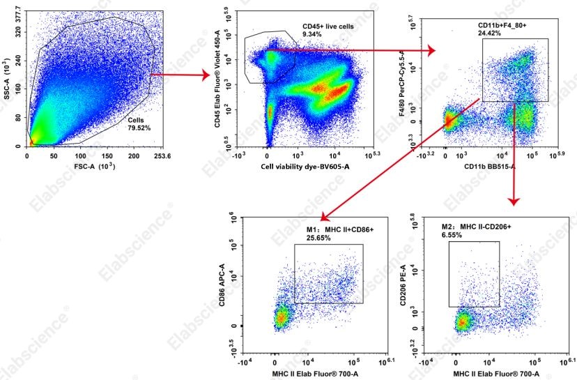
Fig. 1 Detection of macrophages associated with tumors (TAMs) in 4T1 transplanted tumors in BALB/c mice. Twenty days after subcutaneous injection of 4T1 cells into the neck of BALB/c mice, tissues bearing tumors were harvested, and suspensions of single cells were prepared by enzymatic digestion. Flow cytometry antibodies against CD45, CD11b, F4/80, MHC II, CD206, and CD86, along with a viability dye, were used to detect macrophages in the 4T1 transplanted tumors. The results showed that M1 type macrophages accounted for 25.65%, while M2 type macrophages accounted for 6.55%.
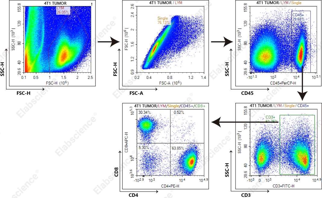
Fig. 2 Detection of T cells associated with tumors in 4T1 transplanted BALB/c mouse models. Thirteen days after subcutaneous injection of 4T1 cells into the neck of BALB/c mice, tissues bearing tumors were harvested, and suspensions of single cells were prepared by enzymatic digestion. A viability dye and flow cytometry antibodies against CD45, CD3, CD4, and CD8 were used to detect T cells in the 4T1 transplanted tumors.
3.4 Experimental Notes
(1) Tumor volume should generally not exceed 1000 mm³, and tumor mass should be between 0.6-0.8 g. (If tumors are larger, double the volumes of all reaction systems described below.)
(2) For enzymatic digestion, the criterion for mincing tumors with scissors is that the tissue can be freely aspirated through a 1 mL pipette tip without obstruction.
(3) During enzymatic digestion, avoid excessive tissue digestion. Remove and inspect the tissue every 20 min, and terminate digestion once no obvious tissue fragments remain. The smaller the cell clumps, the faster the digestion; tissue fragments that can be pipetted with a 1 mL pipette typically require approximately 30-60 min for complete digestion. If the tissue is difficult to mince, it can also be cut into pieces of approximately 1-2 mm³, pipetted gently with a 1 mL pipette every 10 min until the tissue fragments pass smoothly through the 1 mL pipette, with the entire digestion taking approximately 30-60 min.
(4) During grinding, to minimize cell damage, the tissue must be soaked in medium while grinding; avoid dry grinding.
References:
[1] LONG Y, XIE B, SHEN H C, et al. Translation Potential and Challenges of In Vitro and Murine Models in Cancer Clinic[J]. Cells, 2022, 11(23): 3868.
[2] XU C, LI X, LIU P, et al. Patient‑derived xenograft mouse models: A high-fidelity tool for individualized medicine (Review) [J]. Oncology Letters, 2018.
[3] ZENG M, RUAN Z, TANG J, et al. Generation, evolution, interfering factors, applications, and challenges of patient-derived xenograft models in immunodeficient mice[J]. Cancer Cell International, 2023, 23(1). http://dx.doi.org/10.1186/s12935-023-02953-3.
[4] GOTO T. Patient-Derived Tumor Xenograft Models: Toward the Establishment of Precision Cancer Medicine[J]. Journal of Personalized Medicine, 2020, 10(3): 64.
[5] TSUMURA R, KOGA Y, HAMADA A, et al. Report of the use of patient‐derived xenograft models in the development of anticancer drugs in Japan[J]. Cancer Science, 2020, 111(9): 3386-3394.
[6] JIN J, YOSHIMURA K, SEWASTJANOW-SILVA M, et al. Challenges and Prospects of Patient-Derived Xenografts for Cancer Research[J]. Cancers, 2023, 15(17): 4352.
[7] DE VLIEGHERE E, CARLIER C, CEELEN W, et al. Data on in vivo selection of SK-OV-3 Luc ovarian cancer cells and intraperitoneal tumor formation with low inoculation numbers[J]. Data in Brief, 2016, 6: 542-549.
[8] WANG X, PONZIO N M, STUDZINSKI G P. Long-Term Exposure of HL60 Cells to 1,25-Dihydroxyvitamin D3 Reduces Their Tumorigenicity: A Model for Cancer Chemoprevention[J]. Experimental Biology and Medicine, 1997, 215(4): 399-404.
[9] CARRA S, GAUDENZI G, DICITORE A, et al. Modeling Lung Carcinoids with Zebrafish Tumor Xenograft[J]. International Journal of Molecular Sciences, 2022, 23(15): 8126.
[10] PENG J, XU H, CAI J. Establishment and characterization of a new gastric cancer cell line, XGC-1[J]. Cancer Cell International, 2020, 20(1).
[11] KUO W L, UENG S H, WU C H, et al. Establishment of two basal-like breast cancer cell lines with extremely low tumorigenicity from Taiwanese premenopausal women[J]. Human Cell, 2018, 31(2): 154-166.
[12] GEDYE C, SIRSKYJ D, LOBO N C, et al. Cancer stem cells are underestimated by standard experimental methods in clear cell renal cell carcinoma[J]. Scientific Reports, 2016, 6(1).

