

B lymphocytes constitute a functionally and phenotypically heterogeneous population. High-parameter flow cytometry has emerged as the standard strategy to delineate B cell subsets under both physiological and pathological conditions. Nonetheless, an identical marker such as CD38 or CD21 may indicate distinct biological states in a context-dependent manner. This review offers a practical marker-centric framework for the identification of principal human B cell subsets including naive, transitional, germinal center-derived memory, plasmablast, plasma cell, and atypical CD21low/CD11c+ memory B cells. It further summarizes critical distinctions that should be taken into account when adapting gating strategies between human and mouse samples. A consolidated reference table and actionable gating guidelines are included to facilitate flow panel construction and subsequent data interpretation.
Table of Contents
1. Key surface markers for B cell identification
2. Flow cytometry markers for naive and memory B cell differentiation
3. CD24 and CD38 expression in transitional B cells
4. CD38 and CD138 for plasmablast and plasma cell identification
5. CD21 and CD11c in atypical memory B cell identification
6. Human and mouse B cell marker differences
7. Key takeaways
8. Related resources
01 Key surface markers for B cell identification
The initial step of all B cell phenotyping assays is to establish a well-resolved B cell gate. B cells are conventionally separated from total leukocytes through the exclusion of T cells, NK cells, monocytes and granulocytes via a lineage dump cocktail containing CD3, CD14, CD16 and CD56, followed by selection of cells expressing pan‑B markers. This section focuses on core human b cell markers for B cell identification, and related anti cd19 antibody and anti cd20 antibody are widely deployed for such staining[1].
CD19 represents the most commonly adopted pan‑B marker. Its expression spans from the pro‑B stage to mature circulating B cells, and CD19 serves as an integral component of the BCR co‑receptor complex CD19/CD21/CD81. Expression of CD19 is only extinguished in terminally differentiated plasma cells, rendering this marker suitable for gating the full B lineage while retaining plasmablasts. Anti cd19 antibody enables reliable detection of this broad B lineage marker across most developmental stages[1,2].
CD20 is expressed slightly later at the late pro‑B and pre‑B stages. Its expression persists in naive and memory B cells but is absent in plasma cells. CD20 is the target of therapeutic monoclonal antibodies including rituximab and ofatumumab, and anti cd20 antibody is routinely used to resolve this population in flow cytometry panels[1,2].
CD21 also termed CR2 encodes complement receptor 2, which binds C3d‑opsonized antigen and forms the co‑receptor complex together with CD19. Follicular naive B cells exhibit a CD21‑positive phenotype, whereas CD21 downregulation serves as a signature of cellular activation and defines atypical memory B cells[1,2].
CD22 is a B cell‑restricted sialic acid‑binding immunoglobulin‑like lectin that negatively modulates BCR signaling. CD79a and CD79b also known as Igα and Igβ function as the signaling subunits of the BCR. These two markers are stably expressed on the B cell surface and are valuable for confirmatory identification. HLA‑DR corresponding to MHC class II is abundantly expressed by all professional antigen‑presenting B cells[1,2].
Surface immunoglobulin sIg indicates developmental maturation. Immature B cells are sIgM‑positive, while mature naive B cells coexpress sIgM and sIgD. Class‑switch recombination substitutes surface sIgM and sIgD with sIgG, sIgA or sIgE. CD10 also termed CALLA is detected on early B cell precursors as well as immature and transitional B cells in peripheral blood, and its expression declines upon maturation. CD24 and CD38 are ectoenzymes with high expression in immature, transitional, activated and antibody‑secreting cells. Distinctions between human b cell markers and mouse b cell markers should be noted when translating these gating strategies to murine samples[1,2].
A reliable minimal gating hierarchy for B cells is therefore defined as lymphocytes identified by forward and side scatter → singlets → live cells → lineage dump‑negative events → CD19‑positive and/or CD20‑positive events. Subsequent subset analysis is performed on cells prefiltered by this master gate.
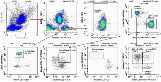
Fig. 1 Flow cytometric analysis of B cell subsets in human peripheral blood cells (PBMCs). Human peripheral blood cells were stained with anti-human CD45 / CD19 / CD20 / CD24/ IgD/ CD3 / CD14 / CD16 / CD69 / CD86 / CD27 / CD38 / CXCR5 / HLA-DR. The B cell subsets were verified by flow cytometry. (The data are provided by Elabscience®)
Elabscience® Quick Overview of Popular Products:
Table 1. Reagents for B cell identification in human peripheral blood cells (PBMCs)
|
Marker |
Fluorescein |
Cat. No |
|
CD45 |
PerCP |
E-AB-F1137F |
|
CD19 |
Elab Fluor® Violet 500 |
E-AB-F1004R |
|
CD20 |
PE-Cy7 |
AN00776H |
|
CD24 |
PE/Elab Fluor® 594 |
AN00645P |
|
IgD |
Elab Fluor® Violet 450 |
E-AB-F1171Q |
|
CD3 |
Elab Fluor® 700 |
E-AB-F1001M |
|
CD14 |
Elab Fluor® 647 |
AN00420M |
|
CD16 |
Elab Fluor® 700 |
E-AB-F1236M |
|
CD69 |
Elab Fluor® Violet 610 |
E-AB-F1138T |
|
CD86 |
PE |
E-AB-F1012D |
|
CD27 |
PerCP-Cy5.5 |
E-AB-F1140J |
|
CD38 |
APC |
E-AB-F1058E |
|
CXCR5 |
FITC |
E-AB-F1287C |
|
HLA-DR |
Elab Fluor® Red 780 |
E-AB-F1111S |
|
Live/Dead dye |
STYX™ Yellow ( AmCyan) |
E-CK-A169 |
|
EasyStain™ Human Fc Receptor Blocking Solution |
/ |
E-CK-A171 |
|
10×ACK Lysis Buffer |
/ |
E-CK-A105 |
Explore the APC Anti-Human CD138/Syndecan-1 Antibody[DL-101] (E-AB-F1154E) Used in Publications →
02 Flow cytometry markers for naive and memory B cell differentiation
After the identification of B cells, the separation of naive and memory populations represents the most informative primary partition within b cell phenotyping workflows. In humans, CD27 serves as the canonical marker for antigen experienced memory B cells[2,3].
Human naive B cells exhibit the phenotype CD19+ CD20+ CD27− IgD+ IgM+ CD10− CD21+ CD23+ CD38low. This profile summarizes core naive b cell markers, and these cells have not undergone class switch recombination or somatic hypermutation[3,4].
Memory B cells can be further stratified according to immunoglobulin isotype and CD27 expression status. Key memory b cell markers include CD27, costimulatory molecules and switched immunoglobulin isotypes for both human and mouse subsets[3,4].
Table 2. Human memory B cell subsets
|
Subset |
Markers |
Notes |
|
Unswitched (IgM) memory |
IgM+ IgD+ CD27+ |
Often called “natural” or T-independent memory |
|
Class-switched memory (CSM) |
IgD− IgG+/IgA+/IgE+ CD27+ |
Post-germinal-center effectors; CD21+ CD23−/+ |
|
IgM-only memory |
IgM+ IgD− CD27+ |
Enriched after infection; somatically mutated |
|
CD27-negative switched |
IgG/IgA+ CD27− |
Accumulates in elderly individuals and in SLE |
Memory B cells typically display elevated expression of CD80/86 and CD95 Fas alongside reduced CD23 relative to naive B cells. One critical practical consideration is that direct staining for IgG IgA or IgE requires rigorous isotype and fluorescence controls. This setup allows clear discrimination between total immunoglobulin signal and the genuine surface isotype expressed by individual cells[3,4].
Mouse B cells are identified using B220/CD45R instead of CD20. Naive follicular B cells in mice show the phenotype B220+ IgM+ IgD+ CD21/35+ CD23+. Memory B cell definition in mice relies far less on CD27, which is not a dependable memory marker in this species. Mouse memory B cells are mainly distinguished by class switching indicated by IgG+ or IgA+ expression, upregulated CD80/86 and CD73, and increased B220 intensity. The age associated memory compartment additionally exhibits T bet expression[3,4].
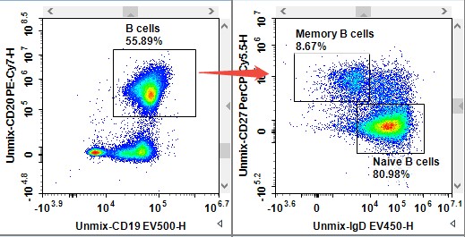
Fig. 2 Flow cytometric analysis of B cells expression in human peripheral blood cells (PBMCs). Human peripheral blood cells were stained with anti-human CD45/CD19/CD20/IgD/CD27 antibodies.Then the naive B cells and memory B cells were verified by flow cytometry. (The data are provided by Elabscience®)
03 CD24 and CD38 expression in transitional B cells
Transitional B cells are newly egressed from the bone marrow and occupy an intermediate developmental stage between bone marrow immature B cells and mature naive B cells. In human peripheral blood, these cells are defined by robust coexpression of CD24 and CD38, together with expression of CD10 and surface immunoglobulin[5].
Table 3. Transitional and mature naive B cell phenotyping
|
Stage |
CD10 |
CD24 |
CD38 |
IgM |
IgD |
CD21 |
CD23 |
|
T1 (transitional 1) |
+ |
++ |
++ |
++ |
+ |
−/lo |
− |
|
T2 (transitional 2) |
+ |
++ |
++ |
++ |
+ |
+ |
+ |
|
T3 (mature transitional) |
lo |
+ |
+ |
++ |
+ |
+ |
+ |
|
Mature naive |
− |
+ |
lo |
+ |
+ |
+ |
+ |
Within the CD19+ CD20+ gate, transitional B cells exhibit a CD10+ CD24++ CD38++ phenotype. T1 and T2 transitional subsets are discriminated based on CD21 and CD23 expression. T1 cells show a CD21−/lo CD23− profile, whereas T2 cells are CD21+ CD23+. Both subsets retain high CD24 and CD38 expression, and these markers are downregulated upon differentiation into naive B cells that acquire a CD24+ CD38low profile[5,6].
Two key interpretive considerations should be noted. First, CD38 lacks specificity for transitional B cells. This marker is also highly expressed on plasmablasts and plasma cells described in Section 4, as well as activated T cells and NK cells. Transitional B cells must therefore be identified within the CD19+ CD20+ CD10+ compartment rather than by CD38 expression alone. Second, transitional B cells display tolerogenic or anergic functional properties. Their frequency increases in systemic lupus erythematosus and during immune reconstitution following B cell depleting therapy, rendering this population a valuable readout of recent bone marrow B cell output[6,7].
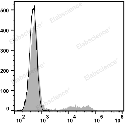
Fig. 3 Flow cytometric analysis of CD24 expression in human peripheral blood lymphocytes. Human peripheral blood lymphocytes are stained with PE Anti-Human CD24 Antibody (filled gray histogram). Unstained lymphocytes (empty black histogram) are used as control. (The data are provided by Elabscience®)
Interpretation: CD24 is a 35-45 kD glycosylphosphatidylinositol (GPI)-linked protein also known as heat stable antigen (HSA), BA-1, Ly-52, and nectadrin. It is expressed on the surface of B cells (but not plasma cells), granulocytes, follicular dendritic cells, and epithelial cells. CD24 may play a role in the regulation of B-cell proliferation and maturation. CD24 crosslinking induces a Ca2+ flux in mature B cells. CD24 has been shown to interact with CD62P (P-selectin).
Assay Used for This Experiment:
PE Anti-Human CD24 Antibody[ML5]
Cat. No.: E-AB-F1147D
Sample Type: cells
Detection Instrument: Flow Cytometer
Fluorophores: PE
Explore the PE Anti-Human CD24 Antibody[ML5] Used in 4 Publications →
04 CD38 and CD138 for plasmablast and plasma cell identification
Antibody secreting cells constitute the terminal effector population of the B lineage and are characterized by markedly elevated CD38 expression together with the loss of canonical B cell markers.
Plasmablasts (PB) represent proliferating predominantly circulating cells that emerge in peripheral blood following infection or vaccination with peak abundance near day 7. These cells exhibit the phenotype CD19+ CD20−/lo CD27++ CD38++ CD138−/lo, alongside intracytoplasmic immunoglobulin and nuclear Blimp 1 and IRF4 expression. Plasmablasts preserve partial B lineage identity through retained CD19 and constitute the antibody secreting subset most easily recovered from peripheral blood[8,9].
Plasma cells PC are terminally differentiated and predominantly tissue resident, localizing within the bone marrow, mucosal compartments and inflamed sites. Their phenotype features CD138 also termed syndecan 1, CD38++, CD19−, CD20−, CD27++ and dim or low CD45, together with abundant intracytoplasmic immunoglobulin. Bone marrow derived plasma cells frequently express CD56. Human bone marrow plasma cells typically display the profile CD38++ CD138+ CD19− CD45lo CD27+ CD56+[8,9].
Discrimination between plasmablasts and plasma cells relies on the retention or loss of CD19 and CD20 as well as CD138 expression. Plasmablasts maintain CD19 and sometimes low CD20 and show a CD138−/lo profile, whereas plasma cells downregulate CD19 and CD20 and acquire CD138 expression. CD138 syndecan 1 serves as the canonical plasma cell marker yet this molecule can undergo shedding, which limits its reliability in circulation and renders it more suitable for tissue specimens[9,10].
In peripheral blood, CD38 high expression combined with CD19 and CD20 loss provides a superior discriminator. One practical gating workflow proceeds as CD19+ events → CD38++ CD27++ events → separation into CD138+ plasma cells and CD138− plasmablasts. For tissue samples, an alternative strategy gates dump negative CD138+ CD38++ CD19− events.
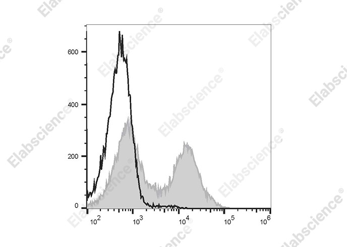
Fig. 4 Flow cytometric analysis of CD38 expression in murine splenocytes. C57BL/6 murine splenocytes are stained with FITC Anti-Mouse CD38 Antibody[NIMR5] (filled gray histogram) or FITC Rat IgG2a, κ Isotype Control (empty black histogram). (The data are provided by Elabscience®)
Interpretation: CD38 is a 42 kD glycoprotein, also known as T10. It is an ADP-ribosyl hydrolase, expressed on B cells, NK cells, a subset of T cells, brain, muscle, and kidney. In mouse, CD38 expression is downregulated on germinal center B cells and plasma cells, whereas this is not the case for humans. By functioning as both a cyclase and a hydrolase, CD38 mediates lymphocyte activation, as well as adhesion and metabolism of cADPR and NAADP. CD31 is the ligand of CD38.
Assay Used for This Experiment:
FITC Anti-Mouse CD38 Antibody[NIMR5]
Cat. No.: E-AB-F1193C
Sample Type: mouse cells
Detection Instrument: Flow Cytometer
Fluorophores: FITC
Explore the FITC Anti-Mouse CD38 Antibody[NIMR5] Used in 5 Publications →
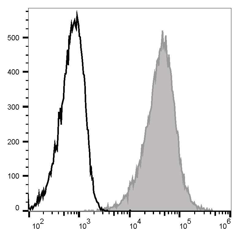
Fig. 5 Flow cytometric analysis of CD138 expression in U266 cells. Staining of U266 cells with APC Anti-Human CD138/Syndecan-1 Antibody[DL-101] (filled gray histogram) or APC Mouse IgG1, κ Isotype Control (empty black histogram). Total viable cells were used for analysis. (The data are provided by Elabscience®)
Interpretation: CD138, a member of the syndecan protein family, is a type I integral membrane heparin sulfate proteoglycan also known as Syndecan-1. Syndecan-1 participates in cell proliferation, cell migration, and cell-matrix adhesion via interaction with collagen, fibronectin, and other soluble molecules (such as FGF-basic). It is expressed on normal and malignant human plasma cells, pre-B cells, epithelial cells, and endothelial cells.
Assay used for this experiment:
APC Anti-Human CD138/Syndecan-1 Antibody[DL-101]
Cat. No.: E-AB-F1154E
Sample Type: cells
Detection Instrument: Flow Cytometer
Fluorophores: APC
Explore the APC Anti-Human CD138/Syndecan-1 Antibody[DL-101] Used in 1 Publications →
05 CD21 and CD11c in atypical memory B cell identification
Accumulating literature has characterized atypical memory B cells (AtM) as a functionally distinct subset that expands under chronic infection and autoimmune conditions. This population is frequently categorized as exhausted or inflammatory[11,12].
Human atypical memory B cells, also referred to as tissue like memory, activated memory or CD21low B cells, display the phenotype CD19+ CD20+ CD27− or CD27+ CD21low/− CD11c+ T-bet+ FCRL5+ CXCR3+, with frequent IgG class switching. These cells exhibit attenuated BCR responsiveness and enhanced production of cytokines including TNF and IFN-γ. Their frequency increases in chronic malaria, HIV infection, viral hepatitis, systemic lupus erythematosus and rheumatoid arthritis[11,12].
The mouse counterpart is age associated B cells (ABCs), which exhibit the phenotype B220+ CD21lo CD23− CD11c+ T-bet+ and often express CD11b. ABC development is driven by combined TLR7 and TLR9 signaling alongside IFN-γ. This subset accumulates with age and is enriched in lupus prone mouse strains[13].
One critical interpretive principle is that CD11c expression alone is insufficient to define atypical memory B cells. CD11c can also be detected on subsets of activated and marginal zone like B cells. The definitive signature of atypical memory B cells relies on the combinatorial profile CD21low plus CD11c+ plus T-bet+, which can be resolved relative to naive B cells with the profile CD21+ CD27− CD11c− and classical memory B cells marked by CD21+ CD27+. This population is best interpreted as a persistent imprint of sustained antigenic and innate immune stimulation rather than a canonical developmental stage[11,14].
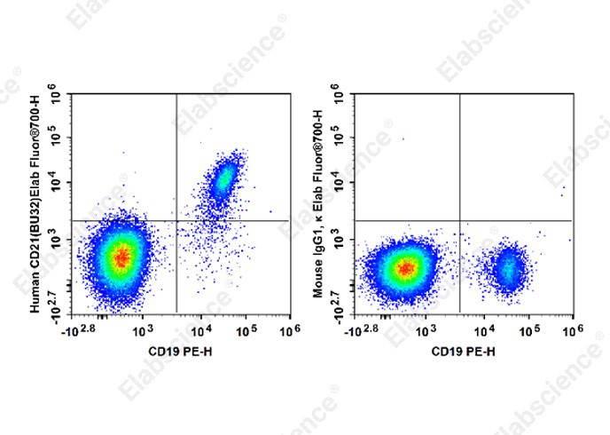
Fig. 6 Flow cytometric analysis of CD21 expression in human peripheral blood cells. Normal human peripheral blood cells were stained with Elab Fluor® 700 Anti-Human CD21 Antibody[BU32] and PE Anti-Human CD19 Antibody[4G7] (left) or Elab Fluor® 700 Mouse IgG1,κ Isotype Control (right).Cells in the lymphocytes gate were used for analysis. (The data are provided by Elabscience®)
Interpretation: CD21 is a 145 kD transmembrane protein also known as complement C3d receptor (C3dR), complement receptor 2 (CR2), and Epstein-Barr virus receptor. CD21 is expressed on B cells, follicular dendritic cells, subsets of normal thymocytes and T cells, and some epithelial cells. CD21 is the receptor used by Epstein-Barr virus to infect B cells and is also the complement receptor for C3d. CD21 has also been shown to interact with a number of proteins, including CD23, CD19, annexin VI, CD81, iC3b, complement receptor 1 (CR1, CD35), and interferon-alpha 1 (IFN-α1).
Assay Used for This Experiment:
Elab Fluor® 700 Anti-Human CD21 Antibody[BU32]
Cat. No.: E-AB-F1046M1
Sample Type: Human peripheral blood cells
Detection Instrument: Flow Cytometer
Fluorophores: Elab Fluor® 700
Explore more applications of CD21 antibodies with various fluorescent labels →
06 Human and mouse B cell marker differences
Markers do not transfer one-to-one between species, and panel design must account for these differences[15-17].
Table 4. Key differences between human and mouse B cell markers[15-17]
|
Feature |
Human |
Mouse |
|
Pan-B gate |
CD19 (primary), CD20 |
B220 (CD45R) primary; CD19 also used; CD20 rarely |
|
B1 cells |
B1-like: CD20+ CD27+ CD43+ IgMhi (CD5+ rare in adults) |
B1a: CD5+ IgMhi IgDlo CD11b+; B1b: CD5− |
|
Marginal zone |
CD21hi CD1c+ IgMhi IgD+ CD27+ |
CD21/35hi IgMhi IgDlo CD1dhi CD23lo |
|
Follicular naive |
IgD+ IgM+ CD23+ CD21+ |
B220+ IgD+ CD21/35+ CD23+ |
|
Memory |
CD27 canonical |
Class-switch + CD80/86 + CD73+ B220hi T-betlo/T-bethi, CD27 unreliable |
|
Immature/transitional |
CD10+ CD24++ CD38++ |
AA4.1 (CD93)+, IgMhi IgDlo, HSA/CD24 |
|
Plasma cell |
CD138+ CD38++ CD19− |
B220− CD138+ CD38++ |
|
Atypical/ABC |
CD21low CD11c+ T-bet+ |
CD21lo CD23− CD11c+ T-bet+ |
Additional practical considerations include the species specificity of numerous antibody clones, which necessitates independent validation. Substantial variation exists across inbred mouse strains, exemplified by differences in marginal zone compartment size between C57BL/6 and BALB/c mice. Most human experiments utilize cryopreserved PBMC, while mouse assays commonly employ fresh spleen or bone marrow samples. Furthermore, limited availability of human tissues such as tonsil and spleen restricts side-by-side comparison of tissue resident subsets.
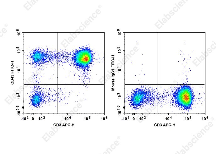
Fig. 7 Flow cytometric analysis of CD43 expression in human peripheral blood mononuclear cells. Normal human peripheral blood mononuclear cells were stained with APC Anti-Human CD3 Antibody and FITC Anti-Human CD43 Antibody[HI161] (left) or FITC Mouse IgG1, κ Isotype Control (right). Cells in the lymphocytes gate were used for analysis. (The data are provided by Elabscience®)
Interpretation: CD43, also known as Ly-48, leukosialin, sialophorin, leukocyte sialoglycoprotein, and W3/13, is a large single chain, type I transmembrane glycoprotein with abundant O-glycosylation and sialylation sites. It has been reported that CD43 binds to CD54 and Siglec-1. CD43 plays dual roles in cell adhesion and anti-adhesion as well as costimulation of T cell activation and survival, and induction of apoptosis of T cells and hematopoietic progenitors.
Assay Used for This Experiment:
FITC Anti-Human CD43 Antibody[HI161]
Cat. No.: E-AB-F1328C
Sample Type: human peripheral blood mononuclear cells
Detection Instrument: Flow Cytometer
Fluorophores: FITC
Explore more applications of CD43 antibodies with various fluorescent labels →
07 Key takeaways
A consolidated marker reference for gating the major human B cell subsets:
Table 5. Summary gating markers for human B cell subsets
|
Subset |
Defining markers |
|
B cell gate |
dump(CD3/CD14/CD16/CD56)− CD19+ CD20+ |
|
Naive |
CD27− IgD+ IgM+ CD10− CD21+ CD23+ CD38lo |
|
Unswitched memory |
IgM+ IgD+ CD27+ |
|
Class-switched memory |
IgG/IgA/IgE+ CD27+ |
|
Transitional (T1/T2) |
CD10+ CD24++ CD38++ (T1: CD21− CD23−; T2: CD21+ CD23+) |
|
Plasmablast |
CD19+ CD20− CD27++ CD38++ CD138−/lo |
|
Plasma cell |
CD138+ CD38++ CD19− CD20− |
|
Atypical memory |
CD21low CD11c+ T-bet+ (CD27−/+) |
Practical recommendations for panel design and analysis:
● Anchor on a clean B cell gate: lineage dump-negative, CD19+ (and/or CD20+), with a live/dead dye and a singlet gate.
● Never rely on a single marker. CD38 is bright on transitional cells, plasmablasts, plasma cells, and activated T/NK cells; its meaning depends entirely on the gate.
● Use proper controls for immunoglobulin isotypes, distinguish total Ig from the surface isotype of each cell with isotype and fluorescence controls.
● Define atypical memory by the combination CD21low + CD11c++ T-bet+, not merely by CD27 negativity.
● For antibody-secreting cells, require CD138 plus CD38 brightness together with loss of CD19/CD20 to separate plasma cells from plasmablasts.
● Account for species and tissue. Translate human panels to mouse with the differences in Section 6 in mind, and validate every antibody.
● Context is everything: the same marker can denote immaturity (transitional), activation (plasmablast), or exhaustion (atypical memory). Always interpret a marker within its gating hierarchy.
Used together, these marker combinations provide a reproducible framework for resolving B cell heterogeneity across human and mouse samples and for tracking subset shifts in infection, vaccination, autoimmunity, and immunotherapy.
08 Related resources
● Explore common immune cell populations and learn about the markers used for immunophenotyping.
Read Phenotyping of Common Immune Cells →
● Simplify flow cytometry panel design with expert support tailored to your research needs.
Explore Flow Cytometry Panel Design Services →
References:
[1] Tucker W ,LeBien, Thomas F ,et al.B lymphocytes: how they develop and function.[J].Blood, 2008.
[2] Sanz I , Wei C , Lee E H ,et al.Phenotypic and functional heterogeneity of human memory B cells.[J].Seminars in Immunology, 2008, 20(1):67-82.
[3] Carsetti R , Rosado M M , Wardmann H .Peripheral development of B cells in mouse and man[J].Immunological Reviews, 2010, 197(1):179-191.
[4] Kurosaki T , Kometani K , Ise W .Memory B cells[J].Nature Reviews Immunology, 2015, 15(3):149.
[5] Taylor J J , Pape K A , Jenkins M K .A germinal center–independent pathway generates unswitched memory B cells early in the primary response[J].Journal of Experimental Medicine, 2012, 209(3):597.
[6] Sims G P , Ettinger R , Shirota Y ,et al.Identification and characterization of circulating human transitional B cells[J].Blood, 2005, 105(11):4390-4398.
[7] Gouw L G , Reading N S , Crockett D K ,et al.RhoF GTPase Transcription Is Upregulated in Germinal Center B-Cells, Shows Altered Expression in Malignant B-Cells, and Interacts with the ATM Pathway[J].Blood, 2005, 106(11):285-285.
[8] Nutt S L , Hodgkin P D , Tarlinton D M ,et al.The generation of antibody-secreting plasma cells[J].Nature Reviews Immunology, 2015, 15(3):160-171.
[9] Odendahl,M.Generation of migratory antigen-specific plasma blasts and mobilization of resident plasma cells in a secondary immune response[J].Blood, 2005, 105(4):1614-21.
[10] Moir S , Fauci A S .Pathogenic mechanisms of B-lymphocyte dysfunction in HIV disease[J].Journal of Allergy & Clinical Immunology, 2008, 122(1):12-19.
[11] Weiss G E , Crompton P D , Li S ,et al.Atypical Memory B Cells Are Greatly Expanded in Individuals Living in a Malaria-Endemic Area[J].Journal of Immunology, 2009, 183(3):2176-2182.
[12] Rubtsov A V , Rubtsova K , Kappler J W ,et al.TLR7 drives accumulation of ABCs and autoantibody production in autoimmune-prone mice[J].Immunologic Research, 2013, 55(1-3):210-216.
[13] Wu C , Li Z , Fu X ,et al.Antigen-specific human NKT cells from tuberculosis patients produce IL-21 to help B cells for the production of immunoglobulins[J].Oncotarget, 2015, 6(30).
[14] Baumgarth N , Tung J W , Herzenberg L A .Inherent specificities in natural antibodies: a key to immune defense against pathogen invasion[J].Springer Seminars in Immunopathology, 2005, 26(4):347.
[15] Griffin, D. O,Holodick, N. E,Rothstein, T. L.Human B1 cells in umbilical cord and adult peripheral blood express the novel phenotype CD20+CD27+CD43+CD70[J].Journal of Experimental Medicine, 2011, 208(4):871-871.
[16] Guidelines for the use of flow cytometry and cell sorting in immunological studies[J].European Journal of Immunology, 2017.
[17] Maecker H T , Mccoy J P , Nussenblatt R .Standardizing immunophenotyping for the Human Immunology Project[J].Nature Reviews Immunology, 2012, 12:191.

