

Conventional IHC can only detect a single biomarker per tissue section[7]. However, the tissue microenvironment is highly complex, and cell-cell interactions involve co-expression of multiple targets. Multiplex Immunohistochemistry (mIHC) enables simultaneous detection of multiple antigens, revealing distinct cell subtypes, functional states and their spatial relationships[1]. It can more accurately reflect disease heterogeneity, providing comprehensive data to support precision diagnosis, immune scoring and discovery of therapeutic targets[2].
Table of Contents
1. Technical principles of multiplex IHC: From single-plex to multiplex staining
2. Key applications of multiplex IHC in tissue and immune profiling
3. ElabPlex™ TSA multiplex fluorescence staining: Step-by-step experimental workflow
4. Reference fluorophore and fluorescence channel configuration
01 Technical principles of multiplex IHC: From single-plex to multiplex staining
Currently, mainstream mIHC technologies are built on the Tyramide Signal Amplification (TSA) system. Its core mechanism is described as follows: in the presence of hydrogen peroxide, HRP-conjugated secondary antibodies catalyze fluorophore-labeled tyramide molecules to form highly reactive intermediates, which instantly covalently bind to tyrosine residues adjacent to target proteins and deposit abundant fluorophores. This achieves a 10–100-fold signal amplification, making the detection of low-abundance target proteins feasible[8].
Another key feature of TSA technology is heat-mediated antibody stripping[4,6]. After each round of staining, heat treatment removes primary and secondary antibodies, while the covalently bound fluorescent signals remain intact. This mechanism supports sequential staining and sequential imaging on the same tissue section, without restrictions on the host species of primary antibodies; antibodies raised in rabbit, mouse, rat and other species can be flexibly combined.
02 Key applications of multiplex IHC in tissue and immune profiling
2.1 Immune Subtyping of Tumor Microenvironment (TME)
The most mature application of mIHC lies in analyzing immune cell infiltration within the TME[3]. This method can simultaneously quantify multiple immune cell populations including CD4⁺ T cells, CD8⁺ T cells, CD20⁺ B cells, CD68⁺/CD163⁺ macrophages on a single section. Nearest-neighbor analysis can further be performed to assess spatial interactions between cells.
2.2 Companion Diagnostics for Immunotherapy
Although single-marker PD-L1 IHC serves as the gold standard for immunotherapy companion diagnostics, a single biomarker cannot fully predict treatment response. mIHC simultaneously detects multiple markers such as PD-1/PD-L1, CD8, FoxP3, LAG3, TIGIT and TIM-3[5]. It can distinguish "hot tumors" from "cold tumors", and identify distinct phenotypes including immune-excluded and Immune-desert tumors.
2.3 Neuroscience and Pathological Tissue Analysis
TSA-based mIHC/multiplex immunofluorescence (mIF) is widely used in three major fields[9]: neuroscience, oncology and immunology. Bibliometric analysis shows that as of 2023, Web of Science has included 873 relevant publications, with a rapid annual increase in output since 2016.
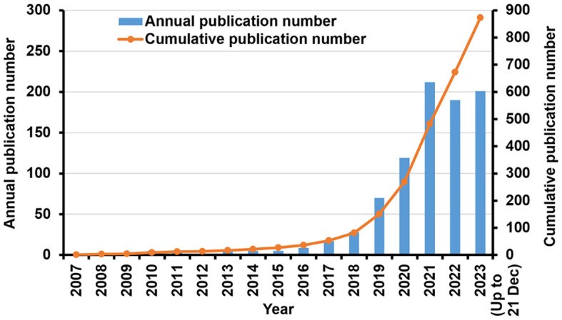
Fig. 1 Research in TSA-based mIHC/IF method: Annual scientific production and cumulative number of publications[1].
03 ElabPlex™ TSA multiplex fluorescence staining: Step-by-step experimental workflow
Standard operating protocol based on the ElabPlex™ TSA Multiplex Fluorescence Staining Kit (Cat. No. E-IR-R335):
Step 1: Sample Preparation
• Paraffin sections: Deparaffinize in xylene, rehydrate through a graded ethanol series, and finally transfer to PBS buffer.
• Frozen sections or cell smears: Fix for 10–30 min. After washing, permeabilize with 0.3% Triton X-100 for 20 min (optional for membrane protein staining), followed by washing and rehydration.
Step 2: Antigen Retrieval
Perform Heat-Induced Epitope Retrieval (HIER). Recommended retrieval buffers: pH 9.0 EDTA buffer (Cat. No. E-IR-R104) or pH 6.0 citrate buffer (Cat. No. E-IR-R105). Heat in microwave to boiling and maintain sub-boiling conditions for 10–20 min, then cool to room temperature for 30 min. This step can be omitted for frozen sections or cell slides. For fragile tissues (e.g., bone tissue), enzymatic retrieval or mild heating retrieval is recommended.
Step 3: Block Endogenous Peroxidase Activity
Add endogenous peroxidase blocking reagent, incubate at room temperature in the dark for 15 min. Wash 3 times with PBS/TBS (pH 7.4), 3 min each time.
Step 4: Block Non-Specific Binding
Add Normal Goat Blocking Buffer (Cat. No. E-IR-R110) or antibody diluent, incubate at room temperature for 30 min.
Step 5: Primary Antibody Incubation
Add diluted primary antibody, incubate at 4 °C overnight or 37 °C for 1–2 h. Wash 3 times with PBS/TBS, 3 min each time.
Step 6: Polymer-HRP Secondary Antibody Incubation
Add polymer HRP-conjugated goat anti-mouse/rabbit secondary antibody, incubate at room temperature for 30 min. Wash 3 times, 3 min each time.
Step 7: TSA Fluorescent Staining
Dilute TSA fluorophores at a ratio of 1:50 to 1:400 (1:200 is recommended). Apply to sections and incubate at room temperature protected from light for 5–10 min. Wash 3 times, 3 min each time.
Step 8: Antibody Stripping
Two alternative stripping methods:
A. Heat-mediated stripping: 100 °C for 20–40 min;
B. Antibody stripping buffer: 37 °C, two rounds of 10-min incubation. Wash 3 times, 3 min each time.
Step 9: Cyclic Multiplex Target Labeling
• For the 2nd to 5th target antigens: Repeat Steps 3–8;
• For the 6th and subsequent target antigens: Repeat Steps 3–7 (As the sixth marker is the last one, no additional antibody removal step is needed).
Step 10: Counterstaining and Mounting
Add Antifade Mounting Medium with DAPI, apply coverslips and incubate for 5 min.
Step 11: Image Acquisition
Capture images using a fluorescence microscope or multispectral imaging system.
Results Presentation:
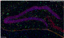
Fig. 2 Mouse brain, 4-marker 5-color.
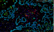
Fig. 3 Human cholangiocarcinoma, 5-marker 6-color.
04 Reference fluorophore and fluorescence channel configuration
Table 1. Reference Fluorescence Channel Configuration
|
Fluorophore |
Excitation Wavelength |
Emission Wavelength |
|
DAPI |
350nm |
420nm |
|
TSA-480 |
440nm |
480nm |
|
TSA-520 |
488nm |
519nm |
|
TSA-570 |
555nm |
570nm |
|
TSA-620 |
594nm |
615nm |
|
TSA-690 |
682nm |
702nm |
|
TSA-780 |
750nm |
780nm |
mIHC technology enables simultaneous detection of multiple targets on a single tissue section while fully preserving spatial information. It has become an essential tool for research on the tumor immune microenvironment and clinical translational medicine. The ElabPlex™ TSA Multiplex Fluorescence Staining Kit provides an all-in-one solution covering antigen retrieval to multiplex imaging, supporting your spatial multi-target analysis workflow.
References:
[1] Yu X, Huang C, Song Y, et al. Research progress and perspectives on the application of tyramide signal amplification-based multiplex immunohistochemistry/immunofluorescence: a bibliometrics analysis[J]. Frontiers in Oncology, 2025, 14: 1473414.
[2] Sun AK, Fan S, Choi SW. Exploring Multiplex Immunohistochemistry (mIHC) Techniques and Histopathology Image Analysis: Current Practice and Potential for Clinical Incorporation[J]. Cancer Medicine, 2025, 14(1): e70523.
[3] Gorris MAJ, et al. Multiplex Immunohistochemical Analysis of the Spatial Immune Cell Landscape of the Tumor Microenvironment[J]. Journal of Visualized Experiments, 2023, (198): e65717.
[4] Multiple immunolabeling with antibodies from the same host species in combination with tyramide signal amplification[J]. Acta Histochemica, 2018, 120(7): 621–631.
[5] A three dimensional immunolabeling method with peroxidase-fused nanobodies and fluorochromized tyramide-glucose oxidase signal amplification[J]. Communications Biology, 2025, 8(1).
[6] Multiplexed immunohistochemistry, imaging, and quantitation: a review, with an assessment of Tyramide signal amplification, multispectral imaging and multiplex analysis[J]. Methods, 2014, 70(1): 46–58.
[7] Spatial analysis of the tumor microenvironment can be achieved via the novel SignalStar™ multiplex immunohistochemistry assay[J]. The Journal of Immunology, 2025, 214(Supplement 1).
[8] Research use only validation of 'Off the Shelf' multiplex immunofluorescence panels with quantitative image analysis for use in retrospective clinical trials[J]. Methods in Molecular Biology, 2023.
[9] Multiplex Immunohistochemistry Analysis of Melanoma Tumor-Infiltrating Lymphocytes[J]. Journal for ImmunoTherapy of Cancer, 2024, 12(Suppl 2).

