Magnetic bead sorting relies on two key mechanisms: antibody-antigen specific binding and magnetic field physical separation. In simple terms, it involves attaching a unique "magnetic tag" that responds to magnetic fields, and then using a strong magnetic field (like a giant "magnet") to efficiently and gently separate the target cells from a complex cell population.
Currently, there are three mainstream sorting methods: positive selection, negative selection, and composite selection. Different experimental needs call for specific sorting strategies; choosing the right method can double the efficiency of your cell experiments. Below, we systematically summarize the key points from four dimensions: selection of sorting strategies, core operational steps, experimental precautions, and common issues with solutions.
Table of Contents
1. Selecting the optimal magnetic bead sorting strategy
2. Optimizing magnetic bead cell sorting workflow and experimental parameters
3. Troubleshooting common challenges in magnetic bead cell sorting
01 Selecting the optimal magnetic bead sorting strategy
The core principles for selecting a sorting strategy are as follows:
Positive selection is the preferred choice when both sorting speed and cell purity are important, and when subsequent experimental detection is not affected by the magnetic beads.
Negative selection is recommended when the target cells will be used for downstream applications such as cell culture or functional activity assays, as it avoids the potential impact of bead attachment on the physiological state of the cells.
Composite selection is recommended for rare cells present at very low frequencies (<1%). This approach first removes unwanted cells through negative selection for pre-enrichment, followed by positive selection to precisely capture the target cells.
The table below provides a comprehensive comparison of the three sorting strategies in terms of their principles, applicable scenarios, advantages, and disadvantages for easy reference.
Table 1. Comparison of magnetic bead sorting strategies for different application scenarios
|
Characteristics |
Positive Selection |
Negative Selection |
Sequential Sorting |
|
Principle |
Directly "capture" the target cells |
Remove all "bystanders," leaving the target cells behind |
Combine multiple strategies to achieve high purity through stepwise processing |
|
Common Scenarios |
1) Enrichment of rare cells (e.g., specific T cell subsets constituting <5% of the population) |
1) Downstream experiments (e.g., cell culture, functional assays) strictly prohibit cells from being labeled with magnetic beads. |
1) The proportion of target cells is extremely low, requiring pre‑enrichment as a first step. |
|
2) Isolation of cells with weakly expressed surface markers |
2) The target cells lack specific surface markers. |
2) Target cells lack specific markers, necessitating an indirect strategy. |
|
|
3) Time‑constrained experiments |
3) A specific cell type (e.g., dead cells) needs to be removed. |
3) The target cells are a rare subset (e.g., regulatory T cells). |
|
|
Advantages |
High purity, fast speed, and simple operation. |
Cells are not labeled, preserving their natural state to the greatest extent. |
Enables extremely high final purity. |
|
Disadvantages |
Cells may carry magnetic beads/antibodies on their surface, which can cause non‑specific activation or affect downstream experiments. |
The amount of magnetic beads required is high; purity may be slightly lower than that of positive selection, and knowledge of the characteristics of all non‑target cells is necessary. |
The process is time‑consuming, and cell loss may be greater. |
02 Optimizing magnetic bead cell sorting workflow and experimental parameters
2.1 Sample Processing Step
After selecting an appropriate sorting strategy, sample processing is the critical first step in magnetic bead sorting, as it directly determines final cell purity, recovery rate, and cell viability. The following details should be carefully controlled during operation.
1) Tissue sample grinding and digestion
Fresh tissue samples are recommended. Whenever possible, complete single‑cell suspension preparation and sorting experiments within 2 hours of tissue isolation. The tissue can be dissociated into a single‑cell suspension using enzymatic digestion (collagenase, trypsin, etc.) or mechanical grinding (tissue grinder, mesh filtration). The core principle of the operation is to balance dissociation efficiency with cell damage.
Precautions
Avoid excessive force during mechanical grinding to prevent cell damage caused by mechanical shearing.
Select the appropriate type of digestive enzyme for enzymatic dissociation; improper enzyme choice may lead to false positives, false negatives, or even loss of target cells. At the same time, strictly optimize the digestion duration to avoid excessive digestion that damages cells or leads to loss of antigen epitopes.
2) Blood sample collection and pretreatment
For peripheral blood samples, it is recommended to use collection tubes containing sodium heparin or EDTA as anticoagulants. Immediately after collection, gently invert the tube 5-8 times to ensure thorough mixing with the anticoagulant. Vigorous shaking should be avoided as it may cause hemolysis. Fresh peripheral blood is preferred, and anticoagulated whole blood should ideally be used for sorting and downstream experiments within 2 hours of collection. This approach ensures high purity and yield of target cells while maximizing cell viability.
3) Sample filtration to remove debris
The prepared single‑cell suspension must be filtered through a 30-70 μm cell strainer prior to sorting. This effectively removes tissue debris and cell clumps, preventing adverse effects on sorting performance.
4) Dead cell removal
Dead cells tend to bind non‑specifically to magnetic beads, significantly reducing sorting purity. If the proportion of dead cells in the sample is high, it is recommended to pretreat the sample with a dead cell removal kit before performing magnetic bead sorting.
5) Cell counting and concentration adjustment
Accurate determination of total cell number and the percentage of target cells is essential. Adjust the cell sorting density appropriately according to the product instructions for different cell types to ensure compatibility with the optimal magnetic bead sorting system. For example, during positive selection, excessively high cell concentrations may clog the sorting column; a concentration range of 10⁶-10⁷ cells/mL is recommended.
6)Experimental Results
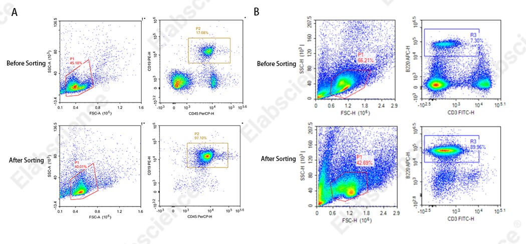
Fig. 1 Sorting results of mouse splenic CD3+ T cells (A: fresh sample; B: sample with poor initial condition).
As shown in the figure, the Elabscience® EasySort™ Mouse CD3+T Cell Isolation Kit (MIM001N) was used to sort mouse splenic tissue samples. In panel B, the purity of the sorted mouse CD3+ T cells was relatively low. Analysis indicates that this was due to the poor initial condition of the cells; the proportion of target cells in the sample was abnormal (as can be seen from the FSC and SSC analysis of cell morphology before sorting). To address such issues, it is recommended to use fresh samples for sorting, minimize mechanical damage during sample preparation, use samples with a normal proportion of target cells, stain with viability dyes before flow cytometry to remove the influence of dead cells on staining results, or use a dead cell removal kit to eliminate dead cells prior to sorting.
%20and%20bone%20marrow%20(right)_.png)
Fig. 2 Sorting results of B cells from mouse spleen (left) and bone marrow (right).
As shown in the figure, the Elabscience® EasySort™ Mouse B Cell Isolation Kit (MIM004N) was used to sort splenocytes and bone marrow cells, respectively. The purity of the sorted cells showed a significant difference between the two tissues. Analysis suggests that the main reason is that the negative selection kit used is not suitable for bone marrow tissue samples. To address this issue, it is recommended to select an appropriate sorting kit and sort only the tissue samples that have been tested and specified as compatible in the product manual.
2.2 Antibody Incubation Step
After preparing the tissue or cell samples for sorting, the following details regarding antibody incubation should be noted:
1) Antibody and magnetic bead incubation time: Typically, incubation at 4°C or room temperature for 5-10 minutes is sufficient, as most antigen-antibody binding is essentially complete within 10 minutes. Prolonging the incubation time unnecessarily does not significantly improve sorting performance. For optimal incubation conditions specific to different cell types, strictly follow the reagent instructions.
2) Incubation temperature control: Incubation is typically performed at 4°C or room temperature. However, low temperature (4°C) may affect antibody binding, requiring optimization of the incubation time. Excessively long incubation increases non-specific binding, so a balance must be struck to achieve efficient antibody binding. For temperature-sensitive cells, it is recommended to perform the entire incubation on ice at 2-8°C. This effectively slows down cellular metabolism, prevents antibody internalization, greatly reduces non-specific binding, and ensures sorting specificity.
3) Sample mixing method: During incubation and resting periods, mix the sample by gentle inversion or low-speed vortexing. Avoid vigorous shaking to prevent mechanical damage to cells. When using automated mechanical sorters, set a uniform low-speed, gentle mixing mode.
4) Cell washing: Washing steps should thoroughly remove unbound components, but excessive washing may lead to loss of target materials.
2.3 Cell Separation Step
After antibody incubation, the following details should be carefully considered during magnetic cell separation:
1) Selection of magnetic beads: Choose appropriate magnetic beads based on the properties of the target, such as particle size and surface‑modified antibodies or ligands. Pay attention to the ratio of beads to sample; excessive beads may cause non‑specific binding, while insufficient beads lead to low separation efficiency. Avoid repeated freeze‑thaw cycles when storing magnetic beads. Some beads require storage at 2-8°C, as freezing can render them ineffective.
2) Magnetic field incubation and standing: The magnetic field incubation time should primarily follow the manufacturer's recommended standard (typically about 5 minutes). Use a magnetic separation device that is compatible with the kit. Avoid excessively prolonged exposure to the magnetic field (e.g., >15 minutes may damage cells). If the magnetic force is relatively weak, the incubation time may be appropriately extended to 1.5 times the standard duration.
3) Washing and purification: Use a gentle elution method to avoid damaging the target structure. Especially in negative selection scenarios, performing two elution steps can improve the yield of target cells.
4) Post‑sorting validation and secondary enrichment: After sorting, it is essential to assess cell purity and recovery rate using flow cytometry. Sorted samples should ideally be analyzed immediately; if temporary storage is necessary, keep at 2-8°C for no more than 24 hours. If the measured purity does not meet the experimental requirements, the collected positive cells can be subjected to an additional round of sorting to further enhance cell purity.
03 Troubleshooting common challenges in magnetic bead cell sorting
Magnetic bead-based cell sorting involves numerous operational details; any deviation in any step may result in suboptimal purity, yield, or cell viability. Below, we have compiled common problems and their corresponding solutions for routine troubleshooting.
Table 2. Summary of common problems and solutions for magnetic bead-based cell sorting
|
Problem Phenomenon |
Possible Cause |
Solution |
|
Low sorting purity |
Dead cells or cell clumps present in the cell suspension |
Remove dead cells or filter the sample before sorting |
|
Insufficient amount of antibodies or magnetic beads |
Thoroughly mix antibodies and magnetic beads before use; add magnetic beads directly to the bottom of the tube when transferring to the sample, avoiding adding along the tube wall |
|
|
No washing or insufficient washing after antibody incubation |
Wash antibodies according to the instructions before adding magnetic beads |
|
|
Insufficient incubation time for antibodies or magnetic beads |
Perform sufficient incubation according to the instructions |
|
|
Magnetic beads not washed or resuspended |
Wash and resuspend magnetic beads using sorting buffer as specified in the instructions |
|
|
Magnetic bead aggregation and precipitation |
Use a room temperature water bath sonication for 2–5 minutes to evenly disperse the magnetic beads; during bead incubation, briefly vortex or shake every 1–2 minutes to keep beads uniformly mixed |
|
|
Low yield (low recovery rate) |
Excessively long magnetic bead incubation time |
Optimize incubation conditions and reduce incubation time |
|
Excessive amount of magnetic beads |
Thoroughly mix magnetic beads before use and pipette the correct volume; appropriately increase the total elution volume and perform a second magnetic separation |
|
|
Cells in the sample aggregate into clumps |
Filter the cell suspension before sorting to remove clumps and impurities; resuspend cells in the recommended sorting buffer to prepare a single‑cell suspension |
|
|
Failure to use fresh samples for sorting, resulting in poor initial cell viability |
Optimize sample handling procedures, maintain low temperature throughout the process, and use freshly prepared samples for sorting |
|
|
Column clogging or cell loss during separation |
Avoid column clogging; switch to a column‑free system if necessary |
|
|
Column clogging |
Cell clumps in the suspension |
Be sure to filter the sample before loading |
|
Cell concentration too high or sample volume too large |
Appropriately dilute the suspension, or switch to a larger‑capacity column |
|
|
Air bubbles in the buffer |
Degas the buffer to reduce air bubbles |
|
|
Poor cell viability |
Excessively vigorous operation or excessive centrifugal force |
Perform all operations gently and use low-speed centrifugation |
|
Overly long incubation or separation process duration |
Optimize the workflow and streamline steps |
|
|
Centrifugation speed too high, or centrifugation not performed at low temperature |
Set centrifuge acceleration to no more than 3 and deceleration to no more than 1; use a refrigerated centrifuge |
|
|
Inappropriate buffer used |
Use a buffer containing 0.5% BSA and 2 mM EDTA |
|
|
Magnetic bead residue |
Insufficient magnetic separation or incomplete washing |
Extend the magnetic separation time to ensure that the magnetic beads are fully captured |
|
Poor resolution of cell populations shown by flow cytometry after sorting |
Inappropriate choice of flow cytometry antibody for detection |
For weakly expressed antibodies, pair them with fluorophores of high fluorescence intensity and select an appropriate clone; for purity assessment in positive selection, choose a detection antibody clone whose epitope does not overlap with that of the sorting antibody. |
The related cell magnetic bead sorting research products are summarized in the table below.
Table 3. List of Elabscience® cell sorting products
|
Product Name |
Cat. No. |
|
EasySort™-5 Magnet |
EC001 |
|
EasySort™ Mouse Naïve CD8+T Cell Isolation Kit |
MIM008N |
|
EasySort™ Mouse Naïve CD4+T Cell Isolation Kit |
MIM007N |
|
EasySort™ Mouse Pan-Naïve T Cell Isolation Kit |
MIM006N |
|
EasySort™ Mouse NK Cell Isolation Kit |
MIM005N |
|
EasySort™ Mouse B Cell Isolation Kit |
MIM004N |
|
EasySort™ Mouse CD8+T Cell Isolation Kit |
MIM003N |
|
EasySort™ Mouse CD4+T Cell Isolation Kit |
MIM002N |
|
EasySort™ Mouse CD3+T Cell Isolation Kit |
MIM001N |
|
EasySort™ Human Memory CD8+T Cell Isolation Kit |
MIH010N |
|
EasySort™ Human Memory CD4+T Cell Isolation Kit |
MIH009N |
|
EasySort™ Human Naïve CD8+T Cell Isolation Kit |
MIH008N |
|
EasySort™ Human Naïve CD4+T Cell Isolation Kit |
MIH007N |
|
EasySort™ Human Naïve Pan T Cell Isolation Kit |
MIH006N |
|
EasySort™ Human B Cell Isolation Kit |
MIH004N |
|
EasySort™ Human CD8+ T Cell Isolation Kit |
MIH003N |
|
EasySort™ Human CD4+ T Cell Isolation Kit |
MIH002N |
|
EasySort™ Human CD3+T Cell Isolation Kit |
MIH001N |
|
Human MSC Analysis Kit |
XJH003 |
|
Mouse Th17 Flow Cytometry Staining Kit |
XJM002 |
|
Mouse Th1/Th2 Flow Cytometry Staining Kit |
XJM001 |
|
Human Th17 Flow Cytometry Staining Kit |
XJH002 |
|
Human Th1/Th2 Flow Cytometry Staining Kit |
XJH001 |
|
RAW 264.7 Polarized M1 Macrophage Induction and Identification Kit |
XJM004 |
|
Mouse Bone Marrow-derived Dendritic Cells (BMDC) Induction and Identification Kit |
XJM003 |

