Ferroptosis is an iron-dependent, non-apoptotic form of regulated cell death, characterized by excessive accumulation of lipid peroxide (LPO).
Erastin, a classic ferroptosis inducer, inhibits the cystine/glutamate antiporter System Xc- (xCT), blocks cystine uptake, depletes glutathione (GSH), inactivates glutathione peroxidase 4 (GPX4), and ultimately triggers lipid peroxidation and cell death.
HepG2 cells (human hepatocellular carcinoma) are highly sensitive to Erastin and serve as an ideal in vitro model for studying Erastin-induced ferroptosis. Multi‑parameter systematic testing of this model relies on reliable and compatible reagent tools. Elabscience® provides a one‑stop solution for ferroptosis research, covering complete tools from model induction to multi‑marker validation.
Table of Contents
1. Reagents and materials for erastin-induced ferroptosis model in HepG2 cells
2. Establishment of erastin-induced ferroptosis model in HepG2 cells
3. Additional ferroptosis-related products
01 Reagents and materials for erastin-induced ferroptosis model in HepG2 cells
Table 1. Reagents and their functions for HepG2 ferroptosis model
|
Reagent |
Function |
|
10 mmol/L Erastin |
Ferroptosis inducer (System Xc⁻ inhibitor) |
|
Lip-1 |
Ferroptosis inhibitor |
|
Lipid Peroxide (LPO) Fluorometric Assay Kit |
Detects lipid peroxidation level |
|
Glutathione Peroxidase 4 (GPX4) Activity Assay Kit |
Measures GPX4 enzyme activity |
|
Cystine Uptake Fluorometric Assay Kit |
Measures cystine uptake capacity |
|
Enhanced Cell Counting Kit 8 (WST-8/CCK8) |
Measures cell viability changes |
02 Establishment of erastin-induced ferroptosis model in HepG2 cells
2.1 Cell Culture
Harvest HepG2 cells in logarithmic growth phase, adjust density to 2×10⁵ cells/mL, and seed into appropriate plates according to experimental requirements. Culture in DMEM complete medium containing 10% FBS and 1% penicillin‑streptomycin at 37°C, 5% CO2 for 24 h. Treat after cell attachment.
Table 2. Seeding parameters of HepG2 cells for different detection assays
|
Plate Type |
Seeding Volume per Well |
Cells per Well |
Application |
|
96‑well |
0.1 mL |
≈2×10⁴ |
CCK‑8, cystine uptake |
|
24‑well |
0.5 mL |
≈1×10⁵ |
LPO |
|
6‑well |
2mL |
≈4×10⁵ |
GPX4 |
Note: Different assays require different cell numbers; select the appropriate plate and seeding density to ensure detection sensitivity and linear range.
2.2 Erastin Treatment and Assay Procedures
CCK‑8 & LPO Assays:
After cell attachment, treat with Erastin at a final concentration of 10 μmol/L for 24 h. Include the following controls:
Negative control: Equal volume of DMSO
Rescue control: 10 μmol/L Erastin + 2 μmol/L Lip‑1
Positive control: 2 μmol/L Lip‑1 for 24 h
After treatment, measure cell viability using CCK‑8 kit and lipid peroxidation using LPO fluorometric assay kit.
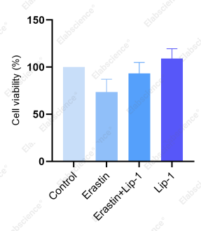
Fig. 1 CCK‑8 cell viability results.
Fig. 2 Lipid peroxidation level results.
Results show that Erastin alone significantly reduces HepG2 cell viability and concomitantly increases LPO levels compared to negative control. Co‑treatment with Lip‑1 partially or fully restores cell viability and reduces LPO levels, indicating that Lip‑1 effectively reverses Erastin‑induced ferroptotic changes.
GPX4 and Cystine Uptake Assays:
Seed cells in 6‑well plates, treat with 10 μmol/L Erastin for 24 h, and add equal volume of DMSO to control. Collect cells and measure using GPX4 colorimetric assay kit and cystine uptake fluorometric assay kit, respectively.
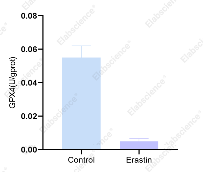
Fig. 3 GPX4 results.
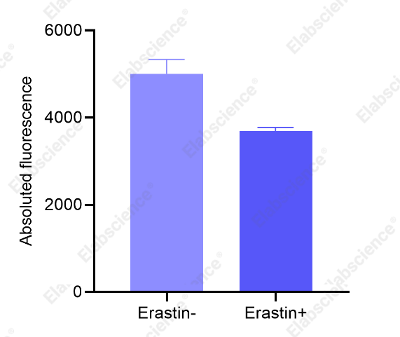
Fig. 4 Cystine uptake results.
Results show that compared to control, Erastin‑treated cells exhibit significantly reduced cystine uptake and markedly decreased GPX4 activity, consistent with the molecular mechanism of ferroptosis. Inhibition of System Xc⁻ reduces cystine supply, impairs GSH synthesis, leads to GPX4 inactivation, and diminishes the capacity to clear lipid peroxides.
2.3 Precautions
① Cells tend to aggregate in the center of the well, affecting growth and drug action. Gently shake the plate in forward‑backward and left‑right directions after seeding to ensure even distribution.
② Erastin should be stored at ‑20℃ protected from light. Prepare working solution fresh before use. Dilution with PBS may cause precipitation; DMSO is recommended. Keep final DMSO concentration ≤1% per well. Include DMSO control groups.
③ LPO fluorescent probes must be handled in the dark to prevent quenching.
④ Different ferroptosis markers have varying abundance in samples; seed cells in appropriate plate formats to obtain the required cell numbers for each assay. Based on the firefly luciferase–luciferin system, ATP serves as the substrate to drive oxidative luminescence. The chemiluminescent signal is proportional to ATP concentration, directly reflecting mitochondrial energy production efficiency.
03 Additional ferroptosis-related products
Table 3. Supplementary ferroptosis-related assay kits and matching detection instruments
|
Plate Type |
Seeding Volume per Well |
Cells per Well |
|
E-BC-F066 |
Cystine Uptake Fluorometric Assay Kit |
Fluorescence microplate reader |
|
E-BC-F140 |
Dihydroorotate Dehydrogenase (DHODH) Activity Fluorometric Assay Kit |
Fluorescence microplate reader |
|
E-BC-K880-M |
Cell Total Iron Colorimetric Assay Kit |
Microplate reader |
|
E-BC-K881-M |
Cell Ferrous Iron Colorimetric Assay Kit |
Microplate reader |
|
E-BC-F108 |
Cell Ferrous (Fe2+) Fluorometric Assay Kit |
Fluorescence microplate reader; Fluorescence microscope; Flow cytometry |
|
E-BC-F022 |
Ferroptosis Suppressor Protein-1 (FSP-1) Activity Fluorometric Assay Kit |
Fluorescence microplate reader |
|
E-BC-K809-M |
Cell Glutathione Peroxidase (GPX) Activity Assay Kit |
Microplate reader |
|
E-BC-K883-M |
Glutathione Peroxidase 4 (GPX4) Activity Assay Kit |
Microplate reader |
|
E-BC-F077 |
Lipoxygenase (LOX) Activity Fluorometric Assay Kit |
Fluorescence microplate reader |
|
E-BC-F003 |
Lipid Peroxide (LPO) Fluorometric Assay Kit |
Fluorescence microplate reader; Fluorescence microscope; Flow cytometry |
|
E-BC-F045 |
Total Glutathione (T-GSH) And Reduced Glutathione (GSH) Fluorometric Assay Kit |
Fluorescence microplate reader |
The Erastin‑induced HepG2 model is a classic system for ferroptosis research, yet experimental success often hinges on subtle details—cell seeding density, proper control design, probe light protection, and more. Only by clarifying the workflow, assembling the right tools, and keeping a close eye on key markers can you ensure that your data stand up to rigorous scrutiny.
For more ferroptosis research protocols, product information, or technical support, follow Elabscience®. We will continue to provide practical experimental insights and cutting‑edge interpretations.

