

Macrophages are core research subjects in hot fields such as oncology, inflammation, neurodegenerative diseases, and tissue repair, and M1/M2 polarization has been a frequent topic in NSFC grants and top-tier journals in recent years. Yet almost everyone has encountered the frustrations of RAW264.7 cells "giving up" after passage, low yields from BMDM isolations, or THP-1 cells floating after induction. Behind these setbacks often lies nothing more than a neglected experimental detail. This article focuses on the most commonly used macrophage cell lines (RAW264.7, THP-1) and primary cell models (BMDM, hMDM), providing a step-by-step guide through their preparation protocols and key considerations, to help you raise well-behaved cells that truly cooperate.
Table of Contents
1. RAW264.7 Cell Polarization: Protocol and Flow Cytometry Analysis
2. THP-1 Cell Differentiation and Polarization: Protocol and Flow Cytometry Analysis
3. Mouse Bone Marrow-Derived Macrophages (BMDM) Isolation and Polarization: Protocol and Flow Cytometry Analysis
4. Human Peripheral Blood Monocyte-Derived Macrophages (hMDM) Differentiation and Polarization: Protocol and Flow Cytometry Analysis
01 RAW264.7 Cell Polarization: Protocol and Flow Cytometry Analysis
1.1 Polarization Culture and Flow Cytometry Detection Procedure for RAW264.7 Cells
General preparation: RAW264.7 cells are thawed, passaged, and expanded to the logarithmic growth phase. After trypsinization and collection, the cell density is adjusted to 2.5 × 10⁵ cells/mL, and the cells are seeded into appropriate culture plates. Cells are cultured in complete medium (high-glucose DMEM supplemented with 10% FBS) and allowed to adhere overnight at 37°C in a 5% CO₂ incubator.
M1 polarization: The old medium is aspirated and replaced with complete medium containing LPS (300 ng/mL) and IFN-γ (200 ng/mL), or alternatively, the M1-type RAW264.7 macrophage polarization culture and detection kit (XJM004) is used directly. Cells are cultured for an additional 24-48 h. After induction, the cells or supernatants are collected for subsequent assays.
M2 polarization: The old medium is aspirated and replaced with serum-free medium for serum starvation for 16-20 h. The medium is then replaced with fresh complete medium for resting culture for 24 h. Finally, IL-4 and IL-13 (50-100 ng/mL) are added to induce polarization for 24-48 h to complete M2 polarization.
Flow cytometry detection:
(1) Collect the cell suspension and centrifuge at 150 × g for 3 min.
(2) Discard the supernatant and resuspend the cell pellet in Cell Staining Buffer or PBS buffer, adjusting the cell density to 1-10 × 10⁶ cells/mL.
(3) Take 100 μL of the cell suspension and add 2 μL of Purified Anti-Mouse CD16/32 Antibody to each tube. Gently pipette to mix, and incubate at room temperature in the dark for 10 min to block Fc receptors on the cell surface.
(4) Add the respective flow cytometry antibodies (5 μL/test), gently pipette to mix, and incubate at 4°C in the dark for 30 min.
(5) After incubation, add 1 mL of Cell Staining Buffer to each tube, gently pipette to mix, centrifuge at 150 × g for 3 min, and discard the supernatant.
(6) Resuspend the cell pellet in 200 μL of Cell Staining Buffer and proceed to flow cytometric analysis.
1.2 Polarization results of RAW264.7 cells toward M1-type macrophages
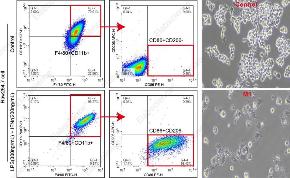
Fig. 1 Flow cytometric detection results of M1 polarization of RAW264.7 cells. The M1-type RAW264.7 macrophage polarization culture and detection kit (XJM004) was used for polarization and subsequent analysis. As illustrated in the figure, M1-type macrophages displayed short, dense pseudopodia and markedly increased cytoplasmic granularity compared to the untreated control group. Flow cytometry confirmed high expression levels of F4/80, CD11b, and CD86.
Precautions:
(1) The optimal conditions for M1 polarization are as follows: plate cells at a density of 2.5 × 10⁵ cells/mL in DMEM high-glucose medium containing 20% heat-inactivated serum and antibiotics, supplemented with LPS (300 ng/mL) and IFN-γ (200 ng/mL), and culture for 48 h.
(2) The induction efficiency is cell-state dependent; higher basal F4/80 expression levels correlate with greater M1 purity.
(3) Morphologically, more highly differentiated M1 macrophages display more pronounced pseudopodia and more evident intracellular vesicles.
(4) RAW264.7 cells readily undergo spontaneous differentiation. When cells transition from a round to a quadrilateral morphology or extend pseudopodia, this indicates an altered cellular state, and timely passage is required.
1.3 Polarization results of RAW264.7 cells toward M2-type macrophages
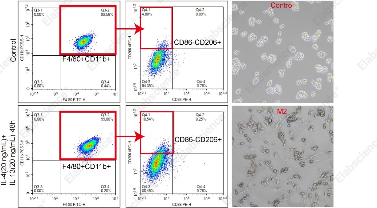
Fig. 2 Flow cytometric detection results of M2 polarization of RAW264.7 cells. The results were analyzed using flow cytometry with specific antibodies. As illustrated in the figure, M2-type macrophages displayed a spindle-like morphology with fine, elongated pseudopodia in contrast to the untreated control group. Flow cytometry confirmed high expression levels of F4/80, CD11b, and CD206.
Precautions:
(1) A serum-free starvation period of 16-20 h followed by a 24 h resting culture in complete medium before the addition of IL-4 and IL-13 promotes more efficient M2-type differentiation.
(2) Higher concentrations of IL-4 and IL-13 enhance M2 phenotypic expression.
(3) When harvesting cells, treat with trypsin for approximately 30 seconds and gently pipette to facilitate detachment, then immediately neutralize with complete medium. Excessive trypsinization may adversely affect CD206 detection.
Elabscience® Recommended Related Products:
Table 1. Recommended reagents and kits for RAW264.7 macrophage polarization and flow cytometry detection
|
Cat. No. |
Product Name |
|
XJM004 |
RAW 264.7 Polarized M1 Macrophage Induction and Identification Kit |
|
E-CK-A107 |
Cell Staining Buffer |
|
E-AB-F0997A |
Purified Anti-Mouse CD16/32 Antibody[2.4G2] |
|
E-AB-F1081F |
PerCP Anti-Mouse/Human/Monkey CD11b Antibody[M1/70] |
|
E-AB-F1081J |
PerCP/Cyanine5.5 Anti-Mouse/Human/Monkey CD11b Antibody[M1/70] |
|
E-AB-F0995C |
FITC Anti-Mouse F4/80 Antibody[CI:A3-1] |
|
E-AB-F0994D |
PE Anti-Mouse CD86 Antibody[GL-1] |
|
E-AB-F1135E |
APC Anti-Mouse CD206/MMR Antibody[C068C2] |
02 THP-1 Cell Differentiation and Polarization: Protocol and Flow Cytometry Analysis
2.1 Polarization Culture and Flow Cytometric Analysis Workflow for THP-1 Cells
General preparation (M0 differentiation stage): THP-1 cells are revived and expanded to logarithmic phase. Since THP-1 cells grow in suspension, they are counted and seeded at a density of 3 × 10⁵-5 × 10⁵ cells/mL in appropriate culture plates. PMA (100-200 ng/mL) is added to complete RPMI-1640 medium containing 10% FBS, and cells are incubated at 37°C in 5% CO₂ for 24 h to differentiate into adherent, quiescent macrophages (M0).
M1 polarization: Following 24 h of PMA treatment, the PMA-containing medium is removed, and cells are gently rinsed 1-2 times with sterile PBS or basal medium. Fresh complete medium supplemented with LPS (1 μg/mL) and IFN-γ (100-200 ng/mL) is then added, and cells are cultured for an additional 24 h at 37°C in 5% CO₂. Upon completion of induction, cells or culture supernatants are harvested for downstream analyses.
M2 polarization: After 24 h of PMA induction, the PMA-containing medium is discarded, and cells are gently washed 1-2 times with sterile PBS or basal medium. Fresh complete medium is added, and cells are allowed to rest for 24–48 h at 37°C in 5% CO₂ to stabilize the M0 state and enhance subsequent polarization efficiency. The medium is then replaced with fresh complete medium containing LPS (1 μg/mL), IL-4 (50-100 ng/mL), and IL-13 (50-100 ng/mL), and cells are cultured for another 24-48 h. After induction, cells or supernatants are collected for further analysis.
Flow cytometric analysis: Follow the same flow cytometry detection protocol as described for RAW264.7 cells.
2.2 Polarization results of THP-1 cells toward M1-type macrophages
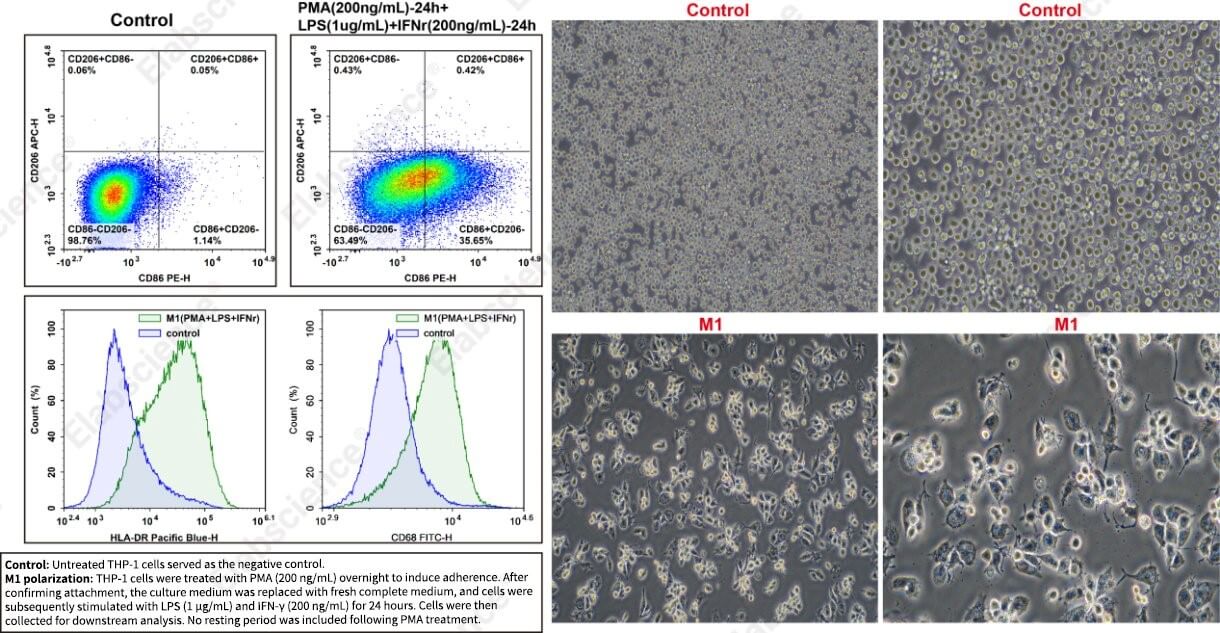
Fig. 3 Flow cytometric detection results of M1 polarization of THP-1 cells. The results were analyzed using flow cytometry with specific antibodies. As illustrated in the figure, M1-polarized cells displayed sparse and short pseudopodia with marked cytoplasmic granulation. In comparison to the untreated control group, M1 cells exhibited elevated expression of HLA-DR, CD86, and CD68, along with reduced expression of CD206. Note: To ensure consistent and reproducible results, it is critical to maintain optimal initial cell conditions, including high cell viability and stable subculturing.
2.3 Polarization results of THP-1 cells toward M2-type macrophages
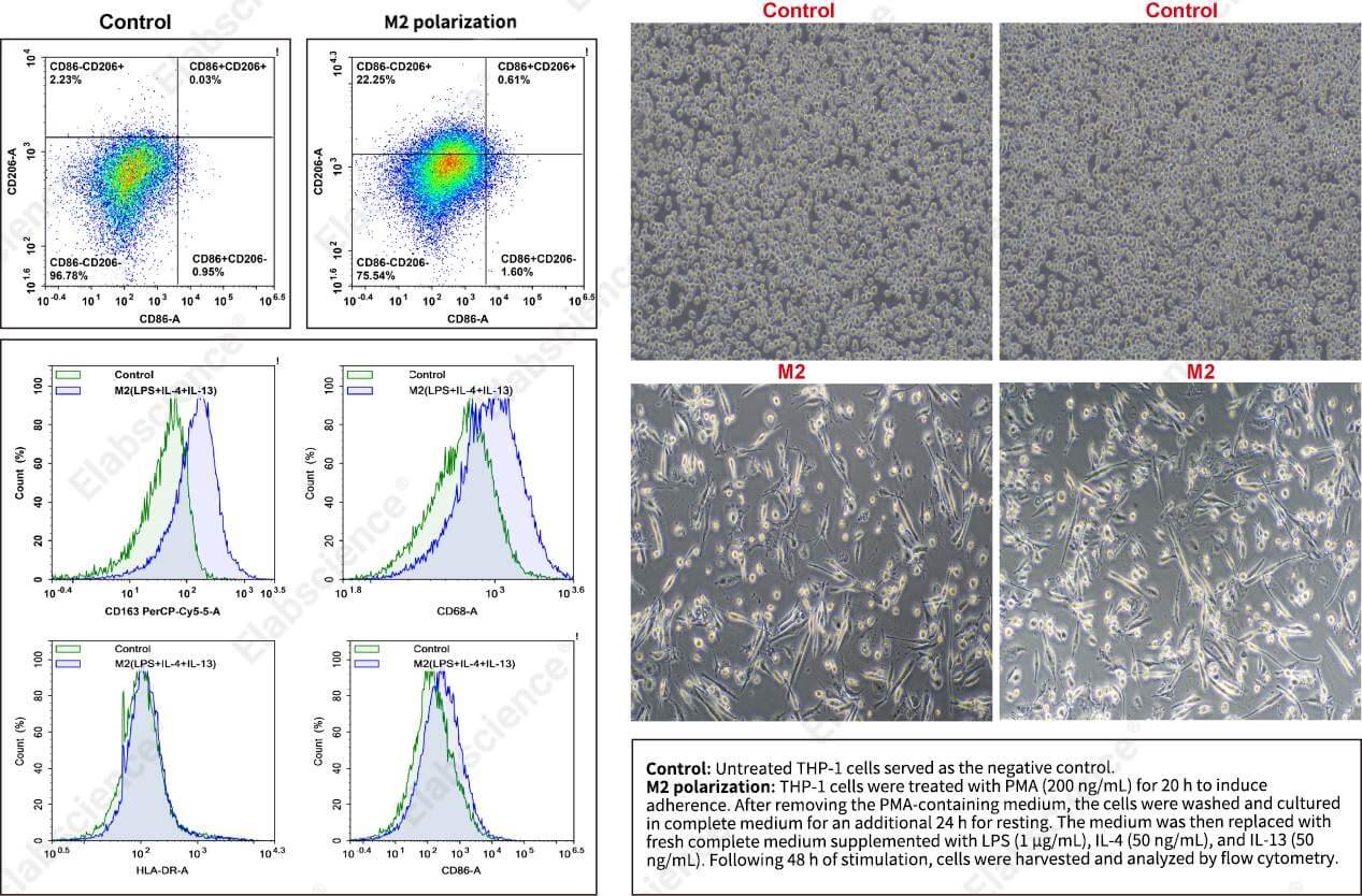
Fig. 4 Flow cytometric detection results of M2 polarization of THP-1 cells. The results were analyzed using flow cytometry with specific antibodies. As illustrated in the figure, M2-polarized macrophages displayed an elongated spindle-shaped, fibroblast-like appearance with irregular morphology. In comparison to the untreated control group, M2 cells demonstrated elevated expression of CD206 and CD163, along with reduced expression of HLA-DR and CD86.
Precautions:
(1) The optimal protocol for M2 polarization of THP-1 cells is as follows: plate cells at a density of 3.5 × 10⁵/mL, treat with PMA (100-200 ng/mL) for 16-20 h, gently wash 2-3 times with complete medium to remove PMA, replenish with fresh complete medium, allow resting culture for 24 h, and then add LPS combined with IL-4 and IL-13 for an additional 24-48 h.
(2) Complete removal of PMA is essential, as any residual PMA may interfere with downstream assays.
(3) For cell harvest using trypsin, pipette gently to facilitate detachment after 30-60 s of trypsinization, and promptly neutralize with FBS-supplemented medium. Over-trypsinization may adversely affect CD206/CD163/CD68 surface marker expression, while having minimal effect on CD86 expression.
Elabscience® Recommended Products:
Table 2. Recommended reagents for THP‑1 macrophage differentiation, polarization and flow cytometry analysis
|
Cat. No. |
Product Name |
|
E-CK-A107 |
Cell Staining Buffer |
|
E-AB-F0997A |
Purified Anti-Mouse CD16/32 Antibody[2.4G2] |
|
E-AB-F1012D |
PE Anti-Human CD86 Antibody[BU63] |
|
E-AB-F1161E |
APC Anti-Human CD206/MMR Antibody[15-2] |
|
E-AB-F1111Q |
Elab Fluor® Violet 450 Anti-Human/Monkey HLA-DR Antibody[L243] |
|
E-AB-F1299L |
Elab Fluor® 488 Anti-Human CD68 Antibody[Y1/82A] |
|
E-AB-F1298J |
PerCP/Cyanine5.5 Anti-Human/Monkey CD163 Antibody[GHI/61] |
03 Mouse Bone Marrow-Derived Macrophages (BMDM) Isolation and Polarization: Protocol and Flow Cytometry Analysis
3.1 Polarization Culture and Flow Cytometric Analysis Workflow for BMDM
Isolation and culture of BMDM:
(1) Following euthanasia, immerse the mouse carcasses in 75% ethanol for 5 min to ensure sterilization.
(2) Carefully harvest both femurs and tibias, and meticulously remove all adhering muscle and connective tissues.
(3) Separate the cleaned femurs from the tibias, and snip off both epiphyses of each bone with scissors. Using a syringe filled with pre-chilled induction medium, flush the bone marrow from the bone cavities. Perform this flushing procedure three times until the bones appear free of red marrow.
(4) Pipet the cell suspension vigorously to break up any clumps.
(5) Pass the cell suspension through a 70 μm cell strainer into a 15 mL conical tube, centrifuge at 300 × g for 5 min, and discard the supernatant.
(6) Resuspend the pellet in red blood cell lysis buffer and mix thoroughly. After a 2-min incubation at room temperature, add 10 mL of PBS to wash the cells.
(7) Centrifuge again at 300 × g for 5 min, discard the supernatant, and collect the cell pellet for subsequent plating and culture. For enhanced purity, the Mouse Monocyte Isolation Kit (MIM010N) is recommended.
General preparation: Bone marrow cells are isolated from mouse femurs and tibias, or previously cryopreserved BMDM precursors are thawed. Cells are resuspended in complete medium (high-glucose DMEM with 10% FBS) containing M-CSF (20 ng/mL), adjusted to the appropriate seeding density, and plated. The cells are cultured at 37°C in 5% CO₂ for 72 h to promote differentiation into adherent, quiescent macrophages (M0), with a half-medium change performed at approximately 48 h to replenish M-CSF.
M1 polarization: The M-CSF-containing differentiation medium is removed, and cells are gently rinsed 1-2 times with sterile PBS or basal medium. Fresh complete medium supplemented with LPS (300 ng/mL) and IFN-γ (200 ng/mL) is then added, and cells are cultured for an additional 24 h. Upon completion of induction, cells or culture supernatants are harvested for downstream analyses.
M2 polarization: The M-CSF-containing differentiation medium is discarded, and cells are gently washed 1-2 times with sterile PBS or basal medium. Fresh complete medium supplemented with M-CSF (20 ng/mL), IL-4 (20 ng/mL), and IL-13 (20 ng/mL) is added, and cells are cultured for a further 24 h. After induction, cells or supernatants are collected for subsequent detection.
Flow cytometric analysis: Follow the same flow cytometry detection protocol as described for RAW264.7 cells.
Note: Throughout the culture period in M-CSF-supplemented medium, cell morphology and medium condition should be monitored. Once the medium becomes acidic (indicated by a yellow color change), fresh medium should be added. Typically, a half-volume medium change is performed at around 48 h of culture.
3.2 Polarization results of BMDM toward M1-type macrophages
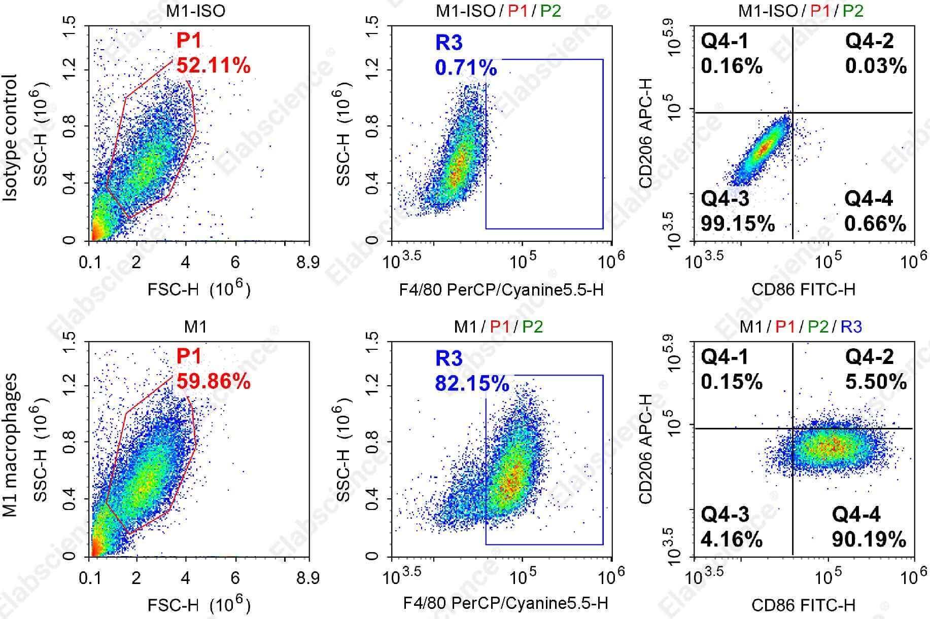
Fig. 5 Flow cytometric detection results of M1 polarization in bone marrow-derived macrophages from C57 mice. The results were analyzed using flow cytometry with specific antibodies. As illustrated in the figure, M1-polarized macrophages exhibited significantly elevated expression of F4/80 and CD86 compared to the isotype control group.
3.3 Polarization results of BMDM toward M2-type macrophages
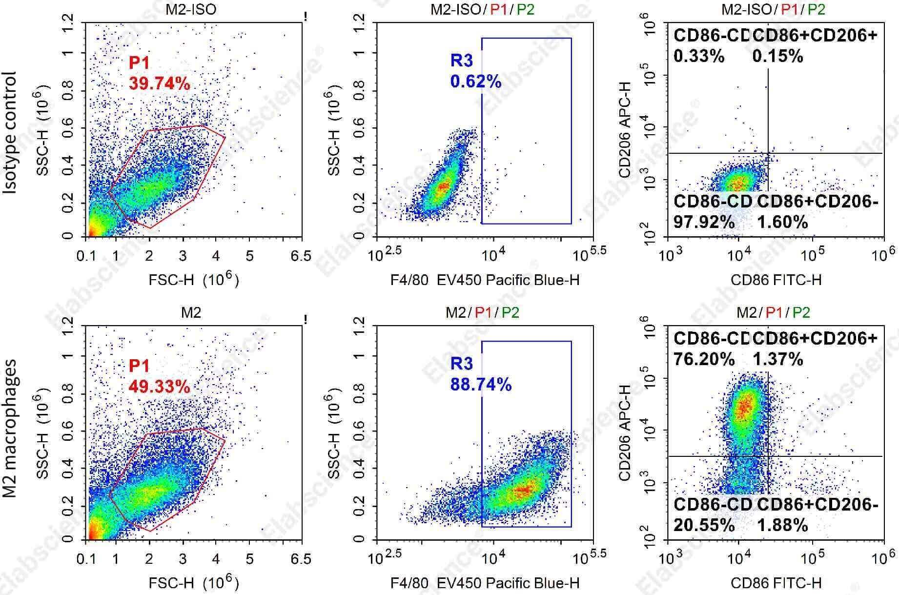
Fig. 6 Detection results of M2 polarization in bone marrow-derived macrophages from C57 mice. Flow cytometric antibodies were used for detection and analysis. As shown in the figure, compared with the isotype control group, M2-polarized macrophages showed high expression of F4/80 and CD206.
3.4 Comparison of morphological characteristics between M1- and M2-polarized BMDM
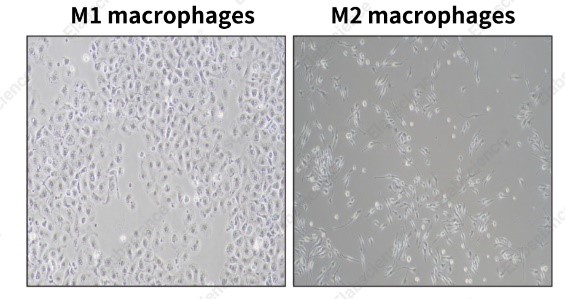
Fig. 7 Morphological comparison between M1- and M2-polarized bone marrow-derived macrophages from C57 mice. As illustrated in the figure, M1-polarized macrophages presented a more rounded shape, scant pseudopodia, and marked cytoplasmic granulation, while M2-polarized macrophages assumed an elongated spindle-shaped, fibroblast-like morphology.
Precautions:
(1) BMDM exhibit strong adherence to the culture substrate, necessitating extended trypsin digestion compared to conventional cell lines. Digestion progress should be closely monitored under a microscope.
(2) Strict aseptic technique must be maintained at all times. When chilling samples on ice, ensure that the ice container is not placed within the sterile work area.
(3) Red blood cell lysis should be limited to a short duration (no longer than 10 min) to prevent damage to nucleated cells.
Elabscience® Recommended Products:
Table 3. Recommended flow cytometry reagents for BMDM isolation and polarization assay
|
Cat. No. |
Product Name |
|
E-CK-A107 |
Cell Staining Buffer |
|
E-AB-F0997A |
Purified Anti-Mouse CD16/32 Antibody[2.4G2] |
|
E-AB-F0995J |
PerCP/Cyanine5.5 Anti-Mouse F4/80 Antibody[CI:A3-1] |
|
E-AB-F0995Q |
Elab Fluor® Violet 450 Anti-Mouse F4/80 Antibody[CI:A3-1] |
|
E-AB-F0994C |
FITC Anti-Mouse CD86 Antibody[GL-1] |
|
E-AB-F1135E |
APC Anti-Mouse CD206/MMR Antibody[C068C2] |
04 Human Peripheral Blood Monocyte-Derived Macrophages (hMDM) Differentiation and Polarization: Protocol and Flow Cytometry Analysis
4.1 Polarization Culture and Flow Cytometric Analysis Workflow for hMDM
Collection and differentiation of monocytes:
(1) Fresh human peripheral blood is collected in sodium heparin anticoagulant tubes. PBMCs are isolated using Human PBMC Separation Solution (P 1.077), resuspended in pre-chilled complete medium, centrifuged at 150 × g for 5 min, and the supernatant is discarded.
(2) The cell pellet is resuspended in pre-chilled complete medium, counted, and adjusted to a density of 1.5-2 × 10⁶ cells/mL. M-CSF is added to a final concentration of 50 ng/mL, and the cells are gently mixed and cultured at 37°C in 5% CO₂ for 24 h to facilitate adherence and macrophage differentiation.
(3) After incubation, the culture dish is gently swirled, and the supernatant along with non-adherent cells is aspirated and discarded. The adherent cells are washed with an appropriate volume of complete medium, and the wash solution is removed. Fresh complete medium supplemented with the appropriate cytokines is then added to induce polarization toward M1- or M2-type macrophages.
M1 polarization: Adherent monocyte-derived macrophages are cultured in complete medium supplemented with LPS (100 ng/mL) and IFN-γ (20 ng/mL) for 48 h. Following induction, cells are harvested and examined microscopically. M1-polarized cells exhibit a flattened, pancake-like morphology with distinct pseudopodia and marked cytoplasmic granulation. Cells are collected for downstream analysis to evaluate polarization efficiency.
M2 polarization: Adherent monocyte-derived macrophages are cultured in complete medium containing IL-4 (20 ng/mL) and IL-13 (20 ng/mL) for 48 h. After induction, cells are harvested and observed under a microscope. M2-polarized cells display an elongated, spindle-shaped, or stellate morphology with prominent cellular extensions. Cells are collected for subsequent analysis to assess the extent of M2 polarization.
Flow cytometric analysis: Follow the same flow cytometry detection protocol as described for RAW264.7 cells.
4.2 Flow cytometric analysis results of monocytes isolated from normal human peripheral blood
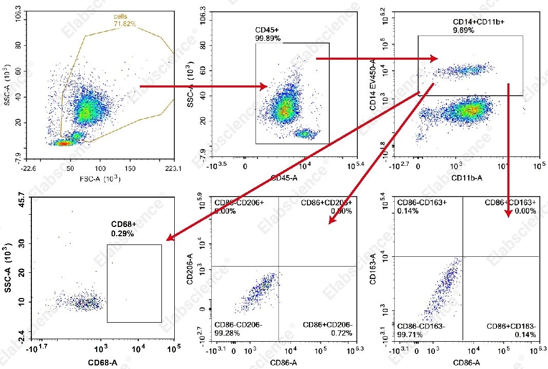
Fig. 8 Representative flow cytometric characterization of monocytes from normal human peripheral blood. Peripheral blood samples from healthy donors were stained with antibodies against human CD45, CD11b, CD14, CD68, CD86, CD206, and CD163, and subsequently analyzed by flow cytometry. As illustrated, the proportion of monocytes (CD45⁺CD11b(mid/high)CD14⁺) in normal peripheral blood was approximately 9.69%, whereas CD68⁺ macrophages were virtually absent.
4.3 Results of M1 and M2 polarization of human monocyte-derived macrophages (hMDM)
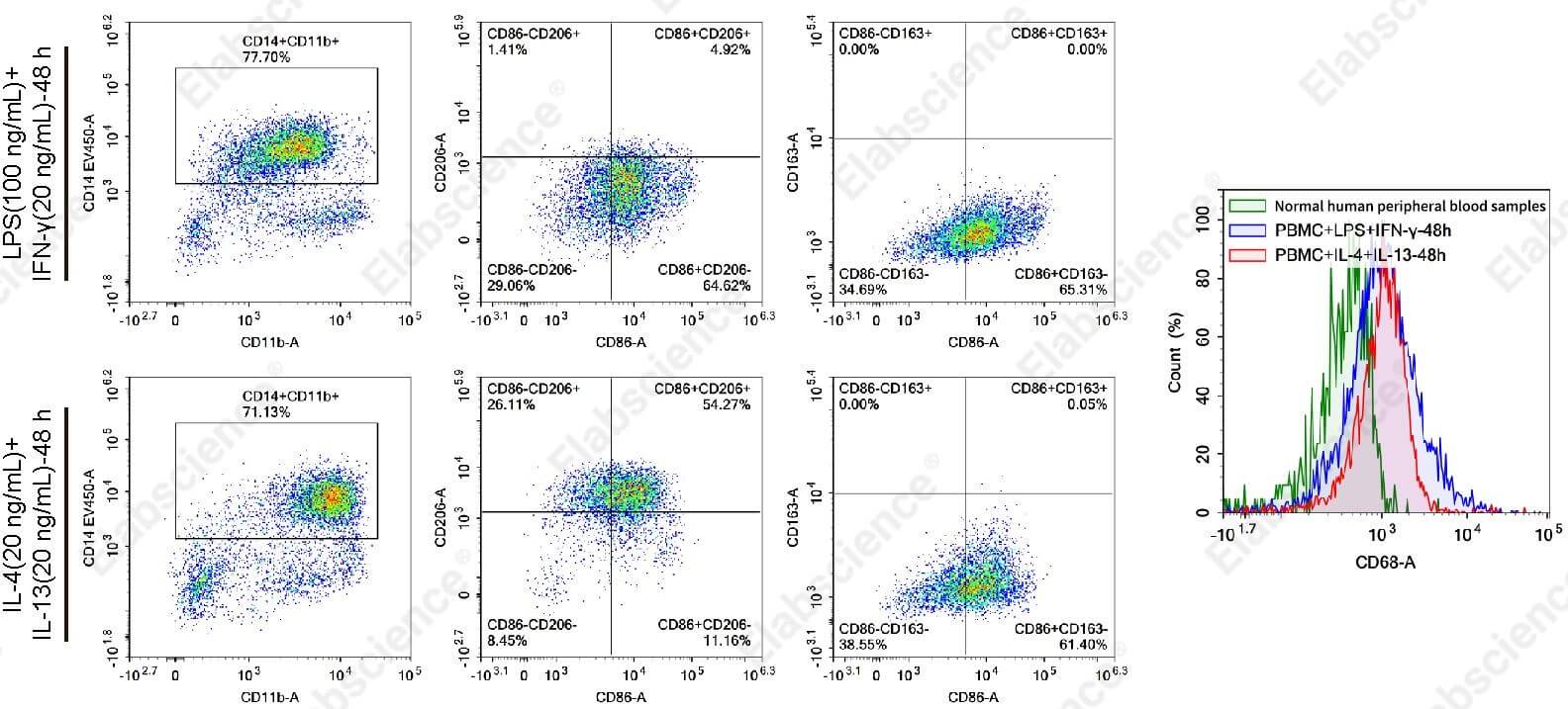
Fig. 9 Representative flow cytometric characterization of normal human peripheral blood and PBMC-derived macrophages following polarization induction. Normal human peripheral blood and PBMCs polarized toward M1 or M2 phenotypes were stained with antibodies against human CD45, CD11b, CD14, CD68, CD86, CD206, and CD163, and analyzed by flow cytometry. The results revealed that:
(1) Following induction culture, CD68⁺ expression in PBMC-derived cells was markedly upregulated, confirming a significant increase in the macrophage population.
(2) In the M1-polarized group, the CD86⁺CD206⁻ subset constituted 64.62% of the monocyte/macrophage gate, indicating a prominent enrichment of M1-type macrophages.
(3) In the M2-polarized group, the CD86⁻CD206⁺ subset accounted for 26.11% of the monocyte/macrophage population. Notably, no CD86⁻CD163⁺ cells were observed, suggesting that this protocol primarily induced M2a-type macrophages (characterized by high CD206 expression), whereas M2c-type macrophages (characterized by high CD163 expression) were not generated under these conditions.
Precautions:
(1) During PBMC isolation, centrifugation should be performed with minimal acceleration and deceleration (set the brake to the lowest setting) to prevent disruption of the density gradient interface. The buffy coat layer should be carefully aspirated with gentle handling to avoid contamination with separation medium or plasma debris.
(2) Do not change the culture medium within the first 24 h of differentiation, as the cells are not yet firmly attached. Early medium replacement may result in detachment and loss of adherent cells.
Elabscience® Recommended Products:
Table 4. Recommended reagents for hMDM differentiation, polarization and flow cytometry characterization
|
Cat. No. |
Product Name |
|
E-CK-A103 |
Human PBMC Separation Solution(P 1.077) |
|
E-AB-F1137C |
FITC Anti-Human CD45 Antibody[HI30] |
|
E-AB-F1081D |
PE Anti-Mouse/Human/Monkey CD11b Antibody[M1/70] |
|
AN00213Q |
Elab Fluor® Violet 450 Anti-Human CD14 Antibody[H332-1B10] |
|
E-AB-F1299M |
Elab Fluor® 647 Anti-Human CD68 Antibody[Y1/82A] |
|
E-AB-F1012H |
PE/Cyanine7 Anti-Human CD86 Antibody[BU63] |
|
E-AB-F1161M1 |
Elab Fluor® 700 Anti-Human CD206/MMR Antibody[15-2] |
|
E-AB-F1298J |
PerCP/Cyanine5.5 Anti-Human/Monkey CD163 Antibody[GHI/61] |
RAW264.7, THP-1, BMDM, and hMDM-each of these four models offers distinct advantages, yet each also comes with its own idiosyncrasies. Truly reliable data can only be achieved by grasping their individual traits and meticulously managing each step of the experimental process. We hope this compilation helps you avoid common stumbling blocks and maintain your cells in optimal condition. Should you have any hard-won experience or unconventional wisdom, we welcome you to share and exchange ideas in the comments below!

