

Not every immunoassay fits into a ready-to-run plate. When your target, sample, or experimental design sits outside the standard workflow, an uncoated ELISA kit hands the protocol back to you. The coating step, the blocking strategy, the sample matrix, and the signal optimization are all yours to tune.
Table of Contents
1. Selecting the right capture and detection antibody pair for ELISA assays
2. Comparing Uncoated ELISA kits with Pre-coated ELISA kits for experimental flexibility
3. How sample type influences Uncoated ELISA kit selection
4. Choosing ELISA kits based on detection sensitivity and dynamic range requirements
5. Optimizing Uncoated ELISA assays for low-abundance biomarkers in immunology research
6. Selecting Uncoated ELISA kits for cell culture supernates analysis
01 Selecting the right capture and detection antibody pair for ELISA assays
Every sandwich ELISA lives or dies on the quality of its ELISA antibody pairs. In an uncoated format the capture antibody is adsorbed directly onto your plate, the analyte binds, and a biotinylated detection antibody completes the sandwich before a streptavidin-HRP conjugate generates signal. The pairing scheme, not the reagent volume, is the single biggest determinant of sensitivity, specificity, and dynamic range.
1.1 What defines a validated matched pair?
● Non-competing epitopes. Capture and detection antibodies must recognize spatially distinct, non-overlapping epitopes so they can bind the same analyte molecule simultaneously. To this end, the uncoated series antibody pairs undergo systematic epitope screening from dozens of candidate antibodies and sandwich pairing tests, with dual validation using both recombinant protein and native samples. This ensures that the selected pairs recognize native conformational epitopes with optimal affinity and specificity, bringing the results closer to the true sample state. Competing pairs silently cut your signal, while native-conformation-validated pairs are the first line of defense for data authenticity.
● Pre-titrated ratio with practical optimization strategy. An optimized kit supplies both antibodies at pre-determined working concentrations. Elabscience® uncoated series have undergone initial condition scouting; we generally recommend starting with both capture and detection antibodies at 500-fold dilution. However, due to variations in assay buffer systems, incubation conditions, and operational environments, the optimal ratio may shift. We suggest:
1) Initial test: Use the 500-fold dilution directly and evaluate the standard curve signal.
2) If signals are too high (e.g., top standard OD saturates beyond the detector's linear range): Increase the dilution – try checkerboard titrations with capture and detection antibodies at 500x, 1000x, and 2000x to reduce background and achieve a better curve range.
3) If signals are too low (e.g., top standard OD is insufficient for sensitivity): Decrease the dilution – try checkerboard titrations at 125x, 250x, and 500x to boost signal output.
This strategy gives you a reliable starting point while preserving room for fine-tuning, helping you quickly lock in the best pairing ratio for your specific system.
● Recombinant, calibrated standard. A matched protein standard lets you build a 4-parameter logistic (4-PL) curve and convert OD into absolute concentration with confidence.
● Demonstrated specificity. Look for cross-reactivity data against related family members (e.g., IL-6 vs. IL-6R, TNF-α vs. TNF-β/lymphotoxin).
1.2 Special considerations for dimeric & multimeric targets
Not every analyte is a clean monomer. Many cytokines, chemokines, and growth factors circulate or are secreted as homo- or hetero-dimers, trimers, or higher-order multimers (e.g., homotrimeric TNF-α / TNF-β[1], dimeric TGF-β[2], hexameric IL-6/IL-6R/gp130 signaling complexes[3], multimeric PD-L1 or galectin family members). For these targets, the pairing scheme carries an extra layer of geometry that monomer logic does not capture. We have added the following validation checkpoints in Elabscience® antibody-pair development specifically for multimeric targets, ensuring that the two antibodies you receive work synergistically in the native multimeric conformation.
● Epitope topology across subunits. For a homodimer, both capture and detection antibodies may each see two identical epitopes. If the two antibodies bind overlapping sites on a single subunit, they will still compete even though a "matched pair" screen on monomer might have passed[4]. Elabscience® antibody pairs for all multimeric targets are additionally validated for epitope compatibility on the native multimeric form (not just recombinant monomer), guaranteeing unobstructed binding under the true conformation.
● Bridging vs. same-subunit detection. For heterodimers, capture and detection antibodies can target the same subunit (cis-pairing) or different subunits (trans-pairing). During development, Elabscience® systematically compares both strategies and prioritize the pairing that delivers stronger signal and lower hook-effect risk, sparing you the need to explore pairing geometry yourself.
● Specificity control under avidity effects. Because multimers present repeated epitopes, bivalent capture leverages avidity to significantly improve sensitivity and specificity – but if the detection antibody cross-reacts with denatured or fragmented monomeric subunits, background will rise. We specifically introduce degraded samples and monomer interference tests in QC to ensure that the detection antibody targets the intact multimeric conformation, not free fragments.
● Strict matching of standard to analyte form. If the oligomeric state of the standard does not match that of the sample analyte, concentration quantification will be systematically biased. Recombinant standards are confirmed by gel filtration or dynamic light scattering to have the same oligomeric state as the native analyte, so your standard curve readings reflect true concentration, not an estimate of epitope density.
Multimer Pairing Tip
For dimeric and multimeric targets, ask the supplier three questions:
(1) Was the matched pair validated on the native oligomeric form of the analyte? (2) Do capture and detection antibodies bind the same or different subunits/epitope clusters? (3) Is the recombinant standard provided in the same oligomeric state? The answers determine whether your ELISA antibody pairs will read true concentration or merely total epitope density.
02 Comparing Uncoated ELISA kits with Pre-coated ELISA kits for experimental flexibility
The uncoated vs pre-coated ELISA kits comparison is at heart, a flexibility trade-off. Pre-coated ELISA kits arrive with capture antibody already onto and validated on the plate. They are fast, consistent, and ideal when throughput and lot-to-lot reproducibility matter more than customization. Uncoated kits ship the same pre-titrated reagents but leave the plate uncoated, returning the coating, blocking, and incubation steps to you.
Table 1. Comparison of Uncoated ELISA kits with Pre-coated ELISA kits for experimental flexibility
|
Attribute |
Pre-Coated ELISA Kit |
Uncoated ELISA Kit |
|
Plate coating |
Done at the factory |
You coat overnight (your plate, your buffer) |
|
Blocking & wash |
Pre-optimized |
Fully adjustable to your matrix |
|
Protocol length |
~4 hours, sample-to-result |
~24 hours (overnight coating included) |
|
Assay variance control |
Supplier-validated (low CV) |
Operator-controlled; best for experienced users |
|
Cost per plate |
Higher |
Lower (built for budget-conscious, high-volume work) |
|
Customization ceiling |
Locked |
Open (swap buffers, substrates, sample volume) |
Why should you consider an uncoated ELISA kit?
● Working with unusual sample types? Whether it's high-lipid serum, tissue lysate, or non-standard species samples, Elabscience® technical support team can help you optimize assay conditions to get your detection up and running smoothly.
● Running large sample cohorts? Coating your own plates significantly reduces cost per plate – ideal for large-scale cohort studies or budget-conscious projects.
● Need flexible assay configurations? Whether you're cross-validating in-house antibodies or testing multiple targets with shared reagents, the uncoated format gives you the customization you need.
● New to uncoated ELISAs? No worries. We provide end-to-end technical guidance from coating and blocking to detection, and support team is just a call away whenever you run into questions.
03 How sample type influences Uncoated ELISA kit selection
Sample matrix is the variable most often underestimated in uncoated ELISA kit selection. The same analyte can behave very differently in serum, plasma, cell lysate, urine, or culture supernates, and because you control the diluent and blocking step in an uncoated format, you can tune the assay to the matrix instead of forcing the matrix into a fixed protocol.
Table 2. The influence of sample type on kit selection
|
Sample type |
Key concern |
What to look for in the kit |
|
Serum / plasma |
Heterophilic antibodies, complement, rheumatoid factors, hook effect at high titer |
Validated sample diluent; dilution-linearity & spike-recovery data (80~120%) |
|
Cell culture supernates |
Phenol red, serum proteins, drug carriers interfering with detection |
Low-background matrix matching; compatible diluent |
|
Cell / tissue lysate |
Detergents (RIPA), protease activity, endogenous biotin |
Detergent-tolerant detection; avidin-HRP robust to lysate |
|
Urine / saliva / CSF |
Low protein, variable pH/salt |
High-sensitivity range; concentratable protocol |
|
Multiplex / scarce sample |
Limited volume (tens of µL) |
Small-volume protocol; wide dynamic range |
Matrix Lesson
Because the uncoated format lets you choose the plate, blocking buffer, and sample dilution freely, it is a natural choice when working with non-standard samples, particularly those that behave differently from conventional serum samples. Always run a spike-recovery (target 80~120%) and parallelism check before committing a full cohort[5].
Elabscience® Uncoated ELISA kits by sample type
Use the catalog below to find the uncoated ELISA kit validated for your sample matrix. Fields are left blank here so you can map in your institution's product numbers and links.
Table 3. Products in the field of Cell culture supernates
|
Cat. No. |
Product Name |
Size |
|
E-UNEL-M0064 |
Uncoated Mouse IL-1β(Interleukin 1 Beta) ELISA Kit |
96T*5/96T*15 |
|
E-UNEL-H0175 |
Uncoated Human TNF-α(Tumor Necrosis Factor Alpha) ELISA Kit |
96T*5/96T*15 |
|
E-UNEL-M0070 |
Uncoated Mouse IL-6(Interleukin 6) ELISA Kit |
96T*5/96T*15 |
|
E-UNEL-M0103 |
Uncoated Mouse TNF-α(Tumor Necrosis Factor Alpha) ELISA Kit |
96T*5/96T*15 |
|
E-UNEL-M0043 |
Uncoated Mouse IFN-γ(Interferon Gamma) ELISA Kit |
96T*5/96T*15 |
|
E-UNEL-H0069 |
Uncoated Human IFN-γ(Interferon Gamma) ELISA Kit |
96T*5/96T*15 |
04 Choosing ELISA kits based on detection sensitivity and dynamic range requirements
Two specifications dominate uncoated ELISA kit selection: analytical sensitivity (the lowest concentration you can reliably distinguish from zero) and dynamic range (the span over which OD tracks concentration linearly enough for a 4-PL fit). Pick them against your expected sample concentration, not in the abstract.
High-abundance analytes, such as secreted cytokines in stimulated cultures, require a wide dynamic range so that both high- and low-concentration samples can be measured on the same plate without additional dilution steps, with a typical range spanning 2–3 log units. For example, PBMCs cultured under untreated or treatment conditions for 0–6 days before collecting conditioned media show widely varying levels of human IL-6 and other cytokines.
Table 4. The cytokines levels of PBMCs treated for 0 or 6 days
|
Cat. No. |
Treated 0d |
Treated 6d |
||
|
Dilution factor |
Optical density |
Dilution factor |
Optical density |
|
|
E-UNEL-H0069 |
1 |
0.417 |
1000 |
2.666 |
|
E-UNEL-H0175 |
1 |
0.279 |
10 |
2.727 |
|
E-UNEL-H0099 |
50 |
0.265 |
1000 |
2.009 |
|
E-UNEL-H0084 |
1 |
0.045 |
1 |
0.709 |
|
E-UNEL-H0097 |
1 |
1.714 |
100 |
3.467 |
Low-abundance analytes (basal cytokines in unstimulated serum) need sensitivity first; look for sub-pg/mL limits of detection and a high-sensitivity formulation.
Widespread cohorts (healthy vs. diseased, dosed vs. vehicle) need both sensitivity at the floor and headroom at the ceiling.
Table 5. Choosing ELISA kits based on different need
|
Need |
Specification to prioritize |
Typical uncoated kit signature |
|
Detect trace cytokines |
Sensitivity (LOD) |
< 1 pg/mL high-sensitivity range |
|
One-pass read across cohort |
Dynamic range |
2–3 log units, e.g. 15.6–1,000 pg/m |
|
Linearity across dilutions |
Parallelism / dilution linearity |
Recovery 80–120% across dilution series |
|
Reproducibility between runs |
Intra/inter-assay CV |
< 10% intra-assay; < 12% inter-assay |
A wider range does not automatically mean better. If your samples sit near the floor, prioritize the lowest LOD; you can always dilute concentrated samples, but you cannot recover signal below the noise.
05 Optimizing Uncoated ELISA assays for low-abundance biomarkers in immunology research
In immunology research, the most interesting biology often sits at the bottom of the detectable range of basal cytokines, early-response mediators, subtle treatment shifts. The uncoated format is a powerful platform here precisely because every signal-amplifying variable is in your hands. A well-optimized uncoated ELISA assay can rival the sensitivity of fixed-format kits when the low-abundance levers are pulled correctly.
Levers that move sensitivity on an uncoated plate
● Capture antibody density. Titrate coating concentration to maximize specific capture without saturating plate capacity, which raises background.
● Blocking reagent. Choose a blocker matched to your matrix (BSA for many cytokines; casein or fish-gelatin for lipid-rich samples) to collapse non-specific binding.
● Detection antibody & streptavidin-HRP titration. Lowering detection concentration often improves signal-to-noise by cutting background faster than signal.
● Substrate choice. TMB standard for colorimetric; switch to a high-sensitivity TMB or chemiluminescent substrate when pushing the LOD.
● Sample diluent & incubation. A matched diluent plus extended sample incubation recovers signal from trace analyte that a fast, fixed protocol would miss.
Worked Example: Il‑6 In Immunology
Interleukin-6 is a benchmark inflammation and immuno-oncology biomarker whose serum levels can sit at the edge of detectability in unstimulated conditions. A validated uncoated human IL-6 ELISA kit provides the matched capture and detection antibody pair, a recombinant standard, and streptavidin-HRP, giving you the freedom to lower the assay floor through coating-density and substrate optimization. This makes it ideal for tracking basal IL-6 levels, monitoring treatment response, or measuring cytokine release derived from the tumor microenvironment, all without committing to a single fixed protocol. The range for detecting treated cell media supernates using the uncoated human IL-6 elisa kit is 1.56-100 pg/mL.
06 Selecting Uncoated ELISA kits for cell culture supernates analysis
Cell culture supernates is the most natural home for the uncoated format. Secreted cytokines, growth factors and inflammatory mediators are released into a complex medium containing phenol red, FBS proteins, and sometimes drug carriers that rewards the buffer-tuning freedom only an uncoated kit allows. The goal is to quantify secreted target cleanly, without the medium itself generating signal.
Checklist for supernates work
● Confirm the kit reacts with your secreted target, rather than only the cell-bound type.
● Matrix-matched diluent that neutralizes phenol red and FBS interference; run an unspiked medium blank to confirm background.
● Range of expected secretion stimulation cultures can spike high; basal cultures can sit near zero.
● Small-volume compatibility when collecting from low-yield wells or primary cells.
● Flexible plate scale (5-plate or 15-plate options) so a 96-well secretion screen doesn't force a fixed-plate purchase.
TNF-α Secretion Monitoring Example
Tumor necrosis factor-alpha is the classic readout of macrophage/T-cell activation and inflammatory drug response in vitro. A validated uncoated mouse TNF-α ELISA kit gives you the matched antibody pair, recombinant standard, and streptavidin-HRP to quantify TNF-α in murine supernatant that across LPS stimulation time courses, knockout vs. wild-type comparisons, or drug-candidate screening with full control over the medium-matching and background-suppression steps that fixed kits lock down.
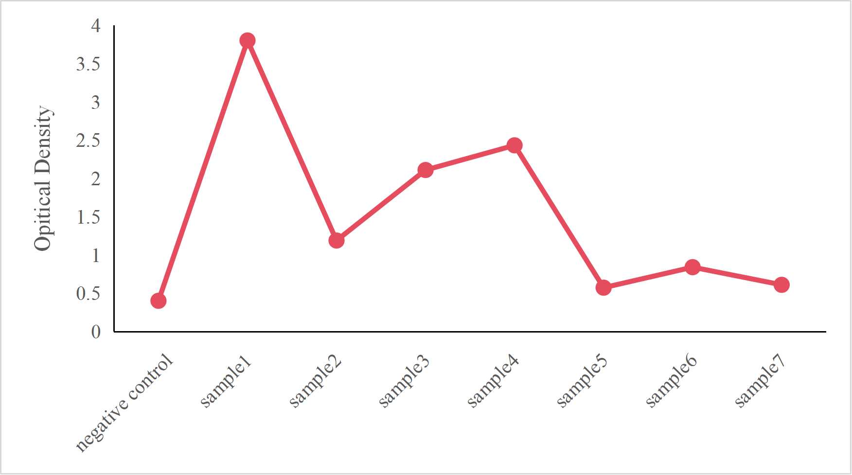
Fig. 1 Results of uncoated mouse TNF-α ELISA kit for different treated PBMCs sample models.
References:
[1] Jones EY, Stuart DI, Walker NPC. Structure of tumour necrosis factor. Nature. 1989;338(6212):225-228.
[2] Daopin S, Piez KA, Ogawa Y, Davies DR. Crystal structure of transforming growth factor-β2: an unusual fold for the superfamily. Science. 1992;257(5071):369-373.
[3] Boulanger MJ, Chow DC, Brevnova EE, Garcia KC. Hexameric structure and assembly of the interleukin-6/IL-6 receptor complex. Science. 2003;300(5628):2101-2104.
[4] Laver WG, Air GM, Webster RG, Smith-Gill SJ. Epitopes on protein antigens: misconceptions and realities. Cell. 1990;61(4):553-556.
[5] Findlay JWA, et al. Validation of immunoassays for bioanalysis: a pharmaceutical industry perspective. J Pharm Biomed Anal. 2000;21(6):1249-1273.

