

Chemokines are a specialized family of small, heparin-binding cytokines, typically 8 to 12 kDa, that orchestrate the directed movement and positioning of immune cells. Unlike broadly acting cytokines, chemokines signal through seven-transmembrane G protein-coupled receptors (GPCRs) to generate spatially restricted concentration gradients, guiding leukocytes from circulation into tissues during homeostasis, inflammation and immune surveillance. Based on the arrangement of conserved cysteine residues, they are classified into CC, CXC, CX3C and XC subfamilies, each with distinct receptor specificity and cellular targets. This system underlies lymphoid organogenesis, wound healing and host defense, and its dysregulation contributes to chronic inflammation, fibrosis, autoimmunity and tumor progression, making precise chemokine detection a central requirement in modern immunology research.
This review article provides an integrated overview of chemokine biology and its experimental interrogation. We first examine how chemokines establish directional cues for immune cell migration and how discrete chemokine-receptor pairs encode trafficking specificity. We then discuss the formation of tissue-specific gradients and spatial localization, followed by practical strategies for quantifying chemokines in cell culture supernatants and biological fluids. Finally, we explore chemokine networks within the tumor microenvironment and the consequences of chemokine dysregulation during chronic inflammation, linking mechanistic insights to flow cytometry, multiplex and ELISA-based workflows.
Table of Contents
1. Chemokines as directional signals for immune cell migration
2. Chemokine–receptor pairs that control immune cell trafficking
3. Chemokine gradients and tissue-specific immune cell localization
4. Quantifying chemokines in cell culture and biological samples
5. Chemokine networks in the tumor microenvironment
6. Chemokine dysregulation during chronic inflammation
01 Chemokines as directional signals for immune cell migration
Chemokines function as directional signals that convert soluble concentration fields into cellular navigation, a concept that distinguishes chemokine function from the pleiotropic actions of classic cytokines. Secreted as soluble or matrix-immobilized gradients, chemokines bind glycosaminoglycans (GAGs) on endothelial surfaces and within extracellular matrix, presenting haptotactic trails that are sensed by leukocyte GPCRs. Ligand engagement triggers Gi protein-dependent activation of phosphoinositide 3-kinase (PI3K), DOCK2 and Rac, leading to actin polymerization, integrin activation and persistent migration along the gradient[1]. In tumors and inflamed tissues, stromal and myeloid cells co-establish overlapping gradients that compete for receptor occupancy, as illustrated by recent mapping of immune cell migration in human cancers[1]. Unlike growth factors that primarily drive proliferation, chemokines prioritize positioning, and unlike acute-phase proteins such as C-reactive protein (CRP), they act locally through receptor-mediated chemotaxis rather than systemic amplification. Natural killer (NK) cell trafficking studies demonstrate that microenvironmental barriers actively reshape chemokine fields, altering the density and directionality of gradients and thereby limiting cytotoxic cell entry into tumor nests[2]. Stromal perineural niches further illustrate this principle, where nerve-associated fibroblasts secrete CXCL12 and CCL2 to create perineural highways that attract CCR2 and CXCR4 expressing leukocytes and tumor cells alike[3]. Sex-specific immune variation also reflects tunable chemokine output, indicating that steady-state chemokine tone defines tissue residency of memory and innate lymphocytes[4].
.png)
Fig. 1 Physical and chemotactic barriers of the tumor microenvironment (TME) that regulate the infiltration and activity of natural killer (NK) cells. The figure illustrates the cellular heterogeneity of the TME composed of tumor cells, endothelial cells, cancer-associated fibroblasts (CAFs), dendritic cells, and macrophages—and its impact on NK cell migration[2].
02 Chemokine–receptor pairs that control immune cell trafficking
The specificity of immune cell trafficking is encoded by discrete chemokine-receptor pairs, each pairing a ligand family to a leukocyte subset through selective GPCR expression. Conventional chemokine receptors such as CC chemokine receptor 2 (CCR2) and CXC chemokine receptor 3 (CXCR3) exhibit high ligand selectivity, whereas atypical chemokine receptors (ACKRs), exemplified by atypical chemokine receptor 4 (ACKR4), scavenge and shape gradients without canonical G protein signaling[1,5]. Phosphorylation of the ACKR4 C-terminal tail by G protein-coupled receptor kinase (GRK) isoforms differentially regulates beta-arrestin recruitment and scavenging efficiency, demonstrating that receptor trafficking and signal desensitization are modulated at the post-translational level[5]. Functionally, CC chemokine ligand 2 (CCL2)-CCR2 drives classical monocyte and inflammatory macrophage egress from bone marrow, CXC chemokine ligand 10 (CXCL10)-CXCR3 recruits effector T helper type 1 (Th1) cells and effector CD8+ T cells into inflamed sites, and CXC chemokine ligand 12 (CXCL12)-CXC chemokine receptor 4 (CXCR4) retains hematopoietic progenitors and directs plasma cell homing; quantification of these axes commonly relies on a chemokine assay combined with receptor flow cytometry, and when precise ligand concentration is required a ccl2 elisa or cxcl12 elisa provides sensitive readouts[1,6]. Vascular reprogramming studies show that CXC ligands remodel tumor endothelium to facilitate leukocyte extravasation, linking ligand-receptor matching to endothelial activation[6]. Comparative analyses of fibroblast-cancer-immune crosstalk reveal that stromal cells produce paired chemokines such as CCL19/CCL21 and CXCL13 to position dendritic cells and lymphocytes within adventitial niches[7], while population-level genetic studies associate distinct immune cell frequencies with chemokine receptor polymorphisms[8].
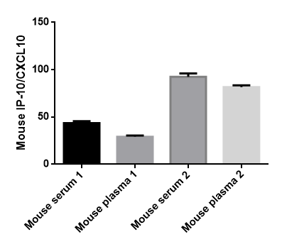
Fig. 2 The presentation of CXCL10 chemokine assay data in mice. (The data are provided by Elabscience®)
03 Chemokine gradients and tissue-specific immune cell localization
Chemokine gradients do not operate as uniform soluble clouds but as spatially anchored fields that encode tissue-specific immune cell localization. Electrostatic interactions with heparan sulfate proteoglycans immobilize chemokines on extracellular matrix and endothelial glycocalyx, generating steep, matrix-bound slopes that persist against diffusion and flow[1]. In tumors and mucosal tissues, the same ligand can produce divergent positioning outcomes depending on local GAG composition and on scavenging by ACKRs, which collapse gradients to prevent ectopic infiltration[1]. Integrated single-cell and bulk transcriptomics of macrophages illustrates this spatial logic, where CC chemokine ligand 3 (CCL3), CC chemokine ligand 4 (CCL4) and JUNB mark inflammatory macrophage niches enriched for gradient-forming chemokines[9]. Studies in solid tumors further show that natural killer (NK) cell exclusion correlates with physical microenvironmental obstacles that disrupt chemokine diffusion and presentation, indicating that architecture itself modulates chemokine availability[2]. Gut-prostate axis investigations reveal that organ-specific chemokine milieus shape regional lymphocyte residency and susceptibility to chronic inflammation, suggesting tissue-imprinted stromal memory[10]. At subcellular resolution, spatial profiling of kidney allografts identifies Fc gamma receptor III positive (FcγRIII+) innate cells clustered around discrete chemokine foci, while nanobody-based epitope mapping provides tools to visualize accessible chemokine deposits without disrupting gradient geometry[11,12]. Skin inflammation models demonstrate that protease activity, including meprin-dependent shedding, can truncate and inactivate chemokines post-secretion, dynamically re-tuning gradient steepness during homeostasis versus psoriasis[13].
04 Quantifying chemokines in cell culture and biological samples
Accurate quantification of chemokines in cell culture supernatants and biological fluids underpins mechanistic studies and biomarker development, with sandwich enzyme-linked immunosorbent assay (ELISA) remaining the reference method for absolute concentration measurement. In a typical chemokine assay, matched capture and biotinylated detection antibodies flank the analyte, and 3,3,5,5 prime-tetramethylbenzidine (TMB) conversion provides a linear dynamic range of approximately 15.6 to 1000 pg/mL, enabling longitudinal monitoring of stimulation-induced secretion[14]. For example, pharmabiotic intervention studies illustrate that reduced circulating CC chemokine ligand 2 (CCL2) and CXC chemokine ligand 1 (CXCL1) levels after treatment are resolved at single-digit pg/mL sensitivity, correlating with improved liver fibrosis indices[14]. Epithelial-mesenchymal signaling studies similarly employ a chemokine elisa kit to link METTL1-mediated N7-methylguanosine (m7G) modification of bradykinin receptor B1 (Bdkrb1) messenger RNA (mRNA) to downstream CXCL5/CXCL8 release during psoriatic inflammation[15]. The nuclear interleukin 33 (IL-33)-nuclear factor kappa B (NF-kappaB) axis, where IL-33 amplifies NF-kappaB signaling by inducing inhibitor of kappa B kinase (IKK)-dependent phosphorylation and degradation of inhibitor of kappa B alpha (IκBα) and subsequent p65/p50 nuclear translocation, was validated by multiplex chemokine detection showing elevated neutrophil-attracting CXCL1/CXCL2 in Mycoplasma-infected endocervix[16]. When investigating analyte-specific workflows, investigators routinely select a cxcl10 elisa for interferon gamma (IFN-gamma)-induced Th1 recruitment panels and a ccl2 elisa for monocyte chemoattraction panels, with assay validation including spike-recovery and dilution-linearity in serum, plasma and conditioned medium[17,18]. Standard operating procedures recommend immediate centrifugation, aliquoting to avoid freeze-thaw cycles, and parallel measurement of related chemokines to control for matrix effects.
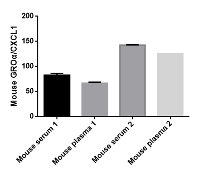
Fig. 3 The presentation of CXCL1 chemokine assay data in mice. (The data are provided by Elabscience®)
05 Chemokine networks in the tumor microenvironment
Within the tumor microenvironment, chemokines form interdependent networks that simultaneously regulate immune cell infiltration, vascular patterning and tertiary lymphoid structure (TLS) organization, defining whether a lesion is immunologically hot or cold. Chemokines in the tumor microenvironment are produced not only by malignant cells but also by cancer-associated fibroblasts, tumor-associated macrophages and endothelial cells, whose collective output determines the balance between effector recruitment and immunosuppression. In triple-negative breast cancer, coordinated expression of CCL2, CCL5, CXCL9 and CXCL10 correlates with CD8+ T cell density and with expression of programmed death-ligand 1 (PD-L1), suggesting that a CXCL9/CXCL10-CXCR3 axis predicts responsiveness to immune checkpoint blockade[19]. Hepatobiliary and pancreatic cancers exemplify immune resistance beyond checkpoints, where exclusion signatures enriched for CXCL12-CXCR4 and CCL20-CCR6 are associated with stromal fibrosis and reduced cytotoxic lymphocyte entry[20]. Complementary analyses of colorectal cancer reveal patient-stratified chemokine patterns, where high CXCL8 and CCL20 associate with myeloid inflammation and poor prognosis, while CXCL13 marks TLS-associated B cell aggregation and improved survival[21]. TLS themselves function as local chemokine factories, producing homeostatic chemokines such as CCL19, CCL21 and CXCL13 that compartmentalize T cell and B cell zones and can be profiled as neoadjuvant immunotherapy biomarkers[22]. Integrating these layers, multiplex chemokine profiling combined with spatial transcriptomics provides a systems-level readout of chemokines in the tumor microenvironment for translational stratification.
06 Chemokine dysregulation during chronic inflammation
Chronic inflammation sustains chemokine dysregulation through persistent transcriptional activation and failure of resolution circuits, converting transient recruitment into a self-reinforcing leukocyte influx. In contrast to acute responses that resolve upon pathogen clearance, low-grade inflammatory states maintain elevated CC chemokine ligand 20 (CCL20)-CC chemokine receptor 6 (CCR6) and CXC chemokine axes, which continuously attract mucosal-associated invariant T (MAIT) cells, M2 macrophages and circulating monocytes[23,24]. Systems-level profiling of circulating immune cells during chronic immune activation demonstrates that an interpretable inflammation landscape, anchored by CXCL10, CCL2 and CXCL12 signatures, distinguishes active versus quiescent disease across heterogeneous cohorts[25]. Mechanistically, crosstalk between type I interferon (IFN) and tumor necrosis factor (TNF) converts epithelial repair programs into pathogenic chemokine production, converting regenerative inflammation into sustained tissue damage in inflammatory bowel disease (IBD)[26]. Substance abuse models further illustrate systemic consequences, where repeated exposure amplifies NF-kappaB-dependent chemokine transcription beyond the central nervous system, propagating peripheral inflammation[27]. Cardiac immunology studies reveal that myocardial immune cells respond to persistent chemokine cues with fibrotic remodeling, while exercise-based interventions show that physical activity can downregulate selected chemokines and restore immune homeostasis[24,28]. Importantly, prolonged chemokine exposure is not interchangeable with acute-phase protein elevation; the former reflects local GPCR-driven cell recruitment, whereas C-reactive protein (CRP), an acute-phase protein synthesized in liver, reflects systemic inflammatory amplification and should not be conflated with chemokine mediators[25,27].
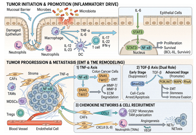
Fig. 4 Cytokine and chemokine networks in colorectal cancer[21].
In summary, chemokines are small, structurally classified cytokines whose spatially confined gradients encode the timing, location, and cellular specificity of immune cell trafficking. From GAG-anchored directional cues and receptor-specific pairings such as CCL2-CCR2, CXCL10-CXCR3 and CXCL12-CXCR4, to tissue-anchored positioning and quantitative detection, each layer provides an experimentally accessible readout. Rigorous chemokine assay workflows, including a validated chemokine elisa kit and analyte-specific kits such as cxcl10 elisa, ccl2 elisa and cxcl12 elisa, enable accurate measurement of chemokine function across culture systems and patient samples. Within tumors, coordinated chemokines in the tumor microenvironment predict immune contexture, TLS formation and responsiveness to immunotherapy, while persistent chemokine dysregulation sustains chronic inflammation when resolution fails. Profiling these networks with complementary GPCR, transcriptomic and multiplex protein approaches offers a practical framework for designing targeted interventions and monitoring therapeutic responses.
Elabscience® Quick Overview of Popular Products:
Table 1. Research Tools for Detection
|
Cat. No. |
Product Name |
|
E-EL-M0018 |
Mouse GROα/CXCL1(Growth Regulated Oncogene Alpha) ELISA Kit |
|
E-EL-M0021 |
Mouse IP-10/CXCL10(Interferon Gamma Induced Protein 10kDa) ELISA Kit |
|
E-EL-M0471 |
Mouse CXCL5(Chemokine C-X-C-Motif Ligand 5) ELISA Kit |
|
E-EL-M0007 |
Mouse MIP-1α(Macrophage Inflammatory Protein 1 Alpha) ELISA Kit |
|
E-EL-M3001 |
Mouse MCP-1(Monocyte Chemotactic Protein 1) ELISA Kit |
|
E-EL-M3046 |
Mouse SDF-1/CXCL12(Stromal Cell Derived Factor 1) ELISA Kit |
|
E-EL-M3063 |
Mouse TNF-α(Tumor Necrosis Factor Alpha) ELISA Kit |
|
E-EL-H6008 |
Human IL-8(Interleukin 8) ELISA Kit |
|
E-EL-R0009 |
Rat IFN-γ(Interferon Gamma) ELISA Kit |
References:
[1] Zhang et al., Migration of immune cells in tumors and inflammation: molecular mechanisms and therapeutic targets. Signal Transduction and Targeted Therapy, 2026. 11(1): 45-72.
[2] Wang et al., Natural killer cell immunotherapy in solid tumors: microenvironmental obstacles and translational 3D models. Biology, 2026. 15(3): 167-189.
[3] Liu et al., Stromal regulation of tumor perineural invasion: a multicellular and neuro-ecological perspective. Advanced Science, 2026. 13(2): 76248.
[4] Chen et al., Sex in immune cells and parasitic diseases - a complex relationship. Immunological Reviews, 2026. 315(1): 88-104.
[5] Smith et al., GPCR kinases shape ACKR4 functions via differential C-terminal phosphorylation. Nature Communications, 2026. 17(1): 73074.
[6] Li et al., CXC chemokine-driven vascular reprogramming: modulating tumor vasculature to boost therapeutic response. Advanced Science, 2026. 13(8): 202523331.
[7] Thompson et al., The cellular triumvirate: fibroblasts entangled in the crosstalk between cancer cells and immune cells. Frontiers in Immunology, 2023. 14: 1337333.
[8] Patel et al., Causal association analysis between 731 immune cells and cystitis: a 2-sample, bidirectional Mendelian randomization study. Medicine, 2026. 105(12): e49051.
[9] Brown et al., Macrophage-centered integration of single-cell and bulk transcriptomic data identifies CCL3, CCL4, and JUNB as inflammatory regulatory signatures. Genes, 2026. 17(7): 70841.
[10] Lee et al., The gut-prostate axis in chronic prostatitis/chronic pelvic pain syndrome: mechanisms, evidence gaps, and biomarker-stratified translation. iScience, 2026. 29(4): 116935.
[11] Garcia et al., Transcriptional and spatial profiling of the kidney allograft unravels a central role for FcγRIII+ innate immune cells in rejection. Nature Communications, 2023. 14: 39859.
[12] Muller et al., Simultaneous selection of nanobodies for accessible epitopes on immune cells in the tumor microenvironment. Nature Communications, 2023. 14: 43038.
[13] Fischer et al., Meprin proteases in cutaneous homeostasis and psoriasis: pathophysiological mechanisms and therapeutic prospects. Frontiers in Immunology, 2026. 17: 1817338.
[14] Zhou et al., Pharmabiotics, Phocaeicola dorei, ameliorates cholestatic liver fibrosis by alleviating macrophage efferocytosis of neutrophils. Nature Communications, 2026. 17(1): 73166.
[15] Huang et al., Epidermal METTL1-mediated m7G modification drives psoriatic inflammation by stabilizing Bdkrb1 and orchestrating neutrophil recruitment. Advanced Science, 2026. 13(4): 75970.
[16] Davis et al., Nuclear IL-33 amplifies NF-kappaB signaling to drive neutrophil recruitment and activation in the endocervical epithelium during Mycoplasma genitalium infection. Frontiers in Immunology, 2026. 17: 1820874.
[17] Anderson et al., Deficient early canonical bone morphogenetic protein 9 (BMP9)-SMAD signaling and dysregulated macrophage microenvironment characterize the failure of bone healing. Frontiers in Endocrinology, 2026. 17: 1815554.
[18] Martinez et al., Upregulated fatty acid synthase (FASN)-mediated lipogenesis in senescent macrophages contributes to liver fibrosis progression. Frontiers in Immunology, 2026. 17: 1863063.
[19] Yang et al., The chemokine network in triple-negative breast cancer: its role in immune microenvironment regulation, and prospects for targeted therapy. Frontiers in Immunology, 2026. 17: 1883663.
[20] Roberts et al., Immune resistance in hepatobiliary and pancreatic cancers: rethinking the tumor microenvironment beyond checkpoint blockade. Frontiers in Immunology, 2026. 17: 1870326.
[21] Kim et al., Cytokine and chemokine networks in colorectal cancer. Frontiers in Immunology, 2026. 17: 1822271.
[22] Nguyen et al., Tertiary lymphoid structures as biomarkers and therapeutic targets in neoadjuvant cancer immunotherapy. Frontiers in Immunology, 2026. 17: 1882195.
[23] Schneider et al., CCL20-CCR6 axis mediates mucosal-associated invariant T-M2 macrophage crosstalk to drive immune dysregulation in liver cirrhosis. Frontiers in Immunology, 2026. 17: 1859932.
[24] Taylor et al., Myocardial immune cells: the basis of cardiac immunology. Journal of Immunology, 2023. 210(8): 2200924.
[25] Jackson et al., Interpretable inflammation landscape of circulating immune cells. Nature Medicine, 2026. 31(6): 04126.
[26] White et al., Regenerative inflammation in inflammatory bowel disease: how type I interferons and tumor necrosis factor cross-talk converts epithelial repair into therapeutic response. Cells, 2026. 15(3): 11344.
[27] Harris et al., Substance abuse and inflammation: consequences beyond the brain. Trends in Immunology, 2026. 47(5): 04.011.
[28] Moore et al., Mechanisms underlying the beneficial effects of physical exercise on multiple sclerosis: focus on immune cells. Frontiers in Immunology, 2023. 14: 1260663.

