

Can't detect cytokines in serum? You're not alone.
After 3 months of mouse work, you collect serum, run the plate, and read the results. Almost every OD value is close to blank. The control group is ND. The model group is ND. No signal, no difference, no clear conclusion. Was the model induction unsuccessful? Did freeze-thaw degrade the proteins? Or was it your technique?
Before doubting yourself, consider a more common cause: insufficient assay sensitivity.
Table of Contents
1. Why cytokine concentrations are low in serum
2. Limitations of traditional ELISA for low-abundance cytokine detection
3. High-sensitivity ELISA for low-abundance cytokine detection
4. How to select a high-sensitivity ELISA kit
5. High-sensitivity ELISA kits for low-abundance cytokine detection
01 Why cytokine concentrations are low in serum
Low concentrations are the physiological norm for cytokines in peripheral blood—this does not indicate sample abnormality.
Proinflammatory cytokines such as TNF-α, IL-1β, IL-6, and IL-17A primarily act through autocrine or paracrine mechanisms. Four characteristics determine that their levels in samples are inherently low:
Table 1. Proinflammatory cytokines commonly measured in serum and their research relevance
|
Target |
Core Function |
Classic Research Areas |
|
TNF-α |
Key initiator of inflammatory cascades |
Sepsis, rheumatoid arthritis, IBD, tumor immunology [1] |
|
IL-1β |
Key effector molecule of inflammasome (NLRP3) pathway |
Pyroptosis research, gout, neuroinflammation |
|
IL-6 |
Acute-phase response, "cytokine storm" biomarker |
Infection, autoimmune diseases [2], critical care research |
|
IL-17A |
Signature effector factor of Th17 cells |
Psoriasis, asthma, multiple sclerosis |
● Physiological concentrations are inherently at pg/mL levels
Cytokines can transmit signals at extremely low concentrations. Under healthy or resting conditions, serum concentrations typically range from pg/mL or even lower [3].
● Extremely short half-lives
Most cytokines have half-lives of only minutes to hours in vivo, being "produced and cleared simultaneously." The window for detection is narrow.
● Primarily local production and local action
Cytokines act mainly through autocrine/paracrine mechanisms. The truly high-concentration sites are locally at the lesion (alveoli, joint cavity, inflamed tissue). Peripheral blood contains only the "overflow" fraction. Even BALF, though sampled locally, is significantly diluted by lavage fluid.
● Interference and binding
Soluble receptors (sTNFR) and antagonist proteins (IL-1ra) can bind and mask antigen epitopes. Heterophilic antibodies, complement, and other matrix components in serum can also interfere with detection.
Therefore, low-abundance cytokine detection demands high assay sensitivity.
02 Limitations of traditional ELISA for low-abundance cytokine detection
Commercially available traditional ELISA kits typically have sensitivity (LOD/LOQ) in the range of 15–60 pg/mL. Healthy human serum TNF-α is approximately 1–10 pg/mL, while traditional ELISA detection limits are around 15 pg/mL. This means most resting-state samples are destined to fall outside the standard curve from the start. Traditional ELISA likely cannot achieve reliable quantification, leading to false negatives or irreproducible data. Note that LOD is the limit of detection, while LOQ is the limit of quantification. When determining whether accurate quantification is possible, compare expected concentrations with LOQ rather than LOD alone.
03 High-sensitivity ELISA for low-abundance cytokine detection
Choose high-sensitivity ELISA kits with improved sensitivity.
High-sensitivity ELISA employs high-affinity paired antibody screening and signal amplification system optimization to achieve sensitivity at the 0.1–1 pg/mL level, 1–2 orders of magnitude lower than traditional ELISA, precisely covering the true concentration range of resting-state to low-inflammation samples.
Table 2. Why low abundance cytokines are hard to detect and how to solve it
|
Customer Pain Point |
Solution |
|
Low-abundance targets, weak/no signal with traditional ELISA |
fg/mL-level sensitivity, 2–100× improvement over traditional kits |
|
Detectable but unstable, poor reproducibility |
Validated with real samples, 30%–100% detection rate in serum/plasma |
|
High-impact publication, reviewers question detection method |
Cited and validated by top journals including Cell and Advanced Materials |
Note: For specific sensitivity and detection rates, please refer to product manuals and official website information.
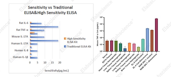
Fig. 1 Sensitivity comparison between traditional and high-sensitivity cytokine kits.
04 How to select a high-sensitivity ELISA kit
Before ordering, consult the literature to confirm the expected concentration of your target in your sample type and disease model, then compare with the kit's LOQ.
If expected concentrations are below the LOQ, choose the high-sensitivity version directly to avoid false negatives due to insufficient sensitivity, which wastes samples and time.
Important note: Under strong stimulation conditions such as LPS, cell culture supernatant cytokine concentrations may reach ng/mL levels, far exceeding the linear range of high-sensitivity kits. In such cases, dilution is required before detection to avoid the "hook effect" causing false negatives. We recommend performing a pilot experiment to determine the appropriate dilution factor.
05 High-sensitivity ELISA kits for low-abundance cytokine detection
Table 3. Selected High Sensitivity ELISA kits for low abundance cytokine targets
|
Cat. No. |
Product Name |
|
E-HSEL-M0001 |
High Sensitivity Mouse IL-1β (Interleukin 1 Beta) ELISA Kit |
|
E-HSEL-M0003 |
High Sensitivity Mouse IL-6 (Interleukin 6) ELISA Kit |
|
E-HSEL-M0009 |
High Sensitivity Mouse TNF-α (Tumor Necrosis Factor Alpha) ELISA Kit |
|
E-HSEL-H0001 |
High Sensitivity Human IL-1β (Interleukin 1 Beta) ELISA Kit |
|
E-HSEL-H0003 |
High Sensitivity Human IL-6 (Interleukin 6) ELISA Kit |
|
E-HSEL-R0002 |
High Sensitivity Rat IL-1β (Interleukin 1 Beta) ELISA Kit |
|
E-HSEL-R0004 |
High Sensitivity Rat IL-6 (Interleukin 6) ELISA Kit |
|
E-HSEL-R0001 |
High Sensitivity Rat TNF-α (Tumor Necrosis Factor Alpha) ELISA Kit |
Note: The table lists only selected best-selling products. For more high-sensitivity ELISA products, please visit www.elabscience.com or contact your local distributor.
The high-sensitivity ELISA series currently covers 23 low-abundance cytokine targets including TNF-α, IL-1β, IL-6, IL-10, IL-17A, and IFN-γ, with sensitivity as low as 0.2 pg/mL. Serum, plasma, and cell culture supernatant sample types have all been validated. The procedure is identical to standard ELISA—no additional learning curve required.
References:
[1] Nakase H, Shimomori Y .Association of Chronic Inflammation-Associated Cancer With Cytokines[J]. Cureus: Journal of Medical Science, 2025, 17(9).
[2] Donniacuo A , Mauro A , Cardamone C ,et al.Comprehensive Profiling of Cytokines and Growth Factors: Pathogenic Roles and Clinical Applications in Autoimmune Diseases[J]. International Journal of Molecular Sciences, 2025, 26(18):8921.
[3] Hui Xie,Guiting Zhang,Yongquan Xia, et al. Establishment of reference intervals for serum TNF-α, IFN-α, IL-1β, IL-2, IL-4, IL-5, IL-6, IL-8, IL-10, IL-12p70, IL-17 and IFN-γ in healthy Chinese adults using flow cytometry[J]. Journal of Immunological Methods, 2026, 547(c):114034.

