

Cytometry is a core single-cell analytical technology in the fields of immunology, tumor biology, clinical diagnosis and drug discovery, which can rapidly perform quantitative detection and population analysis of physicochemical characteristics and biological phenotypes of cell populations.
At present, mainstream laboratory instruments are divided into two categories: conventional flow cytometry and the new-generation spectral flow cytometry. Spectral flow cytometry breaks through the technical bottlenecks of conventional flow cytometry and enables high-dimensional multicolor flow detection, yet both technologies are suitable for distinct application scenarios.
To address the confusion of researchers in experimental design and instrument selection, this paper systematically compares the two flow cytometry technologies from the dimensions of working principle, core performance, technical advantages and disadvantages, and applicable scenarios, so as to provide selection references for practical applications.
Table of Contents
1. Core working principles of the two flow cytometry technologies
2. Multi-dimensional comparison of core performance
3. Comprehensive summary of technical advantages and disadvantages
4. Targeted selection scheme for experimental scenarios
5. Summary and technical outlook
01 Core working principles of the two flow cytometry technologies
All performance differences between conventional flow cytometry and spectral flow cytometry essentially stem from the differences in fluorescence signal acquisition and deconvolution logic.
1.1 Conventional Flow Cytometry
Fixed Bandpass Filter Detection plus Compensation Correction Conventional flow cytometry is a channel-limited detection technology. Fixed-wavelength bandpass filters are used to divide detection intervals, and each channel only receives fluorescence signals in a specific wavelength band. The emission spectra of most fluorophores exhibit tailing and overlap, so a single fluorescence signal leaks into multiple detection channels, resulting in fluorescence spillover. To eliminate signal interference and ensure quantitative accuracy, conventional flow cytometry must rely on compensation adjustment to subtract spillover signals between channels and finally restore the true expression level of each fluorophore (Fig 1).
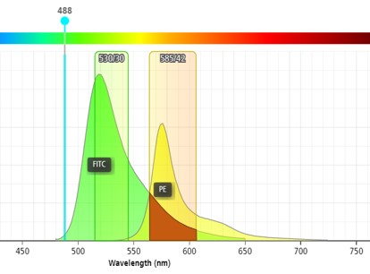
Fig. 1 Conventional flow cytometry.
1.2 Spectral Flow Cytometry
Full-Spectrum Acquisition plus Algorithmic Deconvolution Spectral flow cytometry abandons fixed bandpass filters and compensation mechanisms and adopts full-spectrum acquisition. Through gratings and high-precision optical detectors, the complete emission spectrum waveform of all fluorophores on a single cell is captured without signal truncation or wavelength omission. After detection, the built-in algorithm of the system fits and matches the mixed spectral signals with the reference spectra of each fluorophore, intelligently deconvolving heavily overlapping signals and fundamentally resolving fluorescence crosstalk (Fig 2).
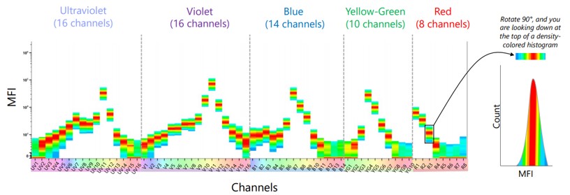
Fig. 2 Spectral flow cytometry.
02 Multi-dimensional comparison of core performance
2.1 Multicolor Detection and High-Dimensional Analysis Capability
Restricted by the number of filters and spectral crosstalk, conventional flow cytometry has a stable detection upper limit of 15 colors for standard instruments, and top-tier models can hardly exceed 20 colors. As the number of fluorescent labels increases, the degree of spectral overlap rises exponentially, compensation difficulty surges, data errors are significantly amplified, and high-dimensional cell subpopulation analysis cannot be supported.
Spectral flow cytometry has no fixed channel limitations. Relying on full-spectrum deconvolution algorithms, it can stably achieve ultra-high-dimensional detection of 20–50 colors. Dozens of cellular markers can be analyzed simultaneously in a single sample, meeting the research requirements for complex cell atlas and refined subpopulation classification.
2.2 Fluorescence Crosstalk Control and Data Reliability
Fluorescence spillover is the core technical shortcoming of conventional flow cytometry. Compensation calibration is highly dependent on operator experience with strong manual intervention. Parameter consistency varies between operators and experimental batches, which easily causes false positives, signal attenuation, loss of weak expression signals and other issues, and interferes significantly with the detection results of low-abundance antigens.
Spectral flow cytometry completes automatic spectral deconvolution through algorithms, avoiding systematic and human errors introduced by manual compensation. It can accurately distinguish subtle differences in antigen expression, with markedly improved data repeatability and quantitative precision compared with conventional flow cytometry. Nevertheless, spectral flow cytometry is heavily dependent on the quality of single-stained control tubes.
2.3 Detection Sensitivity and Sample Compatibility
Conventional flow cytometry is suitable for basic detection of routine cell samples, but it has a relatively high lower limit of detection, insufficient ability to identify trace samples, low-expression antigens and rare cell populations, and tends to lose key information of cell subpopulations.
Spectral flow cytometry completely retains full-band fluorescence signals without weak signal loss, delivering higher detection sensitivity. It can accurately capture low-abundance antigen expression and shows better compatibility with trace samples, complex heterogeneous cell samples and rare cell detection.
2.4 Selection of Flow Antibodies
All flow cytometry antibodies and auxiliary reagents from Elabscience® are applicable to both conventional flow cytometry and spectral flow cytometry.
03 Comprehensive summary of technical advantages and disadvantages
3.1 Conventional Flow Cytometry
Technical advantages: mature technical system, high stability, wide instrument popularity, low operation and maintenance costs; fast detection within 15 colors, well-recognized data, abundant public experimental data and clinical reference standards, suitable for high-throughput screening of routine samples.
Technical shortcomings: limited high-dimensional detection capacity and inability to perform ultra-multicolor analysis; compensation dependence brings inevitable human errors; weak resolution of weak signals and limited precision in distinguishing cell phenotypes; many constraints on fluorophore panel combination and low flexibility in experimental design.
3.2 Spectral Flow Cytometry
Technical advantages: ultra-high-dimensional multicolor detection capability enabling in-depth single-cell multi-parameter analysis; no manual compensation errors with excellent data accuracy and repeatability; outstanding ability to identify weak signals and detect rare cells; high flexibility in fluorophore selection, greatly reducing experimental limitations caused by spectral overlap, enabling mining of in-depth biological information from samples and suitable for cutting-edge refined scientific research.
Technical shortcomings: higher instrument purchase and maintenance costs, lower laboratory penetration compared with conventional flow cytometry; high-dimensional detection generates large datasets, imposing higher requirements on data analysis capabilities and supporting software; its high-performance advantages cannot be exerted in simple routine detection scenarios.
04 Targeted selection scheme for experimental scenarios
4.1 Scenarios suitable for conventional flow cytometry
Basic cell phenotype analysis with ≤15 markers; high-throughput screening of routine samples and standardized clinical detection (such as lymphocyte subset counting); basic research and routine clinical detection prioritizing cost-effectiveness and experimental stability.
4.2 Scenarios suitable for spectral flow cytometry
Complex cell atlas analysis requiring ultra-high-dimensional labeling of more than 20 colors; refined detection of rare cells, trace samples and low-expression antigens; experimental systems with heavily overlapping fluorophore spectra that cannot be effectively compensated by conventional flow cytometry; cutting-edge immunological mechanism research, in-depth single-cell analysis, high-precision research scenarios for innovative drug development.
05 Summary and technical outlook
Overall, conventional flow cytometry is a fundamental and universal technology for cell analysis. With its maturity and low cost, it has long supported basic scientific research and routine clinical testing and serves as essential laboratory equipment. Spectral flow cytometry is an advanced high-precision technology that specifically addresses the core pain points of conventional flow cytometry including severe crosstalk, limited multicolor capacity and insufficient precision, greatly expanding the detection depth and breadth of flow cytometry.
As life science research advances toward refined and high-dimensional single-cell analysis, the application scenarios of spectral flow cytometry will keep expanding and gradually become the core configuration of high-end research platforms. However, restricted by cost and application scenarios, conventional flow cytometry will not be replaced in the short term. The two technologies will complement each other and coexist for a long time, covering full-level demands from basic routine detection to advanced in-depth research.
In the future, spectral flow cytometry will evolve toward miniaturization, low cost and intelligent data analysis, continuously lowering the barriers to use and further expanding its popularity.

