

1. PBMC Sample Preparation (Self-provided Human Peripheral Blood Mononuclear Cell Separation Medium (P 1.077) (E-CK-A103) required)
1) Take 10 mL of fresh human heparin-anticoagulated blood and perform the separation procedure in the cell culture room.
Note: Maintain sterility throughout the entire separation process; the collected fresh human blood must be separated within 1 h. Approximately 1 × 10⁷ PBMCs can be obtained from 10 mL of human blood via separation; the Ficoll separation centrifugation process must be performed at 20 ± 2°C. Excessive temperature may reduce cell yield, while excessively low temperature may result in incomplete separation and reduced cell purity.
2) Prepare a 15 mL centrifuge tube. Add 5 mL of human peripheral blood mononuclear cell separation medium, then slowly add 5 mL of fresh human blood along the tube wall (invert or shake the blood to mix again before adding; control the adding speed to avoid excessive mixing of blood and separation medium. Clear layering indicates that the blood is fresh and in good condition, as shown in the left figure below; if operating multiple tubes of samples, try to improve operational efficiency to reduce natural sedimentation and mixing of blood in the separation medium; do not shake the centrifuge tube to mix the blood and separation medium after adding the blood!!). After adding, do not mix or shake the centrifuge tube. Gently place it into the centrifuge and centrifuge at 500 × g for 20 min.
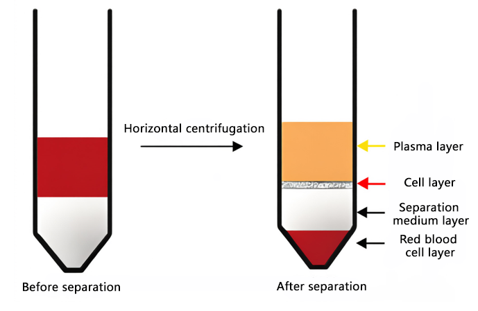
Figure 1. Blood cell separation before and after centrifugation (left: before centrifugation; right: after centrifugation).
3) Use a pipette to aspirate and discard part of the top light-yellow supernatant (indicated by the yellow arrow in the figure above; approximately 2 mL can be aspirated and discarded, mainly plasma components).
4) Use a pipette to carefully aspirate the white membranous layer between the light-yellow plasma layer and the transparent separation medium layer (indicated by the red arrow in the figure above; a small amount of separation medium may be aspirated to improve cell yield), and collect it into a 50 mL centrifuge tube.
5) Add 20 mL of RPMI-1640 basal medium (self-provided), mix well, centrifuge at 250 × g for 5 min, and discard the supernatant.
Note: The cell pellet is generally grayish-white. If it appears red, it indicates a small amount of residual blood cells, and it can be used directly or an appropriate amount of red blood cell lysis buffer can be added to lyse the red blood cells.
6) Add an appropriate amount of RPMI-1640 complete medium (self-provided) to resuspend the cells and count them. Adjust the cell density to 1 × 10⁶ cells/mL with complete medium and inoculate.
2. Stimulation and Blocking (Self-provided Stimulation and Blocking Kit (E-CK-A091) required)
7) Reagent preparation (before preparation, centrifuge the powder at 8000-10000 × g for 1 min to gather the powder at the bottom of the tube before dissolving and preparing):
a) 500× Cell Stimulation MIX: Add 100 μL of Cell Stimulation MIX Solvent to each tube of Cell Stimulation MIX Powder (50 μg), pipette thoroughly to mix, and prepare 500× Cell Stimulation MIX.
b) 1000× Protein Transport Inhibitor MIX: Add 50 μL of 33% DMSO aqueous solution (self-provided) to each tube of Protein Transport Inhibitor MIX Powder (200 μg), pipette thoroughly to mix, and prepare 1000× Protein Transport Inhibitor MIX.
8) Add 500× Cell Stimulation MIX and 1000× Protein Transport Inhibitor MIX according to the table below, and incubate the cells in a 37°C, 5% CO2 incubator for 5-6 h, mixing once every 1-2 h during the incubation.
|
Experimental Grouping |
Experimental Content |
|
Negative Control Group |
No treatment |
|
Experimental Group |
For every 1 mL of cell suspension, add 2 μL of 500× Cell Stimulation MIX and 1 μL of 1000× Protein Transport Inhibitor MIX |
|
Isotype Control Group |
9) After the incubation, collect the cells, centrifuge at 300 × g for 5 min, discard the supernatant, add 1 mL of Cell Staining Buffer [E-CK-A107] or 1× PBS (self-provided), mix well, centrifuge at 300 × g for 5 min, and discard the supernatant.
10) Resuspend the cells with an appropriate amount of 1× PBS, then count the cells and adjust the cell density to approximately 1 × 10⁶ cells/mL.
3. Setting Up Sample Tube Grouping
|
Sample Tube Setup |
Purpose |
Viability Dye |
CD3 |
CD4 |
IFN-γ |
IL-4 |
iso-IL-4 |
|
|
Blank |
Adjust instrument voltage |
|
|
|
|
|
|
|
|
Viability Dye Single Stain (if available) |
Adjust compensation |
✔ |
|
|
|
|
|
|
|
CD3 Single Stain |
Adjust compensation |
|
✔ |
|
|
|
|
|
|
CD4 Single Stain |
Adjust compensation |
|
|
✔ |
|
|
|
|
|
IFN-γ Single Stain |
Adjust compensation |
|
|
|
✔ |
|
|
|
|
IFN-γ Isotype Control |
Eliminate background staining |
✔ |
✔ |
✔ |
|
✔ |
✔ |
|
|
IL-4 Single Stain |
Adjust compensation |
|
|
|
|
|
✔ |
|
|
IL-4 Isotype Control |
Eliminate background staining |
✔ |
✔ |
✔ |
✔ |
|
|
✔ |
|
Fully Stained Tube |
Experimental results |
✔ |
✔ |
✔ |
✔ |
|
✔ |
|
4. (Optional) Amine-based Viability Dye
11) According to the sample tube grouping, add 1 mL of cell suspension to each tube.
12) Add 1 μL of STYX™ Fixable Viability Dye (Note: Select an appropriate amine-based viability dye based on the flow cytometry panel), pipette gently to mix, and incubate at room temperature or 4°C in the dark for 30 min.
13) Centrifuge at 300 × g for 5 min, discard the supernatant, add 1 mL of PBS to resuspend the cell pellet, centrifuge at 300 × g for 5 min, and discard the supernatant.
14) Resuspend the cells in 100 μL of Cell Staining Buffer [E-CK-A107] (the cell density at this point is approximately 1 × 10⁷ cells/mL).
5. Fc Receptor Blocking
15) Add 5 μL of EasyStain™ Human Fc Receptor Blocking Solution [E-CK-A171] and incubate at room temperature for 10 min.
Note: No washing is required after incubation; surface antibodies can be added directly.
6. Surface Antibody Incubation
16) According to the experimental design, except for the blank control, add 5 μL of each corresponding surface staining antibody (CD3, CD4) to the corresponding single-stain tubes and fully stained tubes, mix well, and incubate at 4°C in the dark for 30 min.
7. Fixation and Permeabilization (Self-provided Intracellular Cytokine Fixation and Permeabilization Reagent E-CK-A109 required)
17) Reagent preparation: Permeabilization Buffer (5×) is a 5× concentrate. Before the experiment, dilute it with deionized water to 1× Permeabilization Working Solution, i.e., take 1 mL of Permeabilization Buffer (5×) and add 4 mL of deionized water, mix well.
Note: 1× Permeabilization Working Solution is recommended to be prepared fresh before use, and the prepared working solution should preferably be used within 3 days.
18) Add 2 mL of Cell Staining Buffer [E-CK-A107] to each tube, centrifuge at 300 × g for 5 min, and discard the supernatant.
19) Add 200 μL of Cell Staining Buffer [E-CK-A107] to resuspend the cells.
20) Add 200 μL of Fixation Buffer to each tube, mix gently, and incubate at room temperature in the dark for 30-60 min.
Note: If the entire detection process cannot be completed on the same day, after fixation, wash the cells twice with PBS, resuspend in PBS or basal medium, and store at 4°C for subsequent experiments the next day.
21) Add 1 mL of 1× Permeabilization Working Solution, centrifuge at 600 × g for 5 min, and discard the supernatant.
8. Intracellular Antibody Incubation
22) Add 100 μL of 1× Permeabilization Working Solution to resuspend the cells.
23) Add 5 μL of each corresponding intracellular antibody (IFN-γ, IL-4) to the corresponding single-stain tubes and fully stained tubes, and mix well.
24) Incubate at room temperature in the dark for 30 min.
25) Add 2 mL of Cell Staining Buffer [E-CK-A107] to each tube.
26) Centrifuge at 600 × g for 5 min, and discard the supernatant.
9. Flow Cytometry Detection
27) Add 200 μL of Cell Staining Buffer [E-CK-A107] to resuspend the cells.
28) Adjust the instrument parameters and perform flow cytometry detection.
Note: The voltage and threshold of the flow cytometer need to be adjusted before formal acquisition. Once signal acquisition begins, the voltage and threshold must not be adjusted.

