

What are cytotoxic t cells? Cytotoxic T lymphocytes (CTLs) are central to adaptive immunity, serving as the primary effectors for eliminating virus‑infected cells and tumour cells. This review systematically defines CTLs by their signature surface markers (e.g., CD8, CD62L, KLRG1) and core cytolytic functions, then describes how they discriminate target cells through peptide‑MHC class I recognition. We detail the perforin‑granzyme pathway as the principal lethal mechanism, followed by an examination of T‑cell exhaustion, highlighting key biomarkers such as PD‑1, TIM‑3, and LAG‑3, and their functional implications, and contrast this with the generation and persistence of memory CTLs that sustain long‑term protective immunity. Finally, we discuss two widely used cytotoxic t cell assays, flow cytometry (intracellular cytokine staining and cytotoxicity markers) and ELISPOT, to detect CTL function. Collectively, this review integrates molecular mechanisms, phenotypic states, and detection strategies to offer a holistic perspective on CTL biology.
Table of Contents
1. What are cytotoxic t cells? Key surface markers and core functions
2. How cytotoxic T cells recognize target cells through MHC class I
3. Perforin and granzyme release: the primary cytotoxic mechanism
4. Exhausted cytotoxic T cells: biomarkers and functional consequences
5. Memory cytotoxic T cells and long-term immune protection
6. Measuring cytotoxic T cell function using flow cytometry and ELISPOT
01 what are cytotoxic t cells? Key surface markers and core functions
Cytotoxic T lymphocytes (CTLs) are the core effector cells in adaptive immunity responsible for eliminating virus-infected cells, intracellular bacteria, and tumor cells. Their “identity” is defined by a combination of surface cytotoxic t cell markers and effector functions.
During thymic development, the T cell lineage is divided into two major branches based on the expression of co-receptors: CD4⁺ helper T cells and CD8⁺ cytotoxic T cells. CD8 is a transmembrane disulfide-linked glycoprotein that is specifically expressed as an αβ heterodimer (CD8αβ) on the surface of classical MHC class I-restricted αβ T cells, whereas the CD8αα homodimer is expressed on various cell types, including intestinal intraepithelial lymphocytes, γδ T cells, and NK cells. CD8 is not only the most important phenotypic marker of CTLs, but also a functional co‑receptor in the T‑cell antigen recognition machinery: its extracellular domain binds to the non‑polymorphic region of MHC class I molecules, while its cytoplasmic tail associates with the Src‑family tyrosine kinase p56lck, thereby recruiting the kinase to the vicinity of the TCR signaling complex, catalyzing the phosphorylation of CD3 ITAM motifs, and amplifying TCR‑triggered signals[1]. Therefore, the combination of CD8αβ⁺ and αβTCR⁺ constitutes the core phenotypic signature that defines classical CTLs. In addition, effector/memory CTLs are often distinguished by the differential expression of markers such as CD27, CD28, CD44, CD62L, CCR7, KLRG1, and IL‑7Rα (CD127). Depending on their cytokine secretion profiles, cytotoxic potency, and functional characteristics, effector CD8⁺ T cells can be further subdivided into distinct effector subsets, among which the three major types are Tc1 (Type 1 Cytotoxic T Cells), Tc2 (Type 2 Cytotoxic T Cells), and Tc17 (Type 17 Cytotoxic T Cells). The characteristic cytotoxic t cell markers of each subset are listed in the table below.
Table 1. Characteristic markers of cytotoxic T cell subsets
|
Classifications |
Markers |
Inducer |
Cytokines |
Transcription Factor |
|
Tc1 |
CD49d |
IL-2, IL-12 |
Perforin, granzyme B, IFN-γ, TNF-α |
STAT4, T-bet, EOMES |
|
Tc2 |
CyslT1, BLT-1 |
IL-4 |
IL-4, IL-5, IL-13 |
STAT6, GATA3 |
|
Tc9 |
IL-9R |
IL-4, TGF-β |
IL-9, IFN-γ |
STAT6, IRF4 |
|
TC17 |
CD161, CD26, CD6, CD39, CD69, CD120b, PD-1 |
IL-6, TGF-β, IL-1β, IL-21, IL-23 |
IL-17, IL-22, GM-CSF |
STAT3, RORγt |
|
TC22 |
CD122, Ly49 |
IL-6, IL-21, TNF-α |
IL-22, IL-17 |
AhR, STAT1, STAT3, STAT5 |
|
Tfcs |
CXCR5 |
IL-6, IL-21, IL-23, TGF-β |
IL-4, IL-21, IFN-γ |
TCF-1, BCL-6, E2a, Runx3 |
|
Qa1-restricted CD8+Tregs |
CD122, Ly49 |
IL-15 |
TGF-β, perforin |
Eomes |
|
FOXP3+CD8+ Tregs |
CD103 |
TGF-β |
IL-10, TGF-β |
FOXP3 |
The effector functions of CTLs mature progressively through three phases: clonal expansion, contraction, and memory formation, following acute infection. Effector CTLs (terminal effector cells) express high levels of KLRG1 and low levels of IL‑7Rα and CD27, and possess immediate cytotoxic and cytokine‑secreting capabilities; whereas memory precursor cells with high IL‑7Rα and low KLRG1 retain long‑term survival and self‑renewal potential, and eventually differentiate into subsets such as central memory (TCM, CD62Lhigh CCR7high), effector memory (TEM, CD62Llow CCR7low), and tissue‑resident memory (TRM, CD103high CD69high). The core effector functions of CTLs can be summarized in three aspects: (1) direct killing of target cells via the perforin/granzyme granule exocytosis pathway; (2) induction of target cell apoptosis through death receptor pathways such as FasL‑Fas[2,3]; and (3) secretion of cytokines such as IFN‑γ and TNF to exert immunomodulatory effects.
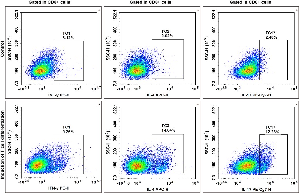
Fig. 1 Results of directed differentiation of naïve CD8+ T cells from mouse spleen. Naïve CD8+ T cells were isolated from C57 mouse splenocytes using a sorting kit to obtain high‑purity populations. The cells were then subjected to directed differentiation using activation beads in combination with cytokines and antibodies, and the differentiation efficiency was assessed by staining with PE‑conjugated anti‑IFN‑γ, APC‑conjugated anti‑IL‑4, and PE‑conjugated anti‑IL‑17 antibodies. The Tc1 cell subset was identified as CD8⁺IFN‑γ⁺, the Tc2 subset as CD8⁺IL‑4⁺, and the Tc17 subset as CD8⁺IL‑17A⁺.
02 How cytotoxic T cells recognize target cells through MHC class I
The molecular basis for CTL recognition of target cells is the specific recognition of peptide–MHC class I (pMHC-I) complexes by TCRs; this recognition event is the first step in cytotoxic t cell activation and also represents the structural manifestation of MHC restriction[4,5].
MHC class I molecules consist of a heavy chain (α1–α3) associated with β2‑microglobulin. The peptide-binding groove is formed by the α1 and α2 domains, is closed at both ends, and typically accommodates short peptides of 8–10 amino acids, with their N- and C-termini and anchor residues embedded in corresponding pockets, while the side chains point toward the TCR[4,5]. MHC class I is highly polymorphic; different alleles possess distinct peptide-binding motifs, which determine their respective presented peptide repertoires[5]. These molecules are ubiquitously expressed on the surface of nucleated cells and present endogenous antigens derived from intracellular proteasomal degradation, thereby enabling CTLs to monitor the internal state of cells.
The TCR is an αβ heterodimer, with variable regions containing CDR1, CDR2, and CDR3. Structural analyses indicate that most MHC‑I‑restricted TCRs bind diagonally across the peptide‑binding groove, with the α chain positioned over the α2 helix and the β chain over the α1 helix; Vα is oriented toward the peptide N‑terminus, while Vβ is oriented toward the C‑terminus[4, 5]. CDR1 and CDR2 mainly contact the MHC helices (reflecting co‑evolution), whereas CDR3 reads the peptide segment, with both chains contributing similarly (α ~52%, β ~48%) [5]. TCR–pMHC affinity is low (Kd ~1–100 μM), and recognition depends on CDR conformational adaptation (induced fit); moreover, T‑cell activation relies more on two‑dimensional affinity and catch‑bond kinetics. In recent years, exceptions such as reversed‑polarity docking and direct peptide reading by CDR2β have also been discovered.
CD8 binds to non-polymorphic regions such as the α3 domain of the MHC class I heavy chain, enhancing the association rate and half‑life of TCR–pMHC binding, and promotes CD3 ITAM phosphorylation via intracellular p56lck, thereby significantly lowering the activation threshold of CTLs[6]. CD8 dependence decreases with increasing TCR–pMHC affinity: weak‑affinity ligands are highly dependent on CD8, whereas high‑affinity ones are not. Recognition of the invariant region by CD8 ensures that peptide specificity is not perturbed; the canonical diagonal docking facilitates cooperative CD8 binding, whereas reversed‑polarity docking hinders effective activation due to impaired CD8 recruitment[5]. Furthermore, CD8 is indispensable for thymic positive selection, and mice lacking CD8α fail to generate mature CTLs and are susceptible to viral infection.
Table 2. Reagents for cytotoxic T cell research
|
Cat. No. |
Product Name |
|
MIM003N |
EasySort™ Mouse CD8+T Cell Isolation Kit |
|
MIH003N |
EasySort™ Human CD8+ T Cell Isolation Kit |
|
MIM008N |
EasySort™ Mouse Naïve CD8+T Cell Isolation Kit |
|
MIH008N |
EasySort™ Human Naïve CD8+T Cell Isolation Kit |
|
E-AB-F1013C |
FITC Anti-Mouse CD3 Antibody[17A2] |
|
E-AB-F1104D |
PE Anti-Mouse CD8a Antibody[53-6.7] |
|
AN00429E |
APC Anti-Mouse MHC I (H-2Kd) Antibody[SF1.1.10] |
|
AN00669D |
PE Anti-Rat TCR α/β Antibody[R73] |
|
E-AB-F1040M1 |
Elab Fluor® 700 Anti-Human CD49d Antibody[BU49] |
|
E-AB-F0987H |
PE/Cyanine7 Anti-Mouse CD161/NK1.1 Antibody[PK136] |
|
AN00418Q |
Elab Fluor® Violet 450 Anti-Mouse CD122 Antibody[TM-Beta 1] |
|
E-AB-F1140C |
FITC Anti-Human/Monkey CD27 Antibody[O323] |
|
E-AB-F1026M |
Elab Fluor® 647 Anti-Mouse CD28 Antibody[37.51] |
03 Perforin and granzyme release: the primary cytotoxic mechanism
The perforin/granzyme pathway constitutes the primary mechanism of CD8⁺ CTL-mediated killing. Upon target cell recognition via the T cell receptor (TCR), an immunological synapse (IS) forms at the effector–target interface within minutes, composed of a central TCR cluster encircled by an LFA‑1 integrin ring. TCR signaling induces polarization of the microtubule‑organizing center (MTOC) toward the synapse, directing lysosome‑like lytic granules, which contain perforin and granzymes, along microtubules via minus‑end‑directed motor proteins to the secretory domain in the central region of the synapse. Fusion of these granules with the plasma membrane results in the directed release of their contents into the synaptic cleft, a process commonly referred to as the “kiss of death.” This mechanism ensures target‑specific killing, spares bystander cells from exposure to toxic mediators, and enables CTLs to engage in serial killing[7].
Perforin is a Ca²⁺-dependent pore-forming protein that is homologous to the complement terminal components C7–C9. It comprises an N‑terminal complement homology domain, which mediates membrane insertion and oligomerization, and a C‑terminal C2 domain responsible for Ca²⁺‑dependent phospholipid binding. In the presence of extracellular Ca²⁺, perforin oligomerizes on the target cell membrane to form pores with diameters of 5–20 nm. Perforin is the only molecule known to deliver granzymes into the cytosol of target cells. Several models have been proposed to explain how perforin facilitates granzyme delivery: the classical plasma‑membrane pore‑formation model, the endolysosomal model put forward by Froelich, and the hybrid model proposed by Lieberman's laboratory. The latter posits that sublytic doses of perforin generate transient small pores in the plasma membrane, which trigger Ca²⁺ influx and activate a membrane repair response (termed the "wound healing" response). Under these conditions, perforin and granzymes are rapidly co‑endocytosed into large endosomes, where perforin subsequently disrupts the endosomal membrane, thereby releasing granzymes into the cytosol[8].
Granzyme B induces apoptosis through two complementary pathways[9-11]: (1) The direct caspase activation pathway: granzyme B directly cleaves procaspase‑3, ‑7, ‑8, and ‑10. Taking caspase‑3 as an example, granzyme B cleaves at Asp175 to generate the p20 fragment, which is subsequently processed via caspase‑mediated autocatalysis into the p19/p17 active forms[10,11]. (2) The Bid‑mitochondrial pathway: granzyme B directly cleaves the BH3‑only protein Bid at Asp75, producing truncated tBid. tBid then translocates to mitochondria and promotes Bax/Bak oligomerization, leading to mitochondrial outer membrane permeabilization (MOMP) and the release of cytochrome c and other pro‑apoptotic factors. This, in turn, activates caspase‑9 through the apoptosome[9,11].
In addition, granzyme A is a trypsin‑like serine protease that acts synergistically with granzyme B in target cell killing and is responsible for “late‑phase” perforin‑dependent apoptosis in the absence of granzyme B. Unlike granzyme B, granzyme A induces apoptosis in a caspase‑independent manner by cleaving substrates such as the SET complex, leading to single‑strand DNA damage[3]. Recent studies have also revealed that granzymes, including granzyme A‑mediated cleavage of GSDMB and granzyme B‑mediated cleavage of GSDME, can activate gasdermin family proteins, thereby inducing pyroptosis‑like cell death in specific target cells, further expanding the repertoire of CTL killing mechanisms[12].
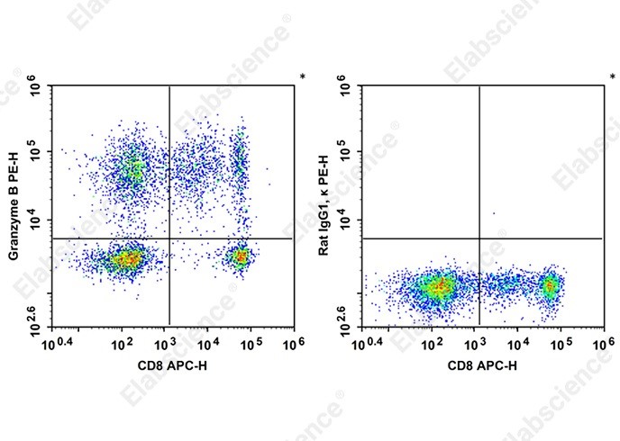
Fig. 2 Staining of normal human peripheral blood mononuclear cells with APC Anti-Human CD8a Antibody [OKT-8] and intracellular stained with PE Anti-Human Granzyme B Antibody [QA18A28] (left) or PE Rat IgG.
04 Exhausted cytotoxic T cells: biomarkers and functional consequences
4.1 The definition and characteristics of T-cell exhaustion
T cell exhaustion is a state of hyporesponsiveness induced by persistent antigen stimulation and is commonly observed in chronic infections, tumors, and autoimmune diseases. Exhausted CD8⁺ T cells (Tex) display transcriptional programs and a unique epigenetic landscape that are distinctly different from those of memory or effector T cells, and these epigenetic alterations become irreversible over time. Tex cells are characteristically marked by high expression of multiple inhibitory receptors (e.g., PD‑1, TIM‑3, LAG‑3, CTLA‑4, and TIGIT), progressive loss of effector functions (reduced production of IL‑2, TNF‑α, and IFN‑γ), diminished proliferative and self‑renewal capacity, and dysregulated metabolic activity[13]. Of note, inhibitory receptors per se are not exclusive to Tex cells, activated effector CD8⁺ T cells also rapidly upregulate various inhibitory molecules, which constitutes a negative feedback regulatory mechanism that limits T cell‑mediated immunopathology[14]. However, under chronic antigen stimulation, these inhibitory receptors remain persistently overexpressed, ultimately leading to a progressive loss of T cell function.
4.2 Heterogeneity and differentiation model of exhausted T cells
The currently accepted model divides Tex cells into two major subsets: progenitor exhausted T cells (Tpex) and terminally differentiated Tex cells (tTex)[13,14]. Tpex are characterized by expression of T cell factor 1 (TCF‑1), retention of costimulatory molecules such as CD28, ICOS, and CD226, and lack of co‑expression of multiple inhibitory receptors[14]. Tpex maintain the Tex cell pool through self‑renewal and differentiation into TCF‑1‑negative effector‑like Tex cells[13,14]. In the context of PD‑1/PD‑L1 blockade therapy, Tpex represent the primary cell population that responds to immune checkpoint inhibition, they proliferate and differentiate upon PD‑1 blockade into effector‑like Tex cells with enhanced cytotoxic capacity and IFN‑γ‑secreting function[14]. These effector‑like Tex cells eventually transition into terminally differentiated Tex cells, which highly express inhibitory receptors and exhibit limited capacity for cytokine production and proliferation, yet retain granzyme B expression[13,14].
4.3 Inhibitory function of exhausted T cells
Of particular interest, recent studies have revealed that terminally differentiated Tex cells are not merely a hypofunctional state, but may also acquire inhibitory functions. Vignali and colleagues demonstrated that CD8⁺ tTex cells exert suppressive activity through CD39, and that hypoxia within the tumor microenvironment (TME) enhances the inhibitory capacity of tTex cells by upregulating CD39 expression, thereby generating adenosine and limiting the efficacy of immunotherapy[15]. This finding redefines exhausted T cells as a cell state characterized by conditional anti‑functional features, rather than a mere “dysfunctional” state.
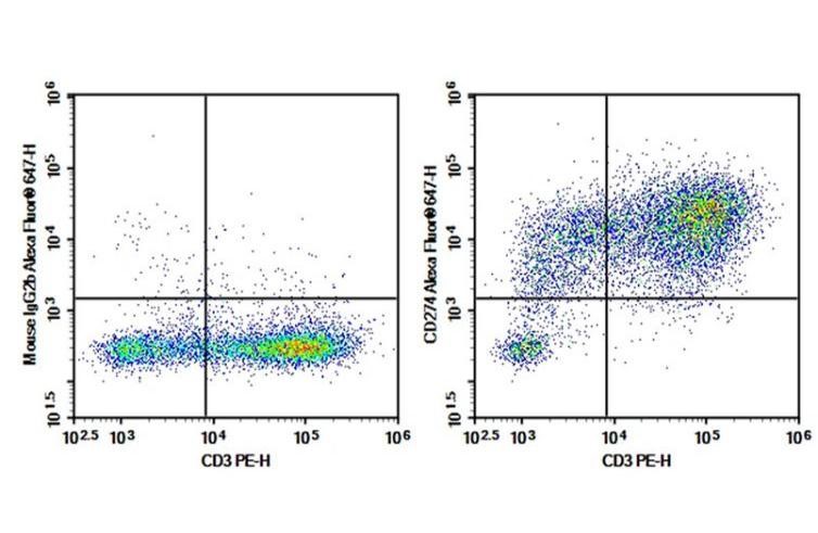
Fig. 3 Detection of exhausted human T cells. Human peripheral blood mononuclear cells (PBMCs) were stimulated with 10 μg/mL phytohemagglutinin (PHA) for 3 days, and the expression of PD‑L1/CD274 in CD3⁺ T cells was analyzed. T cells autonomously regulate their own activity through the expression of PD‑L1, thereby preventing overactivation and functional exhaustion.
Table 3. Reagents for exhausted T cell research
|
Cat. No. |
Product Name |
|
E-AB-F1131D |
PE Mouse anti-PD-1 antibody[29F.1A12] |
|
E-AB-F1192C |
FITC Mouse anti-TIM-3 antibody [RMT3-23] |
|
E-EL-M0398 |
Mouse CTLA4(Cytotoxic T-Lymphocyte Associated Antigen 4) ELISA Kit |
|
AN009730P |
Purified Anti-Human TIGIT Antibody[A15153G] |
|
E-AB-F1026M |
Elab Fluor® 647 Anti-Mouse CD28 Antibody[37.51] |
|
E-AB-F1165H |
PE/Cyanine7 Anti-Human/Monkey CD39 Antibody[A1] |
|
E-AB-F1369E |
APC Anti-Human CD226/DNAM-1 Antibody[11A8] |
05 Memory cytotoxic T cells and long-term immune protection
Memory CD8⁺ T cells are derived from the clonal expansion of naïve T cells following antigen stimulation, giving rise to a residual antigen‑specific cell population accounting for approximately 5–10% after antigen clearance. Based on differences in homing receptor expression and functional characteristics, circulating memory CD8⁺ T cells can be further divided into multiple subsets[16,17].
Central memory T cells (TCM) express CCR7 and CD62L, enabling their entry into secondary lymphoid organs. Upon re‑exposure to antigen, TCM cells rapidly proliferate, produce large amounts of IL‑2, and possess long‑term survival and self‑renewal capacities. TCM cells highly express IL‑7R (CD127) and IL‑15R (CD122), and the integrated survival signals from these receptors contribute to the long‑term stability of the memory T cell pool[16].
Stem cell‑like memory T cells (TSCM) also express CCR7 and CD62L but retain expression of CD45RA, displaying a phenotype reminiscent of naïve T cells. TSCM cells have limited effector functions but exhibit extensive proliferative and self‑renewal capacities upon antigenic stimulation, representing the most stem‑cell‑like subset within the memory T cell differentiation hierarchy[16].
Effector memory T cells (TEM) lack expression of CCR7 and CD62L, but instead express various chemokine receptors (e.g., CX3CR1), which enable their migration to non‑lymphoid tissues. TEM cells possess robust effector functions, including the production of IFN‑γ, TNF‑α, β‑chemokines, and cytotoxic molecules such as perforin and granzymes. Of note, recent studies have further subdivided TEM cells into two functionally distinct subsets: CX3CR1⁻ precursor TEM cells, which express CD127 and TCF‑1 and retain proliferative and recirculation capacities; and CX3CR1⁺ terminally differentiated TEM cells, which exhibit high cytotoxicity but are confined to the intravascular compartment[16].
Tissue-resident memory T cells (TRM) do not recirculate but instead persist long‑term in tissues, forming a distinct network for immune surveillance. TRM cells are characteristically marked by constitutive expression of CD69, which downregulates S1PR1 to prevent lymphocyte egress from tissues, as well as expression of CD103 (integrin αEβ7). CD103 promotes TRM retention and survival at barrier sites through binding to its epithelial‑specific ligand E‑cadherin[16,17]. Studies have demonstrated that CD69⁺ CD8⁺ TRM cells are present in nearly all human organs[16]. Functionally, TRM cells are capable of rapid responses upon antigen re‑exposure, recruiting innate immune cells through the release of cytokines such as IFN‑γ and directly killing infected cells.
Table 4. Reagents for memory T cell research
|
Cat. No. |
Product Name |
|
MIH010N |
EasySort™ Human Memory CD8+T Cell Isolation Kit |
|
E-AB-F1159D |
PE Anti-Human CD197/CCR7 Antibody[G043H7] |
|
E-AB-F1011E |
APC Anti-Mouse CD62L Antibody[MEL-14] |
|
E-AB-F1152H |
PE/Cyanine7 Anti-Human CD127/IL-7RA Antibody[A019D5] |
|
E-AB-F1151M1 |
Elab Fluor® 700 Anti-Human CD122/IL-2RB Antibody[TU27] |
|
AN01022G |
PE/Cyanine 5 Anti-Mouse CX3CR1 Antibody[SA011F11] |
|
E-AB-F1187S |
Elab Fluor® Red 780 Anti-Mouse CD69 Antibody[H1.2F3] |
|
E-AB-F1090L |
Elab Fluor® 488 Anti-Mouse CD103 Antibody[M290] |
|
E-AB-F1052C |
FITC Anti-Human CD45RA Antibody[HI100] |
06 Measuring cytotoxic T cell function using flow cytometry and ELISPOT
6.1 Detection of CTL function by flow cytometry
(1) Intracellular perforin detection: Reliable detection of perforin in mouse lymphocytes has long been a technical challenge in the field of immunology. Brennan et al. developed a novel intracellular staining method for perforin, in which Bouin's fixative is used in place of conventional paraformaldehyde fixation, combined with a prolonged (16‑hour) incubation with the primary antibody. This approach achieved, for the first time, highly sensitive and specific detection of perforin in mouse CTLs. This method is applicable not only to flow cytometry but also to confocal microscopy for visualizing the co‑localization of perforin and granzyme B at the immunological synapse. The dynamic range of this technique is sufficient to distinguish perforin levels between CTLs from Prf1⁺/⁺ and Prf1⁺/⁻ mice[18].
(2) Perforin release assay: Weren et al. established a flow cytometry‑based perforin release assay to quantitatively evaluate degranulation kinetics by monitoring the change in the percentage of perforin‑positive cells within CD8ʰⁱ⁺ CTLs over time. This assay is performed in the absence of target cells, with CTLs stimulated via PMA/ionomycin or CD3 cross‑linking. The decline in the percentage of perforin‑positive cells was highly consistent with the kinetics of BLT‑esterase release and CD63 upregulation, confirming that the reduction in perforin indeed reflects the secretion of perforin‑containing lytic granules[19].
(3) CD107a degranulation assay: CD107a (LAMP‑1) and CD107b (LAMP‑2) are transmembrane proteins stored within lytic granules and are translocated to the cell surface during degranulation. Detection of CD107a surface expression on CTLs by flow cytometry allows quantitative assessment of the degranulation frequency of antigen‑specific CTLs. However, a limitation of this approach is that CD107a upregulation can occur in the absence of stored functional cytotoxic effector proteins; therefore, combined assessment with cytotoxic mediators is required for comprehensive evaluation[20].
%20or%20with%20an%20FITC%20Rat%20IgG2a,%20%CE%BA%20Isotype%20Control%20(right)_.jpg)
Fig. 4 C57BL/6 murine splenocytes are stained with APC Anti-Mouse CD49b Antibody and FITC Anti-Mouse Perforin Antibody (Left). Splenocytes are stained with APC Anti-Mouse CD49b Antibody and FITC Rat IgG2a, κ Isotype Control (Right).
6.2 Detection of CTL function by ELISPOT
(1) IFN‑γ ELISPOT: ifn gamma elispot is one of the most commonly used methods for assessing CTL responses and has been widely applied in immune monitoring for cancer vaccine clinical trials. However, IFN‑γ secretion is not unique to cytotoxic cells, non‑cytotoxic cells can also secrete IFN‑γ, and CTLs with lytic activity do not invariably secrete IFN‑γ.
(2) Granzyme B ELISPOT: The GrB ELISPOT assay established by Shafer‑Weaver et al. quantitatively evaluates CTL cytotoxic activity by capturing and detecting GrB released during effector–target cell interactions. Compared with the conventional ⁵¹Cr release assay, the GrB ELISPOT offers several notable advantages: (1) it does not require radioactive isotopes; (2) it requires 10‑ to 100‑fold fewer effector cells; (3) it can detect GrB release within 10 minutes, substantially earlier than IFN‑γ secretion (which takes approximately 1 hour); and (4) it enables direct quantitation of the precursor frequency of antigen‑specific CTLs[21].
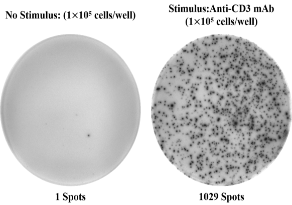
Fig. 5 PBMCs (1 × 10⁵ cells/well) were incubated for 20 h in the absence or presence of anti‑CD3 monoclonal antibody (1:500) as a stimulant. The number of spots corresponds to the number of cells secreting human IFN‑γ.
Table 5. Reagents for cytotoxic T cell function research
|
Cat. No. |
Product Name |
|
ESP-H0002 |
Human IFN-γ (Interferon Gamma) ELISPOT Kit |
|
ESP-H0010 |
Human TNF-α (Tumor Necrosis Factor Alpha) ELISPOT Kit |
|
ESP-H0003 |
Human IL-10 (Interleukin 10) ELISPOT Kit |
|
ESP-H0007 |
Human IL-4 (Interleukin 4) ELISPOT Kit |
|
ESP-H0004 |
Human IL-17A (Interleukin 17A) ELISPOT Kit |
|
E-MSEL-H0019 |
Mini Sample Human GzmB ( Granzyme B ) ELISA Kit |
|
E-EL-H1617 |
Human GzmB(Granzyme B) ELISA Kit |
|
E-EL-H1123 |
Human PRF1(Perforin 1) ELISA Kit |
References:
[1] KAECH S M, CUI W. Transcriptional control of effector and memory CD8+ T cell differentiation [J]. Nat Rev Immunol, 2012, 12(11): 749-61.
[2] KäGI D, VIGNAUX F, LEDERMANN B, et al. Fas and perforin pathways as major mechanisms of T cell-mediated cytotoxicity [J]. Science, 1994, 265(5171): 528-30.
[3] SHRESTA S, PHAM C T, THOMAS D A, et al. How do cytotoxic lymphocytes kill their targets? [J]. Curr Opin Immunol, 1998, 10(5): 581-7.
[4] RUDOLPH M G, STANFIELD R L, WILSON I A. How TCRs bind MHCs, peptides, and coreceptors [J]. Annu Rev Immunol, 2006, 24: 419-66.
[5] SZETO C, LOBOS C A, NGUYEN A T, GRAS S. TCR Recognition of Peptide-MHC-I: Rule Makers and Breakers [J]. Int J Mol Sci, 2020, 22(1).
[6] LAUGEL B, COLE DK FAU - CLEMENT M, CLEMENT M FAU - WOOLDRIDGE L, et al. The multiple roles of the CD8 coreceptor in T cell biology: opportunities for the selective modulation of self-reactive cytotoxic T cells [J]. J Leukoc Biol, 2011, 90(6).
[7] HUSE M, QUANN E J, DAVIS M M. Shouts, whispers and the kiss of death: directional secretion in T cells [J]. Nat Immunol, 2008, 9(10): 1105-11.
[8] PIPKIN M E, LIEBERMAN J. Delivering the kiss of death: progress on understanding how perforin works [J]. Curr Opin Immunol, 2007, 19(3): 301-8.
[9] SUTTON V R, DAVIS J E, CANCILLA M, et al. Initiation of apoptosis by granzyme B requires direct cleavage of bid, but not direct granzyme B-mediated caspase activation [J]. J Exp Med, 2000, 192(10): 1403-14.
[10] GOPING I S, BARRY M, LISTON P, et al. Granzyme B-induced apoptosis requires both direct caspase activation and relief of caspase inhibition [J]. Immunity, 2003, 18(3): 355-65.
[11] AFONINA I S, CULLEN S P, MARTIN S J. Cytotoxic and non-cytotoxic roles of the CTL/NK protease granzyme B [J]. Immunol Rev, 2010, 235(1): 105-16.
[12] VANDENABEELE P, BULTYNCK G, SAVVIDES S N. Pore-forming proteins as drivers of membrane permeabilization in cell death pathways [J]. Nat Rev Mol Cell Biol, 2023, 24(5): 312-33.
[13] CHENG H, MA K, ZHANG L, LI G. The tumor microenvironment shapes the molecular characteristics of exhausted CD8(+) T cells [J]. Cancer Lett, 2021, 506: 55-66.
[14] VAN DER HEIDE V, HUMBLIN E, VAIDYA A, KAMPHORST A O. Advancing beyond the twists and turns of T cell exhaustion in cancer [J]. Sci Transl Med, 2022, 14(670): eabo4997.
[15] SHARMA A, SCHMIDT-WOLF I G H. Tempering of exhausted T cells to comprehend their adaptive response for suitable clinical translation [J]. Cell Mol Immunol, 2023, 20(12): 1401-2.
[16] BUGGERT M, PRICE D A, MACKAY L K, BETTS M R. Human circulating and tissue-resident memory CD8(+) T cells [J]. Nat Immunol, 2023, 24(7): 1076-86.
[17] WU A, JIANG W. Progenitor exhausted T cells contribute to the formation of immunological memory [J]. Nat Rev Immunol, 2025, 25(6): 405.
[18] WEREN A, BONNEKOH B, SCHRAVEN B, et al. A novel flow cytometric assay focusing on perforin release mechanisms of cytotoxic T lymphocytes [J]. J Immunol Methods, 2004, 289(1-2): 17-26.
[19] WATERHOUSE N J, SEDELIES K A, CLARKE C J. Granzyme B; the chalk-mark of a cytotoxic lymphocyte [J]. J Transl Med, 2004, 2(1): 36.
[20] SHAFER-WEAVER K, SAYERS T, STROBL S, et al. The Granzyme B ELISPOT assay: an alternative to the 51Cr-release assay for monitoring cell-mediated cytotoxicity [J]. J Transl Med, 2003, 1(1): 14.
[21] EWEN C L, RONG J, KOKAJI A I, et al. Evaluating antigen-specific cytotoxic T lymphocyte responses by a novel mouse granzyme B ELISPOT assay [J]. J Immunol Methods, 2006, 308(1-2): 156-66.

