

Immune checkpoint inhibitors (ICIs) have profoundly transformed the landscape of tumor therapy, yet objective response rates still fall short of clinical needs. Emerging studies indicate that metabolic reprogramming in the tumor microenvironment (TME) not only supports tumor cell proliferation but also serves as a key driver of immune evasion and therapeutic resistance. Extensive bidirectional regulation exists between metabolic enzymes and immune checkpoints, giving rise to the emerging research paradigm of "immunometabolic checkpoints," which has become an important breakthrough point in next-generation tumor therapy research. This article systematically reviews the latest advances in novel immune checkpoint and metabolic enzyme targets, focuses on their crosstalk mechanisms, and discusses combination therapeutic strategies based on these findings.
Table of Contents
1. Immune checkpoint targets
2. Metabolic enzyme targets
3. Crosstalk between immune checkpoints and metabolic enzymes
4. Therapeutic and research strategies
5. Experimental approaches for immune checkpoint and metabolic enzyme research
01 Immune checkpoint targets
1.1 Classical checkpoints
PD-1/PD-L1 and CTLA-4 are representative classical immune checkpoint targets.
PD-1/PD-L1: The PD-1/PD-L1 axis remains the cornerstone of immunotherapy. By binding to its ligands PD-L1/PD-L2, PD-1 inhibits TCR downstream signaling pathways and negatively regulates T cell activity [1,2,3]. The expression of PD-L1 is regulated by multiple factors, including the IFN-γ/JAK/STAT pathway, oncogenic signaling pathways, and metabolites such as lactate and adenosine [1,4]. Notably, PD-L1 itself also possesses intrinsic signaling functions—studies have found that PD-L1 can upregulate HK2 expression through the PI3K/Akt and Erk pathways, enhancing aerobic glycolysis in tumor cells, thereby suppressing T cell function at the metabolic level [5].
CTLA-4: CTLA-4 primarily acts during the initiation phase of T cell activation, inhibiting T cell activation by competing with CD28 for binding to CD80/CD86 [1,3].
The combination of anti-CTLA-4 and anti-PD-1 regimens has shown superior efficacy over monotherapy in melanoma, with a 5-year overall survival rate of up to 52% [1].
1.2 Emerging immune checkpoint targets
At the membrane protein level:
● LAG-3: LAG-3 has diverse ligands (MHC-II, FGL1) and is expressed on the surface of activated T cells, Tregs, and NK cells, negatively regulating proliferation and cytokine secretion; notably, anti-LAG-3 combined with anti-PD-1 has shown superior efficacy to monotherapy [6].
● TIGIT: It inhibits immunity by binding to CD155/CD112 and regulates NK metabolism, showing synergistic effects with PD-1/PD-L1 blockade [6,3].
● TIM-3: Highly expressed on terminally exhausted T cells, it synergizes with PD-1 to mediate exhaustion, and combined blockade exerts synergistic effects.
● CD47: It inhibits phagocytosis by binding to SIRPα and delivering a "don't eat me" signal [3].
In addition, members such as VISTA, BTLA, ILT4, and B7-H3 continue to expand and enter early-stage clinical trials [2,6], while the CD73-mediated CD39-CD73-adenosine axis creates an immunosuppressive microenvironment in the TME [2].
At the intracellular checkpoint level:
CISH regulates TCR signaling independently of tumor ligands and can be targeted for intervention through CRISPR engineering.
DGKζ negatively regulates TCR signaling, and its selective inhibitors have entered clinical development. PTPN1/PTPN2 synergistically regulate the threshold of interferon responses and represent novel regulatory nodes in immunotherapy [6].
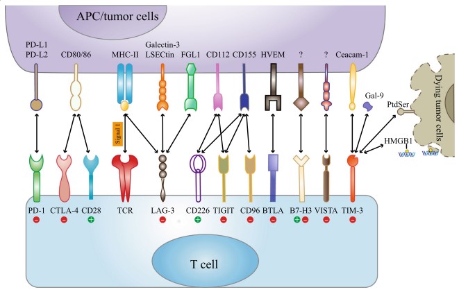
Fig. 1 Immune checkpoint receptors and their corresponding ligands [6].
02 Metabolic enzyme targets
2.1 Key enzymes in the glycolysis pathway
● HK2: HK2 is a rate-limiting enzyme of glycolysis. PD-L1 upregulates HK2 expression through the PI3K/Akt and Erk pathways, and high HK2 expression is associated with reduced CD8⁺ T cell infiltration and decreased effector gene expression [5]; HK2 inhibitors combined with anti-PD-1 antibodies hold broad therapeutic promise.
● PKM2: It promotes aerobic glycolysis in its low-activity dimeric form. Its agonists can enhance oxidative phosphorylation, improve ICB efficacy, and reduce Treg infiltration.
● GLUT1: It mediates glycolysis in "cold tumors." Blocking GLUT1 in combination with anti-PD-1 plus anti-CTLA-4 can induce tumor regression [4].
● LDHA: It catalyzes lactate production, and lactate directly activates the transcription of immune-related genes such as PD-L1 through histone lactylation [7].
2.2 Key enzymes in amino acid and lipid metabolism
● IDO1 catalyzes the tryptophan-kynurenine pathway, suppressing immunity and promoting Treg differentiation by depleting tryptophan or through the kynurenine-AhR axis. Its inhibitors show synergistic effects with PD-1/PD-L1 or CTLA-4 blockade [8].
● CPT1A is the rate-limiting enzyme of fatty acid oxidation. It also succinylates PD-L1 at the K129 site to induce its lysosomal degradation, and CPT1A levels can serve as a predictive biomarker for ICB efficacy [4].
● Inactivation of DHODH (a pyrimidine synthesis enzyme) can modulate ICB efficacy;
Combination targeting strategies involving MAOA, PDK1, GLS, MCT-1, NAMPT, and others are also being explored [4,5].
03 Crosstalk between immune checkpoints and metabolic enzymes
3.1 Metabolic enzymes regulate immune checkpoints
Positive regulation: HK2 promotes PD-L1 expression via IκBα/NF-κB; lactate activates PD-L1 transcription through histone lactylation [7]; tumors with high LDHA expression simultaneously overexpress PD-L1 and IDO1 [5]; PRMT3 regulates glycolysis through PDHK1 to drive PD-L1-mediated immune evasion.
Negative regulation: CPT1A succinylates PD-L1 at the K129 site, inducing its degradation and reducing PD-L1 protein levels [4].
3.2 Immune checkpoints regulate metabolism
● The TIGIT/CD155 axis inhibits GLUT1 and HK1/2 expression, impairing glycolysis in T/NK cells [4];
● LAG-3 and CD39/CD73 cross-regulate each other, leading to T cell metabolic exhaustion;
● PD-1 signaling inhibits T cell glycolysis and promotes fatty acid oxidation [4,5].
3.3 Microenvironmental perspective
In the tumor microenvironment (TME), high glycolysis in tumor cells leads to glucose depletion and lactate accumulation, directly suppressing T cell function and forming a "cold" tumor niche [4,5]. In addition, lactate reshapes the immune landscape through multiple pathways. On the one hand, lactate can inhibit NK activation and DC differentiation/antigen presentation, promote MDSC expansion, and support the FAO-dependent metabolic adaptation of Tregs; on the other hand, lactate also induces TAM polarization toward the M2 phenotype, reinforcing immunosuppression through HIF-1α and arginase [9].
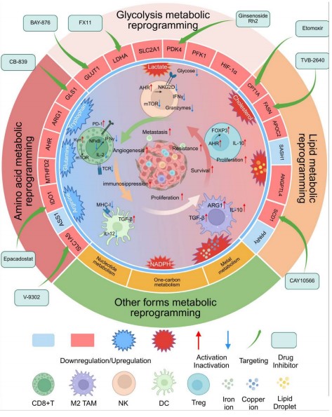
Fig. 2 Interaction between metabolic reprogramming and immunosuppression in tumor cells [9].
04 Therapeutic and research strategies
Combination therapeutic strategies based on immune-metabolic crosstalk are becoming a frontier direction in tumor research.
4.1 Combination treatment strategies
At the metabolic intervention level, immune checkpoint blockade (ICB) can be combined with inhibitors targeting multiple metabolic targets, including glucose transporter GLUT1, hexokinase HK2, indoleamine 2,3-dioxygenase IDO1, and dihydroorotate dehydrogenase DHODH [4,5,8], as well as with novel immune checkpoint blockers such as anti-LAG-3 and anti-TIGIT [3,6]. In addition, IDO1 inhibition or PKM2 agonists can enhance the antitumor function of CAR-T cells [8]; in terms of dual-targeting combination strategies, the combined application of CA IX/XII and NAMPT is also being explored [4]. In hepatocellular carcinoma (HCC), ICB combined with local therapy or neoantigen vaccines has shown synergistic effects, with the objective response rate reaching 30% in the vaccine combination group [3].
4.2 Biomarkers for predicting treatment efficacy
CPT1A, GLUT1, HK2, succinyl-CoA, and others can predict ICB response; in non-small cell lung cancer (NSCLC), patients with PD-L1⁺/low HK2 expression have better efficacy [5]; whereas in HCC, a high Treg/effector T cell ratio and β-catenin mutations are significantly associated with reduced efficacy [3].
4.3 Emerging technologies enabling advances
Regarding new technologies, PROTAC technology can degrade PD-L1 and IDO1 proteins, CRISPR gene editing is used to knock out intracellular checkpoint molecules such as CISH [6], ImmunoPET imaging enables non-invasive visualization of checkpoints, and multi-omics integration provides systematic tool support for immunometabolic research [4].
05 Experimental approaches for immune checkpoint and metabolic enzyme research
Research on immune checkpoints and metabolic enzymes typically requires accompanying cell phenotype and function assays. The following presents detection data on PD-L1 expression levels and glucose uptake capacity obtained using Elabscience® products:
5.1 Immune phenotype analysis: Detection of cell surface PD-L1 expression
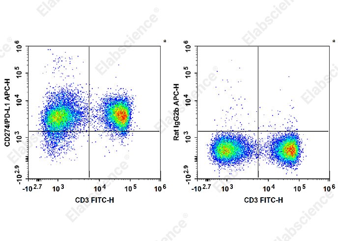
Fig. 3 Experimental results of PD-L1 detection in C57BL/6 mouse splenocytes. As shown, C57BL/6 mouse splenocytes were stained with FITC Anti-Mouse CD3 Antibody [17A2] (E-AB-F1013C) and APC Anti-Mouse CD274/PD-L1 Antibody [10F.9G2] (E-AB-F1132E) (left panel) or APC Rat IgG2b, κ Isotype Control [LTF-2] (E-AB-F09842E) (right panel), followed by flow cytometric analysis.
5.2 Metabolic function validation: Detection of cellular glucose uptake
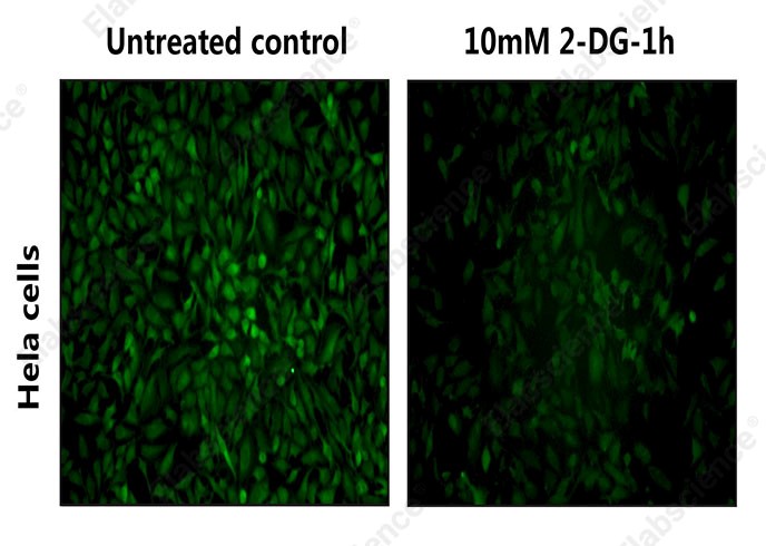
Fig. 4 Detection results of the effect of 2-DG on glucose uptake in HeLa cells. GLUT1-mediated glucose uptake is the rate-limiting step of cellular glycolysis. In this experiment, the 2-NBDG Glucose Uptake Cell-Based Kit (E-CK-A441) was used to detect glucose uptake in HeLa cells after incubation for 60 min without (left) or with 10 mM 2-DG (right). Fluorescence microscopy results showed that incubation with 2-DG inhibited glucose uptake in HeLa cells.
From immune checkpoints to metabolic reprogramming, every breakthrough discovery is underpinned by high-quality research tools. Elabscience® continues to delve into immunoassay research tools, empowering researchers to accelerate their progress on the frontier of tumor research.
Product Recommendations:
Table 1. Elabscience® products for immune checkpoint and metabolic enzyme research
|
Cat. No. |
Product Name |
|
E-BC-F069 |
Extracellular Acidification Rate (ECAR) Fluorometric Assay Kit |
|
E-BC-F070 |
Enhanced Oxygen Consumption Rate (OCR)Fluorometric Assay Kit |
|
E-BC-F201 |
Enhanced ATP Chemiluminescence Assay Kit |
|
E-BC-K784-M |
Fatty Acid Oxidation (FAO) Colorimetric Assay Kit |
|
E-BC-F084 |
Glycolysis Stress Fluorometric Assay Kit |
|
E-BC-F041 |
Glucose Uptake Fluorometric Assay Kit |
|
E-BC-F078 |
Mitochondrial Stress Fluorometric Assay Kit |
|
E-BC-F004 |
ATP/ADP Ratio Chemiluminescence Assay kit |
|
E-BC-K853-M |
Glutamine (Gln) Colorimetric Assay Kit |
|
E-CK-A441 |
2-NBDG Glucose Uptake Cell-Based Kit |
|
E-BC-F037 |
Glucose (GLU) Fluorometric Assay Kit |
|
E-BC-K1107-M |
Carnitine Palmitoyl Transferase-I (CPT-I) Activity Colorimetric Assay Kit |
|
E-BC-K611-M |
Pyruvate Kinase (PK) Activity Assay Kit |
|
E-EL-H0866 |
Human D-LDH(D-Lactate Dehydrogenase) ELISA Kit |
|
E-EL-H0556 |
Human LDHA(Lactate Dehydrogenase A) ELISA Kit |
|
E-BC-K766-M |
Lactate Dehydrogenase (LDH) Activity Assay Kit (WST-8 Method) |
|
E-EL-H2069 |
Human CTLA4(Cytotoxic T-Lymphocyte Associated Antigen 4) ELISA Kit |
|
E-EL-M0398 |
Mouse CTLA4(Cytotoxic T-Lymphocyte Associated Antigen 4) ELISA Kit |
|
E-EL-H1547 |
Human PD-L1(Programmed Cell Death Protein 1 Ligand 1) ELISA Kit |
|
E-AB-F1133C |
FITC Anti-Human CD274/PD-L1 Antibody[29E.2A3] |
|
E-AB-F1132E |
APC Anti-Mouse CD274/PD-L1 Antibody[10F.9G2] |
References:
[1] Relecom A, Merhi M, Inchakalody V, et al. Emerging dynamics pathways of response and resistance to PD-1 and CTLA-4 blockade: tackling uncertainty by confronting complexity. Journal of Experimental & Clinical Cancer Research, 2021. DOI: 10.1186/s13046-021-01872-3
[2] Hargadon KM, et al. Next generation of immune checkpoint inhibitors and beyond. Journal of Hematology & Oncology, 2021. DOI: 10.1186/s13045-021-01056-8
[3] Desert R, Giannone F, Saviano A, et al. Improving immunotherapy for the treatment of hepatocellular carcinoma: learning from patients and preclinical models. npj Gut and Liver, 2025. DOI: 10.1038/s44355-025-00018-y
[4] Kao KC, Vilbois S, Tsai CH, Ho PC. Metabolic communication in the tumour–immune microenvironment. Nature Cell Biology, 2022. DOI: 10.1038/s41556-022-01002-x
[5] Kim S, Jang JY, Koh J, et al. Programmed cell death ligand-1-mediated enhancement of hexokinase 2 expression is inversely related to T-cell effector gene expression in non-small-cell lung cancer. Journal of Experimental & Clinical Cancer Research, 2019. DOI: 10.1186/s13046-019-1407-5
[6] Qin S, Xu L, Yi M, et al. Novel immune checkpoint targets: moving beyond PD-1 and CTLA-4. Molecular Cancer, 2019. DOI: 10.1186/s12943-019-1091-2
[7] Zhang D, et al. Lactylation in cancer: Current understanding and challenges. Cancer Cell, 2024. DOI: 10.1016/j.ccell.2024.09.006
[8] Zhai L, Ladomersky E, Lenzen A, et al. IDO1 in cancer: a Gemini of immune checkpoints. Cellular & Molecular Immunology, 2018. DOI: 10.1038/cmi.2017.143
[9] Chen Y, Bai M, Liu M, et al. Metabolic Reprogramming in Lung Cancer: Hallmarks, Mechanisms, and Targeted Strategies to Overcome Immune Resistance. Cancer Medicine, 2025, 14(21): e71317. DOI: 10.1002/cam4.71317

