

Isotype controls are among the most widely used and most frequently misunderstood controls in flow cytometry. Matched to the antibody of interest in species, isotype, fluorophore and concentration but directed against an antigen absent from the sample, an isotype control provides a baseline for the fluorescence contributed by antigen-independent antibody–cell binding, and thereby indicates whether a staining signal reflects specific recognition. This article reviews that purpose and then examines, in turn, the relationship between flow cytometry isotype controls and unstained controls, their division of labour with fluorescence-minus-one (FMO) controls in gate setting, their role in multicolor panels, and the reference controls required for spectral flow cytometry and unmixing. The central message is that the isotype control is a specificity and troubleshooting control rather than a gating control: in multicolor panels gates should be set with FMO controls, the available controls are additive rather than substitutable, and in spectral flow cytometry the unmixing matrix depends on spectral reference controls (SRCs) and autofluorescence references that an isotype control can never replace.
Table of Contents
1. What is the purpose of an isotype control in flow cytometry?
2. Isotype controls versus unstained controls.
3. Isotype controls versus FMO controls for gating.
4. Isotype controls in multicolor flow cytometry panels.
5. Controls for spectral flow cytometry and unmixing.
6. Key takeaways
7. Related resources
01 What is the purpose of an isotype control in flow cytometry?
An isotype control is an antibody that is "labelled but target-less": its host species, immunoglobulin isotype (for example, mouse IgG1) and fluorophore are all matched to those of the antibody of interest, but it is directed against an antigen that is absent from the sample (such as KLH or DNP) and is used at the same concentration. Its fundamental purpose is to act as a specificity (gating) control, providing a comparable baseline for the fluorescence contributed by non-specific binding, and thereby allowing one to judge whether the staining signal of the antibody of interest originates from antigen-specific recognition[1].
Background fluorescence in flow cytometry has three principal sources: cellular autofluorescence, spectral overlap between fluorophores, and unintended antibody binding. The isotype control addresses the last of these, namely antigen-independent antibody–cell binding[2]. The most typical and most widely discussed case is binding of the antibody Fc region to Fc receptors (FcγR); monocytes/macrophages, dendritic cells and B cells are rich in surface FcγR and are therefore highly prone to false-positive signals (Andersen et al. demonstrated that mouse IgG1 and IgG2a isotype controls bind strongly and non-specifically to human monocytes). In addition, certain fluorophores (such as RPE and FITC) may themselves adhere to cellular components[2,3]. A distinction should be drawn here: a flow cytometry isotype control does not assess autofluorescence (which is monitored with unstained cells), nor does it assess spectral overlap (which is handled by single-stain/compensation controls); the three classes of control play different roles within an experiment[1,2].
An isotype control is, however, not an ideal negative control. Owing to variation in hybridoma origin, purification and conjugation procedures, fluorophore-to-protein (F/P) ratio and lot-to-lot differences, its background binding capacity may remain inconsistent with that of the antibody of interest even when the isotype is matched[4,5]. Current mainstream opinion therefore favours replacing "gating with an isotype control" by FMO controls, unstained cells and multiparameter gating of rare events; nevertheless, in newly established laboratories and during the development of a new antibody or a new protocol, and when verifying whether Fc blocking is sufficient, the isotype control protocol in flow cytometry retains clear reference value, and this holds equally for isotype controls for intracellular staining[3,4,5].

Fig. 1 Antigen-specific binding versus non-specific binding.
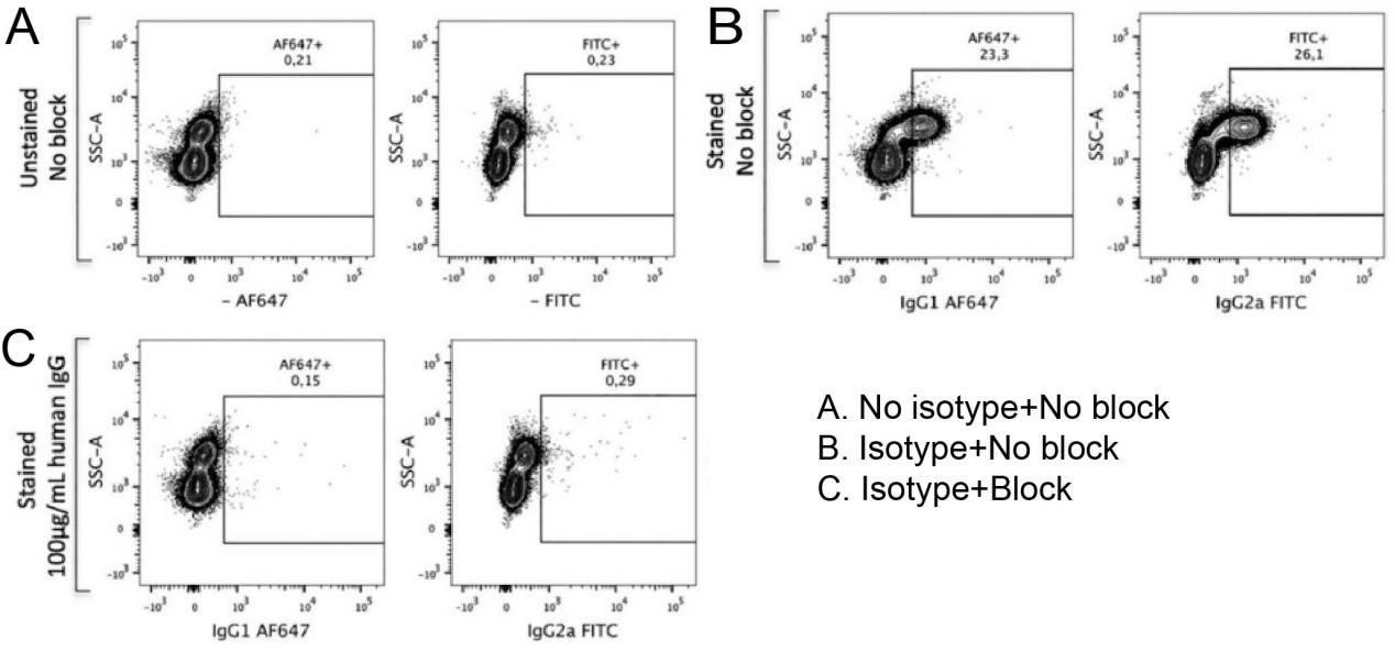
Fig. 2 The isotype control is used to eliminate the background staining caused by non-specific antibody–antigen binding. (A) In a CD45+ leukocyte sample, after dead cells and doublets have been excluded, a blank sample that has been neither stained nor Fc receptor-blocked shows low fluorescence in all channels. (B) After the isotype control is applied, background fluorescence is increased. (C) After FcR blocking, application of the isotype control restores background fluorescence to a level similar to that of the blank (The data are provided by Elabscience®).
02 Isotype controls versus unstained controls
The unstained control and the isotype control answer two different questions. The unstained tube contains no antibody and no dye, and can therefore reflect only cellular autofluorescence and instrument background; it is used to set the PMT voltage and the lowest signal baseline, but it cannot reflect the component of background that appears only after antibody is added[1,2]. The isotype control, by contrast, replaces the antibody of interest with a target-less antibody under the same antibody concentration and fluorophore conditions, and thus captures antigen-independent antibody–cell binding, which arises mainly from Fc receptor (FcγR)-mediated binding and is most frequent in monocytes/macrophages, dendritic cells and B cells[2,4].
Situations in which unstained cells are sufficient: the sample has low FcγR expression (for example, purified T cells or cultured cell lines), a high proportion of viable cells, antibodies that have already been titrated and optimised, and a clearly defined endogenous negative population (such as lymphocytes in CD34 analysis). In these circumstances an unstained tube for setting the baseline, single-stain tubes for compensation and FMO controls for gating are sufficient, and no additional isotype control is needed[2,4].
Situations in which an isotype control is required: the sample is rich in monocytes/macrophages or granulocytes; rare events or low-expression markers are being analysed; an antibody is being used for the first time or a new protocol is being established and it is necessary to determine whether the signal arises from specific binding or from non-specific adherence of the antibody; and when verifying whether Fc blocking is sufficient, the isotype control flow cytometry protocol still provides irreplaceable reference information, a more rigorous approach being to compare isotype controls before and after blocking[3,4,5].
Two further points deserve mention.
● First, neither the isotype control nor the unstained control can replace FMO controls for gating in a multicolor panel: the former carries no information on spillover and spread from other fluorophores, while the latter contains no antibody at all[1,3].
● Second, the isotype control must match the antibody of interest in species, isotype, fluorophore and concentration, and should preferably be obtained from the same supplier; otherwise its background level is not comparable[3,5].
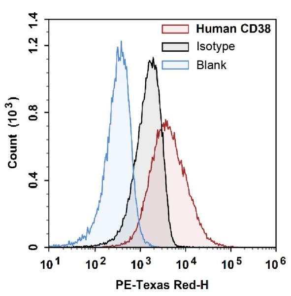
Fig. 3 Use of the isotype control to determine gate placement. Human CD38 is a continuously expressed marker on human peripheral blood lymphocytes. Gating on the basis of a blank tube with no antibody staining (blue) is compared with gating on the basis of the isotype control for the corresponding antibody (black) (The data are provided by Elabscience®).
Assay Used for This Experiment:
PE Anti-Human CD38 Antibody[HIT2]
Cat. No.: E-AB-F1058D
Clone: HIT2
Sample Type: Cell samples
Detection Instrument: Flow cytometer
A flow cytometry antibody for detecting the cell surface glycoprotein CD38.
Explore the PE Anti-Human CD38 Antibody[HIT2] →
03 Isotype controls versus FMO controls for gating
The flow cytometry isotype control and the FMO control likewise answer two different questions. An FMO (fluorescence minus one) control contains every antibody in the panel except the antibody of interest; it therefore measures both the spillover of other fluorophores into that channel and the spread of the negative population after compensation, and it tells you where the negative population lies and where the gate should be drawn[6,7]. An isotype control retains the antibody and withdraws only the antigen specificity; it measures non-specific binding of the antibody itself (in particular the Fc region) to cells, and it tells you whether the signal reflects specific recognition or merely antibody "sticking" to the cells[3,8].
These two definitions mean that the controls cannot be interchanged: by definition an FMO control does not contain the antibody of interest and therefore cannot reflect non-specific antibody binding, whereas an isotype control contains none of the other fluorophores and therefore cannot reflect spillover or spread at all[7]. Using an isotype control to set a positive gate will almost inevitably place that gate incorrectly in a multicolor panel.
The division of labour is therefore unambiguous. The positive/negative boundary should be set with FMO controls, particularly for multicolor panels, weakly expressed antigens and continuously distributed markers; even when compensation has been completed, spread from other fluorophores still raises the background of the negative population, and the FMO control is precisely the tool that quantifies this spread and thereby maximises channel resolution[7,9]. Whether staining is specific, and the troubleshooting of staining problems, should be judged with isotype controls[8].
Three practical points should be emphasised for FMO vs isotype control.
● First, in a multicolor panel every fluorophore requires its own FMO control, and a single isotype control tube cannot substitute for all of them[7].
● Second, if isotype controls are retained, they must match the antibody of interest in species, isotype, fluorophore and concentration; because the conjugation ratio (F/P) can hardly ever be matched exactly, their background need not equal that of the antibody of interest[3,7].
● Third, the recommendation given in the guidelines is precisely that the two controls each perform their own function: FMO controls set the gates, while isotype controls are used to identify staining problems, including non-specific binding[8].
04 Isotype controls in multicolor flow cytometry panels
In a multicolor flow cytometry antibody panel, background arises from two mutually independent routes: spillover of other fluorophores into the channel of interest, which forms spread, and non-specific binding of the antibody itself to cells. The facs isotype control reflects only the latter, and reflects it incompletely; it provides no coverage of the former[6,7]. The more complex the panel, therefore, the less reliable gating based on an isotype control alone becomes.
The cost of this complexity has a quantitative basis: spread error in multicolor measurements arises mainly in the red and far-red regions of the spectrum and markedly limits the detection of weakly expressed markers[10]. Bright fluorophores "spread out" the distribution of the negative population within the same channel, so the more weakly expressed and the more continuously distributed an antigen is, the more necessary it becomes to generate an FMO control channel by channel and for the panel actually in use in order to set the gate[7,10]; an isotype control tube, which contains none of the other fluorophores, cannot by its nature reflect this layer of background[7].
The role of the facs isotype control in a multicolor flow cytometry antibody panel has therefore shifted from "gating tool" to "troubleshooting tool": gating is delegated to FMO controls, while isotype controls are used to judge whether staining is specific, to troubleshoot non-specific binding, and to verify whether Fc blocking is sufficient[6,8]. A more thorough approach dispenses with them altogether, eliminating Fc receptor-mediated background directly with Fc blocking reagents or with Fc-removed engineered antibodies[3,6].
These controls are additive rather than substitutable: a multicolor flow cytometry antibody panel will normally require, at the same time, an unstained tube (to set voltages and read autofluorescence), single-stain tubes (to calculate compensation), channel-by-channel FMO controls (to set positive gates), an isotype control or Fc blocking (to check for non-specific binding), a viability dye (to exclude dead cells, which are highly autofluorescent and readily stained non-specifically), and biological controls[6,7,8].
Three practical points should be noted.
● First, an isotype control need not be prepared for every channel; it is used only where non-specific binding is suspected, for example with cell populations that highly express FcγR, with intracellular staining, with rare or uncommon markers, or with a new antibody or a new lot[8].
● Second, isotype controls cannot be used to calculate compensation: compensation is determined by the fluorophore and is independent of antibody specificity[7].
● Third, once the panel is modified, all FMO controls must be regenerated, whereas isotype control tubes will not "update" accordingly[10].
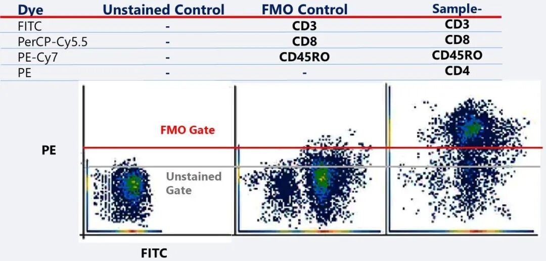
Fig. 4 FMO controls give more accurate gating: comparison of negative-population determination using a blank tube (grey line) and using an FMO control (red line) (The data are provided by Elabscience®).
Table 1. Comparison of unstained, isotype, FMO and single-stain controls
|
Control |
What it actually measures |
When to use it |
What it cannot tell you |
|
Unstained |
Cellular autofluorescence + instrument background |
Always retained; used mainly to set the voltage and the lowest baseline |
Non-specific antibody binding; spectral spillover |
|
Isotype |
Antigen-independent antibody–cell binding (mainly FcγR-mediated) |
Samples with high FcγR expression; rare events/low-expression markers; new antibodies or protocols; verification of Fc blocking |
Spillover and spread from other fluorophores; cannot serve as a gating standard |
|
FMO |
Spillover and spread of the other fluorophores of the panel into that channel |
Gating in multicolor panels (first choice) |
Autofluorescence itself; non-specific antibody binding |
|
Single stain (compensation) |
Spectral spillover of a single fluorophore |
Mandatory in multicolor experiments |
Gate boundaries |
05 Controls for spectral flow cytometry and unmixing
Spectral flow cytometry no longer reads only the emission peak of a fluorophore; it acquires the complete spectrum of that fluorophore across lasers and then uses an unmixing algorithm to recover the abundance of each fluorophore. Because the input to unmixing is a set of known reference spectra, the control system changes accordingly: the single-stain tubes used to calculate compensation in conventional flow cytometry are upgraded here to spectral reference controls (SRCs). Every fluorophore in the panel, including antibodies, fluorescent proteins and viability dyes, requires its own SRC, and the signal intensity of its positive population should not be lower than that of the positive population in the experimental sample, since otherwise unmixing will be distorted[11]. SRCs may be prepared from compensation beads or from cells, but because the normalised spectrum of the same fluorophore on beads and on cells is not necessarily identical, an SRC established with beads must be validated on real samples[11,12].
An autofluorescence reference is also required: unstained cells, which must match the experimental sample in tissue and in processing workflow[13]. When autofluorescence is strongly heterogeneous (tissue digests, cultured cells, intracellular staining), it should be split into several autofluorescence references that are incorporated into the unmixing separately, rather than being represented by a single unstained tube[13,14]. Fully stained samples and FMO controls are still used to check the quality of unmixing, to inspect the morphology of the negative population and to set positive gates[11,13].
The difference from the conventional isotype control is one of dimension. An isotype control is defined by antibody specificity ("labelled but target-less") and is used to assess the background caused by non-specific antibody binding, answering a biological question[6]. An SRC, by contrast, is defined by the spectrum of the fluorophore; it need only carry the same fluorophore, irrespective of the antibody's target, isotype or supplier, or indeed of whether it is directed against the sample in question, and it answers a mathematical question: it is one known spectrum in the unmixing matrix[11,12]. The two are therefore not interchangeable: a missing or inadequate SRC will directly cause unmixing errors (abnormal morphology of the negative population, appearance of over-negative values), whereas an isotype control, however rigorously prepared, can never serve as an input to the unmixing matrix; conversely, an SRC does not assess whether staining is specific. In spectral flow cytometry the isotype control plays the same role as in conventional flow cytometry: it is used only to judge staining specificity, to troubleshoot non-specific binding or to verify whether Fc blocking is sufficient, and it is combined with Fc blocking reagents, viability dyes and biological controls[6,13].
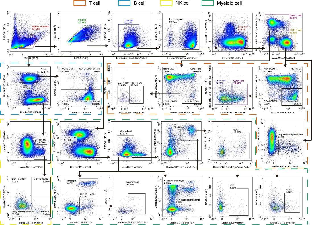
Fig. 5 Spectral flow cytometry results for a 20-color panel of mouse splenic immune cells. In this experiment, Fc receptors were blocked and both a blank control and FMO controls for CD25, CD127, CD62L, Ly6C, NK1.1 and CD44 were included to facilitate gating (The data are provided by Elabscience®).
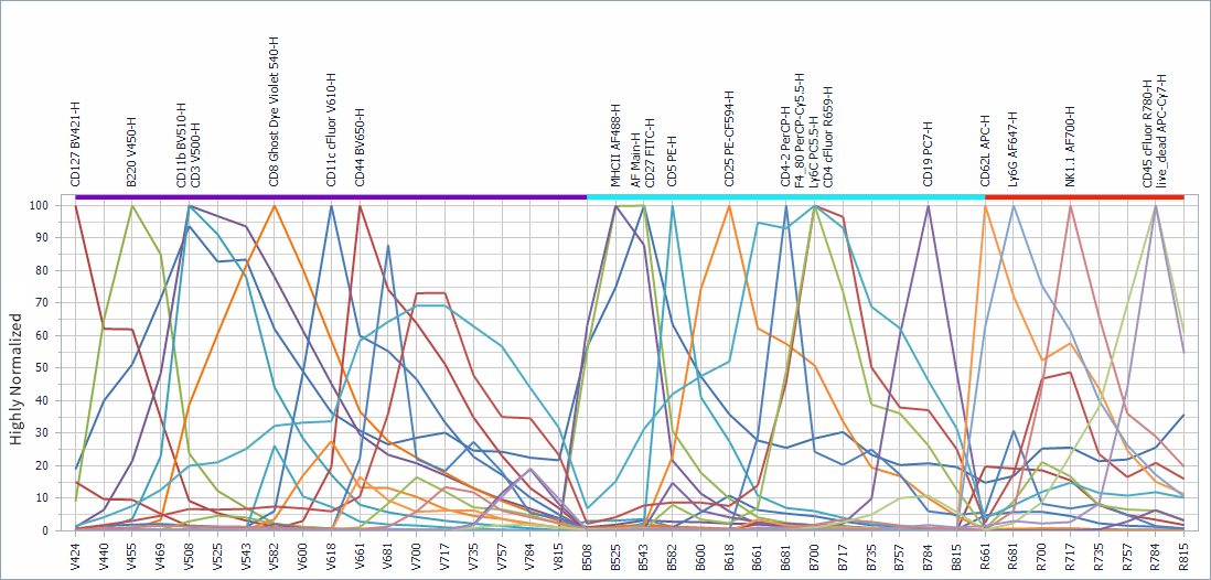
Fig. 6 Spectral reference controls for the 20-color panel of mouse splenic immune cells. In this experiment, a single-stain tube was prepared for each fluorophore in order to generate the reference spectra (The data are provided by Elabscience®).
The isotype control answers a single question: does the signal from the antibody of interest arise from specific recognition, or from antigen-independent binding of the antibody to the cells? It is therefore a specificity and troubleshooting control rather than a gate-setting one. Unstained cells set voltages and reveal autofluorescence, single-stain and spectral reference controls establish compensation and unmixing, FMO controls set the positive/negative gates, and isotype controls, together with Fc blocking, are used to judge whether staining is specific; in a multicolor panel these controls are additive rather than substitutable. Two limitations should be kept in mind. Because of differences in hybridoma origin, purification, conjugation and lot, an isotype control need not reproduce the background of the antibody of interest even when the isotype is matched; and in spectral flow cytometry the controls on which unmixing depends are the SRCs and the autofluorescence references, which an isotype control cannot replace. Used within that framework, in newly established laboratories, during antibody or protocol development, in FcγR-rich samples, in rare-event and low-expression analysis, and when verifying Fc blocking, the isotype control flow cytometry protocol contributes what it is uniquely able to contribute.
Elabscience® Quick Overview of Popular Products
Table 2. A 20-color panel for mouse splenic immune cells
|
Markers |
Antibodies |
Cat.No. |
|
CD45 |
Elab Fluor® Red 780 Anti-Mouse CD45 Antibody[30-F11] |
E-AB-F1136S |
|
CD3 |
Elab Fluor® Violet 500 Anti-Mouse CD3 Antibody[17A2] |
E-AB-F1013R |
|
CD4 |
PerCP Anti-Mouse CD4 Antibody[GK1.5] |
E-AB-F1097F |
|
CD8 |
Elab Fluor® Violet 540 Anti-Mouse CD8a Antibody[53-6.7] |
E-AB-F1104T3 |
|
CD25 |
PE/Elab Fluor® 594 Anti-Mouse CD25 Antibody[PC-61.5.3] |
E-AB-F1102P |
|
NK1.1 |
Elab Fluor® 700 Anti-Mouse CD161/NK1.1 Antibody[PK136] |
E-AB-F0987M1 |
|
CD44 |
Elab Bright™ Violet 650 Anti-Human/Mouse CD44 Antibody[IM7] |
E-AB-F1100U |
|
CD62L |
APC Anti-Mouse CD62L Antibody[MEL-14] |
E-AB-F1011E |
|
CD127 |
Elab Bright™ Violet 421 Anti-Mouse CD127/IL-7RA Antibody[A7R34] |
E-AB-F1023Q2 |
|
B220 |
Elab Fluor® Violet 450 Anti-Mouse CD45R/B220 Antibody[RA3.3A 1/6.1] |
E-AB-F1112Q |
|
CD19 |
PE/Cyanine7 Anti-Mouse CD19 Antibody[1D3] |
E-AB-F0986H |
|
CD5 |
PE Anti-Mouse CD5 Antibody[53-7.3] |
E-AB-F1185D |
|
CD27 |
FITC Anti-Mouse CD27 Antibody[LG.3A10] |
AN00322C |
|
CD11b |
Elab Bright™ Violet 510 Anti-Mouse/Human/Monkey CD11b Antibody[M1/70] |
E-AB-F1081R1 |
|
CD11c |
Elab Fluor® Violet 610 Anti-Mouse CD11c Antibody[N418] |
E-AB-F0991T |
|
MHC-II |
Elab Fluor® 488 Anti-Mouse MHC II (I-A/I-E) Antibody[M5/114] |
E-AB-F0990L |
|
F4/80 |
PerCP/Cyanine5.5 Anti-Mouse F4/80 Antibody[CI:A3-1] |
E-AB-F0995J |
|
Ly6G |
Elab Fluor® 647 Anti-Mouse Ly6G Antibody[1A8] |
E-AB-F1108M |
|
Ly6C |
PE/Cyanine5.5 Anti-Mouse Ly6C Antibody[Monts 1] |
E-AB-F1121I |
|
Cell Viability Dye |
STYX™ Near-IR Fixable Viability Kit |
E-CK-A168 |
Table 3. Isotype control products (part)
|
Fluorescent Dye of Isotype |
Human(Cat. No.) |
Mouse(Cat. No.) |
Rat(Cat. No.) |
|
IgG1, κ |
IgG2a, κ |
IgG2a, κ |
|
|
Elab Bright™ Violet 421 |
\ |
\ |
AN00822Q2 |
|
Elab Fluor® Violet 450 |
AN00840Q |
E-AB-F09802Q |
E-AB-F09832Q |
|
Elab Fluor® Violet 500 |
\ |
E-AB-F09802R |
E-AB-F09832R |
|
Elab Bright™ Violet 510 |
\ |
\ |
AN00822R1 |
|
Elab Fluor® 488 |
AN00840L |
E-AB-F09802L |
E-AB-F09832L |
|
FITC |
AN00840C |
E-AB-F09802C |
E-AB-F09832C |
|
Elab Fluor® Violet 540 |
\ |
E-AB-F09802T3 |
E-AB-F09832T3 |
|
PE |
AN00840D |
E-AB-F09802D |
E-AB-F09832D |
|
Elab Fluor® Violet 610 |
\ |
E-AB-F09802T |
E-AB-F09832T |
|
PE/Elab Fluor® 594 |
AN00840P |
E-AB-F09802P |
E-AB-F09832P |
|
Elab Bright™ Violet 650 |
\ |
\ |
AN00822U |
|
APC |
AN00840E |
E-AB-F09802E |
E-AB-F09832E |
|
Elab Fluor® 647 |
AN00840M |
E-AB-F09802M |
E-AB-F09832M |
|
PE/Cyanine5 |
AN00840G |
E-AB-F09802G |
E-AB-F09832G |
|
PerCP |
AN00840F |
\ |
E-AB-F09832F |
|
PerCP/Cyanine5.5 |
AN00840J |
E-AB-F09802J |
E-AB-F09832J |
|
PE/Cyanine5.5 |
AN00840I |
E-AB-F09802I |
E-AB-F09832I |
|
Elab Fluor® 700 |
\ |
E-AB-F09802M1 |
E-AB-F09832M1 |
|
PE/Cyanine7 |
AN00840G |
E-AB-F09802H |
E-AB-F09832H |
|
Elab Fluor® Red 780 |
\ |
E-AB-F09802S |
E-AB-F09832S |
06 Key takeaways
● An isotype control is a "labelled but target-less" antibody matched to the antibody of interest, used to baseline antigen-independent binding and judge staining specificity.
● It covers only antibody-derived background (mainly FcγR-mediated), not autofluorescence or spectral spillover.
● It is not an ideal negative control, since hybridoma, purification, conjugation, F/P ratio and lot variation can all change its background.
● An unstained tube shows only autofluorescence and instrument background, so an isotype control is unnecessary when FcγR is low and the negative population is clear.
● FMO controls set the gate from spillover and spread, while isotype controls judge specificity, and the two cannot be interchanged.
● In multicolor panels the isotype control becomes a troubleshooting rather than a gating tool, and the controls a panel requires are additive, not substitutable.
● In spectral flow cytometry every fluorophore needs its own SRC plus sample-matched autofluorescence references, which an isotype control can never replace.
07 Related resources
● A successful flow cytometry experiment cannot be achieved without the establishment of control groups. Blank, isotype, single staining and FMO controls should be complementary rather than substitutive, and the choice should be made based on the experimental setup.
Download Your Free Application of Flow Cytometry Guide →
● Learn how to conduct spectral flow cytometry multi-color experiments and select the appropriate fluorescent dyes for your experiments.
Read How to Choose Fluorescent Dyes for Spectral Multicolor Flow Cytometry →
● Learn about the purpose and applications of isotype controls in flow cytometry and how to use them appropriately in your experiments.
Watch the Introduction and Application of Isotype Controls Tutorial Video →
● Learn about the role of fluorescence minus one (FMO) controls in flow cytometry and how they help establish appropriate gating boundaries in multicolor experiments.
Watch the Introduction and Functions of FMO Control Tutorial Video →
References:
[1] Maecker HT, Trotter J. Flow cytometry controls, instrument setup, and the determination of positivity. Cytometry A. 2006;69(9):1037-1042. doi:10.1002/cyto.a.20333. PMID: 16888771.
[2] Hulspas R, O'Gorman MRG, Wood BL, Gratama JW, Sutherland DR. Considerations for the control of background fluorescence in clinical flow cytometry. Cytometry B Clin Cytom. 2009;76(6):355-364. doi:10.1002/cyto.b.20485. PMID: 19575390.
[3] Andersen MN, Al-Karradi SNH, Kragstrup TW, Hokland M. Elimination of erroneous results in flow cytometry caused by antibody binding to Fc receptors on human monocytes and macrophages. Cytometry A. 2016;89(11):1001-1009. doi:10.1002/cyto.a.22995. PMID: 27731950.
[4] Keeney M, Gratama JW, Chin-Yee IH, Sutherland DR. Isotype controls in the analysis of lymphocytes and CD34+ stem and progenitor cells by flow cytometry — time to let go! Cytometry. 1998;34(6):280-283. doi:10.1002/(SICI)1097-0320(19981215)34:6<280::AID-CYTO6>3.0.CO;2-H.
[5] O'Gorman MRG, Thomas J. Isotype controls — time to let go? Cytometry. 1999;38(2):78-80. doi:10.1002/(SICI)1097-0320(19990415)38:2<78::AID-CYTO6>3.0.CO;2-E.
[6] Cossarizza A, Chang HD, Radbruch A, et al. Guidelines for the use of flow cytometry and cell sorting in immunological studies (third edition). Eur J Immunol. 2021;51(12):2708-3145. doi:10.1002/eji.202170126.
[7] Mair F, Tyznik AJ. High-dimensional immunophenotyping with fluorescence-based cytometry: a practical guidebook. Methods Mol Biol. 2019;2032:1-29. doi:10.1007/978-1-4939-9650-6_1.
[8] Brooks HL, de Castro Brás LE, Brunt KR, et al. Guidelines on antibody use in physiology research. Am J Physiol Renal Physiol. 2024;326(3):F511-F533. doi:10.1152/ajprenal.00347.2023. PMID: 38234298.
[9] Maciorowski Z, Chattopadhyay PK, Jain P. Basic multicolor flow cytometry. Curr Protoc Immunol. 2017;117:5.4.1-5.4.38. doi:10.1002/cpim.26. PMID: 28369683.
[10] Brummelman J, Haftmann C, Núñez NG, Alvisi G, Mazza EMC, Becher B, Lugli E. Development, application and computational analysis of high-dimensional fluorescent antibody panels for single-cell flow cytometry. Nat Protoc. 2019;14(7):1946-1969. doi:10.1038/s41596-019-0166-2. PMID: 31160786.
[11] Ferrer-Font L, Pellefigues C, Mayer JU, Small SJ, Jaimes MC, Price KM. Panel design and optimization for high-dimensional immunophenotyping assays using spectral flow cytometry. Curr Protoc Cytom. 2020;92(1):e70. doi:10.1002/cpcy.70.
[12] Shevchenko Y, Lurje I, Tacke F, Hammerich L. Fluorochrome-dependent specific changes in spectral profiles using different compensation beads or primary cells in full spectrum cytometry. Cytometry A. 2024;105(6):458-463. doi:10.1002/cyto.a.24836. PMID: 38511720.
[13] Czechowska K, Bonilla DL, Cotty A, Dankar A, Mead PE, Nash V. Beyond the limits: how is spectral flow cytometry reshaping the clinical landscape and what is coming next? Cells. 2025;14(13):997. doi:10.3390/cells14130997. PMID: 40643518.
[14] Roet JEG, Mikula AM, de Kok M, et al. Unbiased method for spectral analysis of cells with great diversity of autofluorescence spectra. Cytometry A. 2024;105(8):595-606. doi:10.1002/cyto.a.24856.

