

Steroid hormones are a family of lipophilic molecules derived from cholesterol, sharing a common sterane backbone. Functionally, they fall into three categories: glucocorticoids, mineralocorticoids, and sex steroids.
This post breaks down key analytes: Corticosterone (CORT), Cortisol, Estradiol (E2), Estriol (E3), Progesterone (Pg), Testosterone (T), AMH, and FSH, covering their structure, biological roles, and typical panel designs, along with technical considerations for small-molecule ELISA.
Table of Contents
1. Steroid hormone definition and classification
2. Key steroid hormone analytes: structure, function, and research applications
3. Key challenges in steroid hormone detection by ELISA
4. Steroid hormone panel design for different research applications
5. QuicKey Pro™ ELISA kits for rapid steroid hormone detection
6. Frequently asked questions about steroid hormone ELISA
01 Steroid hormone definition and classification
Steroid hormones are a large family of lipophilic molecules derived from cholesterol. Although they have many members, they share a similar sterane skeleton. Based on physiological function, they fall into three major categories.
Table 1. Classification and Functions of Steroid Hormones
|
Class |
Secretion Site |
Core Function |
Representative Hormones |
|
Glucocorticoids |
Adrenal zona fasciculata |
Regulates glucose/lipid/protein metabolism; anti‑inflammatory, immunosuppressive, anti‑stress |
Cortisol (human & large mammals); CORT (main in rodents) |
|
Mineralocorticoids |
Adrenal zona glomerulosa |
Regulates water‑salt balance; maintains blood pressure and electrolyte homeostasis |
Aldosterone (ALD) |
|
Sex steroids |
Gonads (testes/ovaries), adrenal zona reticularis, placenta |
Controls reproduction, secondary sex characteristics, menstrual cycle, pregnancy maintenance |
T, E2, Pg, E3 |
Memory aid: “Sweet, Salt, Sex” – Sweet (glucose metabolism) = glucocorticoids; Salt (Na+/K+ balance) = mineralocorticoids; Sex = sex steroids.
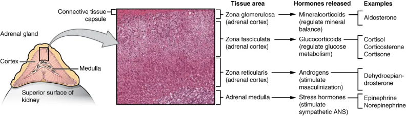
Fig. 1 Hormones produced by the adrenal gland. (Source: OpenStax, Anatomy and Physiology 2e, Figure 17.17, CC BY 4.0)
02 Key steroid hormone analytes: structure, function, and research applications
2.1 Glucocorticoid Core Markers
Glucocorticoids are central to stress and metabolism. In humans and most large mammals, cortisol predominates; rodents (mice/rats) use CORT because their adrenals lack CYP17A1, the enzyme needed for cortisol synthesis[1].
Table 2. Glucocorticoid Core Markers
|
Marker |
MW |
Core Function |
Clinical / Research Significance |
|
362 Da |
Primary stress hormone; raises blood glucose (gluconeogenesis); suppresses immunity |
Cushing’s/Addison’s disease research; chronic stress assessment; tumor immune microenvironment studies |
|
|
346 Da |
Main glucocorticoid in rodents; regulates metabolism and immunity |
Animal stress models (mouse/rat); HPA axis function evaluation |
|
|
Cortisone |
360 Da |
Inactive form of cortisol; generated from cortisol by 11β‑HSD2 in kidney, and can be converted back by 11β‑HSD1 in liver |
11β‑HSD2 activity assessment; kidney disease research |
2.2 Mineralocorticoid Core Marker
Mineralocorticoids are represented by aldosterone, the key effector hormone of the renin‑angiotensin‑aldosterone system (RAAS).
Table 3. Mineralocorticoid Core Marker
|
Marker |
MW |
Core Function |
Clinical / Research Significance |
|
360 Da |
Regulates Na+/K+ balance; maintains blood volume and pressure |
Primary aldosteronism (Conn’s syndrome); hypertension etiology; heart failure prognosis |
2.3 Sex Steroid Core Markers
Sex steroids are secreted by the gonads, adrenal zona reticularis, and placenta, and are involved in reproduction, development, and metabolism.
Table 4. Sex Steroid Core Markers
|
Marker |
MW |
Core Function |
Clinical / Research Significance |
|
288 Da |
Main androgen; promotes muscle growth and spermatogenesis |
Male reproductive function; PCOS (Polycystic Ovary Syndrome); tumor immune microenvironment |
|
|
272 Da |
Major estrogen; regulates follicular development and endometrial proliferation |
IVF follicular monitoring; breast cancer endocrine therapy; ovarian reserve assessment |
|
|
288 Da |
Major pregnancy estrogen, synthesized by the fetal‑placental unit; reflects placental function |
Placental function evaluation; prenatal screening |
|
|
314 Da |
Maintains pregnancy; prepares endometrium for implantation (decidualisation) |
Ovulation confirmation; luteal function assessment; threatened miscarriage research |
|
|
Androstenedione |
286 Da |
Common precursor for androgens and estrogens |
Adrenal cortex function; PCOS research |
2.4 Pituitary‑Gonadal Axis Regulators (Non‑steroid, Commonly Co‑detected with Steroids)
AMH, FSH, and LH are glycoprotein hormones with larger molecular weights, allowing sandwich ELISA. They are the three most frequently combined markers in steroid hormone studies.
Table 5. Pituitary-Gonadal Axis Regulators
|
Marker |
MW |
Core Function |
Clinical / Research Significance |
|
~140 kDa |
Secreted by small ovarian follicles; prevents premature primordial follicle depletion; reflects ovarian reserve |
IVF protocol planning; PCOS; menopause prediction |
|
|
~30 kDa |
Stimulates follicle growth and estrogen synthesis |
Ovarian responsiveness assessment; perimenopausal transition |
|
|
~30 kDa |
Triggers ovulation; promotes corpus luteum formation and progesterone secretion |
Ovulation prediction; PCOS adjunctive evaluation (LH/FSH ratio often elevated, but not diagnostic) |
03 Key challenges in steroid hormone detection by ELISA
Steroid hormone detection via ELISA faces three major hurdles:
(1) Extremely small molecular size (272–363 Da)
Steroid hormones (272–363 Da) offer only one epitope, making sandwich ELISA impossible.
Solutions: Competitive ELISA + high-specificity antibodies.
(2) Cross‑reactivity with structural analogues
Cortisol/cortisone and E1/E2/E3 differ by minor groups, risking antibody cross-reaction.
Solutions: Stringent antibody screening + LC‑MS/MS cross-validation.
(3) Binding protein interference
Over 90% of steroids are bound to CBG/SHBG/albumin, reducing free hormone detection[2–4].
Solutions: Ethanol extraction or charcoal stripping; or directly measure free fraction[5-7].
04 Steroid hormone panel design for different research applications
Table 6. Recommended Combined Detection Panels by Research Scenario
|
Research Scenario |
Recommended Panel |
Purpose |
|
HPA axis function (human/large animals) |
Cortisol (with ACTH for feedback assessment) |
Evaluate stress response and adrenal cortex function |
|
HPA axis function (rodents) |
CORT (with ACTH for feedback assessment) |
Core readout for mouse/rat stress models |
|
Ovarian reserve assessment |
AMH + FSH + LH + E2 |
Predict IVF oocyte yield; POI/PCOS research |
|
Ovulation prediction |
LH + E2 + Pg + FSH |
Dynamic monitoring of LH surge to predict ovulation window; provide timing basis for hCG “trigger” |
|
Pregnancy monitoring |
E3 + Pg |
Assess placental function and fetal‑placental unit status; for preeclampsia risk, combine with sFlt‑1/PlGF |
|
Tumour immune microenvironment |
Cortisol + T + E2 |
Explore the impact of hormone levels on immune checkpoint inhibitor (ICB) therapy efficacy |
05 QuicKey Pro™ ELISA kits for rapid steroid hormone detection
● High efficiency & time‑saving
90‑min rapid assay – 3× faster than conventional kits
● Simple operation
Single‑step addition, single incubation/wash – minimal hands‑on time
● Reliable data
Validated with genuine native samples (serum/plasma/tissue/cells); six performance parameters verified through three‑step quality control
● High‑impact citations
Cited in Cell, Nature Communications, and other top journals
● Selected high‑impact references
1. A Skin‑hypothalamus Axis Couples Heat Stress and Metabolic Dysfunction. Cell, 2026.
DOI: 10.1016/j.cell.2026.03.045
Product: QuicKey Pro Mouse CORT ELISA Kit (E‑OSEL‑M0001)
2. Polylactic Acid Micro/Nanoplastic Exposure Induces Male Reproductive Toxicity by Disrupting Spermatogenesis and Mitochondrial Dysfunction in Mice. ACS Nano, 2025.
DOI: 10.1021/acsnano.4c15112
Product: QuicKey Pro Mouse T ELISA Kit (E‑OSEL‑M0003), Mouse FSH ELISA Kit (E‑EL‑M0511), Mouse LH ELISA Kit (E‑EL‑M3053)
QuicKey Pro™ ELISA Kits Steroid hormone series now covers 11 species: Human, Mouse, Rat, Monkey, Bovine, Canine, Chicken, Porcine, Rabbit, Sheep, Horse – providing differentiated options for model animal research, veterinary studies, and comparative endocrinology.
06 Frequently asked questions about steroid hormone ELISA
Q1: Rodent stress models – cortisol or corticosterone?
A: Measure CORT. Rodents lack adrenal CYP17A1 and cannot synthesise cortisol.
Q2: ELISA vs. LC‑MS/MS – which to choose?
A: LC‑MS/MS is the gold standard for specificity but requires expensive equipment and is low‑throughput. Competitive ELISA is practical, cost‑effective, and shows good correlation with MS – ideal for routine batch screening.
Q3: Why only competitive ELISA for steroids?
A: Steroids are small (272–363 Da) haptens with a single epitope, making sandwich format impossible.
Q4: Cross‑reactivity between cortisol and cortisone?
A: Our cortisol kits are validated for <10% cross‑reactivity with cortisone.
Q5: Do I need to extract serum/plasma samples?
A: Most kits measure total hormone directly. For free‑fraction measurement, use ethanol extraction or ultrafiltration.
References:
[1] Keeney DS, et al. Developmentally regulated expression of adrenal 17 alpha-hydroxylase cytochrome P450 in the mouse embryo, Endocrinology, 1995;136(11):4872-4879.
[2] Hammond GL. Plasma steroid-binding proteins: primary gatekeepers of steroid hormone action, J Endocrinol, 2016;230(1):R13-R25.
[3] Dunn JF, Nisula BC, Rodbard D. Transport of steroid hormones: binding of 21 endogenous steroids to both testosterone-binding globulin and corticosteroid-binding globulin in human plasma, J Clin Endocrinol Metab, 1981;53(1):58-68.
[4] Cizza G, et al. Cortisol Binding Globulin: More Than Just a Carrier? 2012.
[5] Faix JD. Principles and pitfalls of free hormone measurements, Best Pract Res Clin Endocrinol Metab, 2013;27(5):631-645.
[6] Stanczyk FZ, et al. Limitations of direct estradiol and testosterone immunoassay kits, Steroids, 2003;68(14):1173-1178.
[7] Hammond GL, Lähteenmäki PL. A versatile method for the determination of serum cortisol binding globulin and sex hormone binding globulin binding capacities, Clin Chim Acta, 1983;132:101-110.

