

Inflammation is a fundamental host defense mechanism that, when dysregulated, becomes a central driver of numerous chronic diseases, including metabolic disorders, neurodegeneration, and cancer. At the molecular level, this process is orchestrated by a diverse family of soluble mediators known as cytokines, which coordinate communication among immune cells and between the immune system and peripheral tissues. Pro-inflammatory cytokines such as interleukin-1β (IL-1β), interleukin-6 (IL-6), and tumor necrosis factor-α (TNF-α) are rapidly induced upon infection or tissue injury and act through conserved signaling cascades, including the nuclear factor-κB (NF-κB), mitogen-activated protein kinase (MAPK), and Janus kinase/signal transducer and activator of transcription (JAK/STAT) pathways. When these pathways are persistently activated, they sustain a chronic inflammatory state that promotes tissue damage, fibrosis, and malignant transformation. Understanding which cytokines drive inflammation, how they signal, and how they can be reliably measured is therefore essential for both mechanistic research and the development of targeted therapeutic and diagnostic strategies.
This review article provides a comprehensive overview of the pro-inflammatory cytokines that drive inflammation and their roles in disease progression. We first introduce the general biology of inflammatory cytokines and their contribution to immune activation and disease development. We then examine the functions of the major pro-inflammatory cytokines IL-1β, IL-6, and TNF-α in immune activation, followed by a discussion of the signaling pathways that sustain chronic inflammation. The review also covers the principal methods used to quantify these mediators, including enzyme-linked immunosorbent assay (ELISA) and flow cytometry-based intracellular cytokine staining, and concludes by exploring how cytokine changes within the tumor microenvironment shape cancer-associated inflammation and influence therapeutic responses.
Table of Contents
1. Overview of inflammatory cytokines and their roles in disease progression
2. Major pro-inflammatory cytokines: IL-1β, IL-6, and TNF-α functions in immune activation
3. Cytokine signaling pathways driving chronic inflammation
4. Cytokine measurement using ELISA for inflammatory research
5. Cytokine detection by Flow cytometry and intracellular cytokine staining
6. Cytokine changes in the tumor microenvironment and cancer-associated inflammation
01 Overview of inflammatory cytokines and their roles in disease progression
Inflammatory cytokines constitute a family of soluble polypeptide mediators that coordinate innate and adaptive immune responses and, when produced chronically, drive the tissue damage underlying many diseases. The principal inflammatory mediators include pro-inflammatory cytokines such as interleukin-1β (IL-1β), interleukin-6 (IL-6), tumor necrosis factor-α (TNF-α), and interferon-γ (IFN-γ), together with the acute-phase protein C-reactive protein (CRP), link peripheral immune activation to organ dysfunction across diverse pathologies[1]. Their induction is initiated when pattern-recognition receptors (PRRs), including Toll-like receptors (TLRs) and NOD-like receptors (NLRs), recognize pathogen-associated and damage-associated molecular patterns (PAMPs and DAMPs). Engagement of TLR4 by lipopolysaccharide (LPS) triggers NF-κB and MAPK cascades that transcriptionally activate IL-1β, IL-6, and TNF-α, whereas NLRP3 inflammasome assembly promotes caspase-1-mediated maturation and secretion of IL-1β and IL-18[1,2]. Once released, these inflammatory cytokines propagate disease progression through tissue-specific mechanisms. In the central nervous system, they activate indoleamine 2,3-dioxygenase 1 (IDO1) and tryptophan 2,3-dioxygenase (TDO2), diverting tryptophan toward the kynurenine pathway, suppressing brain-derived neurotrophic factor (BDNF) synthesis, and promoting microglia-mediated neuroinflammation, thereby contributing to the pathophysiology of depression[1]. In the tumor microenvironment, bacterial components and microbial metabolites activate the same innate immune receptors and oncogenic pathways, including MAPK, PI3K/AKT, and NF-κB, to sustain chronic inflammation, which is estimated to underlie approximately 20% of epithelial cancers[2]. Macrophages are central effectors of this process, and their polarization between pro-inflammatory M1 and immunosuppressive M2 states is finely regulated by long non-coding RNAs that converge on NF-κB and STAT signaling[3]. Similarly, in metabolic dysfunction-associated steatohepatitis (MASH), lipotoxic stress and gut-derived LPS activate Kupffer cells through the TLR4-MD2 complex, triggering reactive oxygen species (ROS) production and secretion of IL-1β and TNF-α that drive hepatocyte injury[4]. Collectively, these findings establish inflammatory cytokines as both biomarkers of disease activity and central nodes linking immune activation to disease progression.
02 Major pro-inflammatory cytokines: IL-1β, IL-6, and TNF-α functions in immune activation
Among the many mediators of inflammation, three cytokines, namely interleukin-1β (IL-1β), interleukin-6 (IL-6), and tumor necrosis factor-α (TNF-α), are the principal pro-inflammatory cytokines driving immune activation. TNF-α, produced chiefly by activated macrophages, is a potent inflammatory signal that engages tumor necrosis factor receptor 1 (TNFR1) to assemble a FADD/procaspase-8 complex, triggering caspase-3-mediated apoptosis, and upregulates myosin light chain kinase (MLCK) to disassemble epithelial tight junctions[5]. IL-1β, generated by inflammasome-dependent cleavage of pro-IL-1β, activates NF-κB signaling by inducing IKK-dependent phosphorylation and degradation of IκBα, thereby enabling nuclear translocation of p65/p50 and driving transcription of MLCK and additional inflammatory genes; it also downregulates occludin through microRNA-200c-3p, weakening apical junctions[5]. IL-6 is a pleiotropic cytokine that amplifies permeability by upregulating the pore-forming protein claudin-2 via the JNK/AP-1 pathway and sustains inflammatory T-cell survival through STAT3-dependent Bcl-2/Bcl-xL expression[5]. These three cytokines act in a highly interconnected, self-reinforcing network: IL-1β drives de novo IL-6 production, and both promote Th17 polarization, converting acute injury into chronic inflammation[5]. Beyond the gut, IL-1β and TNF-α serve as inflammatory "licensing" signals that activate mesenchymal stromal cells (MSCs) through NF-κB, upregulating immunosuppressive molecules such as indoleamine 2,3-dioxygenase (IDO) and programmed death-ligand 1 (PD-L1)[6]. In the central nervous system, misfolded amyloid-β triggers microglial pattern-recognition receptors and the NLRP3 inflammasome, leading to chronic production of IL-1β, TNF-α, and IL-6 that injure neurons and impair synaptic plasticity[7]. The same cytokines are central to mucosal inflammation in inflammatory bowel disease, where TLR4/NOD2, NF-κB, JAK/STAT, and NLRP3 signaling sustain their release[8]. Because these pro-inflammatory cytokines converge on shared NF-κB, MAPK, and JAK/STAT pathways, therapeutic strategies that simultaneously suppress multiple nodes, such as the flavonoid luteolin, which inhibits NF-κB, NLRP3, and MAPK while enhancing Nrf2, are more effective than single-cytokine blockade[9].
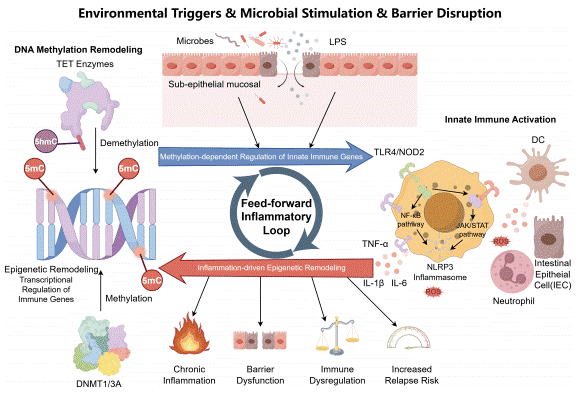
Fig. 1 DNA methylation–innate immunity crosstalk axis in inflammatory bowel disease. Environmental triggers, microbial stimulation, LPS exposure, and intestinal barrier disruption facilitate the translocation of microbial products into the subepithelial mucosal compartment, thereby activating innate immune cells, including macrophages, dendritic cells, neutrophils, and intestinal epithelial cells. Activation of TLR4/NOD2, NF-κB, JAK/STAT, and NLRP3 inflammasome signaling promotes the release of TNF-α, IL-1β, IL-6, and reactive oxygen species (ROS), amplifying mucosal inflammation[8].
03 Cytokine signaling pathways driving chronic inflammation
Chronic inflammation is sustained by a limited set of conserved signaling pathways that convert transient cytokine signals into persistent transcriptional programs. The nuclear factor-κB (NF-κB) and Janus kinase/signal transducer and activator of transcription (JAK/STAT) pathways are the two principal axes driving this process. NF-κB and JAK/STAT signaling are the major pathways that stimulate the senescence-associated secretory phenotype (SASP), in which senescent cells persistently secrete transforming growth factor-β (TGF-β), TNF-α, and IFN-γ to establish a pro-inflammatory "inflammaging" environment that promotes precancerous lesion development[10]. The JAK/STAT cascade is activated when cytokine receptors engage JAK kinases, which then recruit STAT transcription factors; in solid tumors this pathway is hyperactive, driving uncontrolled proliferation and immune suppression, whereas in cardiovascular and metabolic disease it exacerbates vascular remodeling and inflammation[11]. In the liver, chronic activation of the IL-6/JAK/STAT and TNF-α/NF-κB axes is central to hepatocellular carcinoma (HCC) development, with more than 90% of HCC cases arising from a background of chronic inflammation[12]. IL-6 binds its receptor and activates JAK/STAT3, promoting hepatocyte proliferation, inhibiting apoptosis, and increasing genomic instability, while TNF-α/NF-κB acts as a molecular switch that induces a broad panel of pro-inflammatory cytokines and chemokines[12]. These two pathways are synergistically activated and engage in negative crosstalk, forming a positive-feedback network that amplifies IL-6 and TNF-α release and drives malignant transformation[3,4]. Notably, NF-κB p65 is aberrantly activated in HCC tissues, and its inhibition reduces proliferation and invasion, while JAK1 mutations identified by whole-exome sequencing of HCC patient-derived xenografts render tumors sensitive to the JAK1/2 inhibitor ruxolitinib[13]. Because NF-κB and JAK/STAT are shared hubs linking inflammation to malignant transformation across tissues, they represent attractive targets for anti-inflammatory and nanocarrier-based therapeutic strategies[1,3].
04 Cytokine measurement using ELISA for inflammatory research
Accurate quantification of cytokines is essential for understanding inflammatory responses, and the enzyme-linked immunosorbent assay (ELISA) remains the most widely used platform for this purpose. The cytokine elisa assay provides a sensitive, quantitative readout of secreted proteins and is routinely applied to measure the NF-κB-driven pro-inflammatory cytokines IL-6, IL-1β, and TNF-α in both cell-culture supernatants and clinical samples[14]. For example, ELISA-based profiling of M1 and M2 macrophage polarization demonstrated that M1 cells secrete significantly higher levels of TNF-α and IL-12, whereas M2 cells produce more TGF-β and IL-10[15]. The IL-1β elisa and IL-6 elisa are among the most commonly used assays, and their performance depends critically on sample preparation; studies of skin tape-strip extracts showed that buffer type, volume, and sonication time markedly affect cytokine detection, with sonication reducing recombinant cytokine concentrations by approximately 15% and low-volume/high-sonication protocols altering biological conclusions[16]. In disease models, ELISA has been used to quantify serum IgE and epithelial cytokines such as thymic stromal lymphopoietin (TSLP), IL-33, and IL-31, demonstrating dose-dependent reductions in atopic dermatitis[17], and to confirm at the protein level that a plant extract suppresses IL-8 and TNF-α in a psoriasis model[18]. For studies requiring simultaneous measurement of many analytes, the multiplex cytokine assay offers a complementary high-throughput approach, and meta-analyses of chronic-disease cohorts have combined ELISA with multiplex magnetic-bead immunoassays, flow cytometry, and Meso Scale Discovery to track IL-6, IL-1β, and TNF-α across intervention studies[14]. Regardless of platform, the choice of assay must be matched to the biological question, and results should be normalized to total protein to ensure reliable comparisons[16].
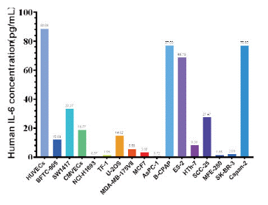
Fig. 2 The presentation of cytokine assay data in various common cell lines. (The data are provided by Elabscience®)
05 Cytokine detection by Flow cytometry and intracellular cytokine staining
While ELISA measures cytokines in solution, flow cytometry enables the detection of cytokines at the single-cell level and, crucially, identifies which cell subset produces them. Intracellular cytokine staining is the cornerstone of this approach: cells are stimulated, treated with a protein transport inhibitor such as brefeldin A or monensin to retain cytokines within the Golgi, fixed, permeabilized, and stained with fluorochrome-conjugated antibodies before analysis by cytokine flow cytometry. This technique was used to show that PMA/ionomycin-stimulated invariant natural killer T (iNKT) cells produce IFN-γ, IL-4, and IL-17, and that loss of the RNA- and DNA-binding protein YB-1 selectively abolishes IL-17-producing iNKT17 cells[19]. A direct comparison of intracellular cytokine staining with ELISPOT for cytomegalovirus-specific T cells found that the two assays correlate well (r = 0.77 for whole-virus stimulation) but are not interchangeable, and that only flow cytometry can resolve the cell source of IFN-γ among CD3+, CD4+, CD8+, and NK cells[20]. In sepsis-related liver injury, intracellular cytokine staining revealed that mucosa-associated invariant T (MAIT) cells shift toward a Th17-like profile with elevated granzyme B, TNF-α, and IL-17A secretion, while multiplex bead-based immunoassays quantified serum IL-1β, IL-6, IL-17A, IFN-γ, and TNF-α[21]. Similarly, intracellular staining for IFN-γ and granzyme B demonstrated that membrane-bound CD100 provides an intrinsic co-stimulatory signal required for the activation and effector differentiation of hepatitis B virus-specific CD8+ T cells[22]. More recently, intracellular CITE-seq approaches such as CIPHER-seq have combined intracellular protein and transcriptome profiling in single cells, revealing that mRNA abundance is an unreliable surrogate for intracellular cytokine protein levels and that coordinated IFN-γ and TNF-α induction expands polyfunctional cells[23]. Together, these methods make flow cytometry an indispensable tool for dissecting the cellular sources and functional states of cytokine-producing cells in inflammatory and infectious diseases.
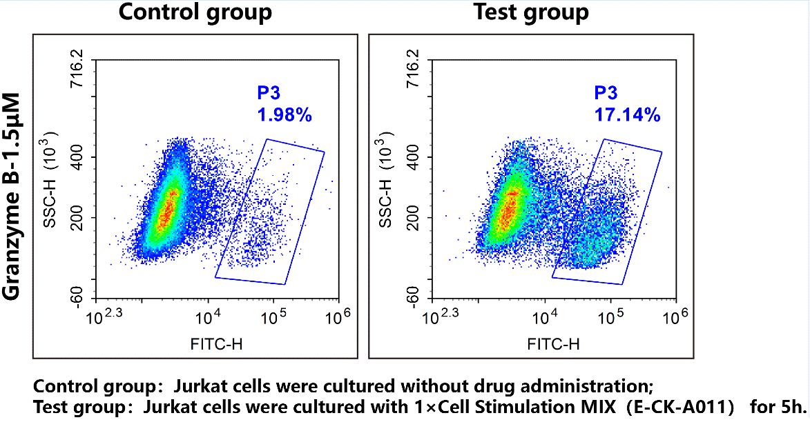
Fig. 3 Jurkat cells were stained with Human Granzyme B Activity Detection Substrates (Green, E-CK-A480) and analyzed by flow cytometry. (The data are provided by Elabscience®)
06 Cytokine changes in the tumor microenvironment and cancer-associated inflammation
The tumor microenvironment (TME) is shaped by a complex network of cytokines that can either promote or restrain cancer, and chronic inflammation is now recognized as a hallmark of malignancy. Pro-inflammatory cytokines such as IL-6, TGF-β, and TNF-α are overexpressed in tumor tissues and are strongly associated with tumor progression, metastasis, and treatment resistance[24]. The tumor-associated microbiome amplifies these circuits: microbial components activate TLR/NF-κB signaling to induce TNF-α, IL-6, and IL-8, and Fusobacterium nucleatum regulates CCL20 expression and, during PD-L1 blockade, activates STING/NF-κB to upregulate PD-L1 and enhance checkpoint responsiveness[24]. Chronic inflammation also drives cancer through the senescence-associated secretory phenotype (SASP), in which senescent immune cells secrete IL-6, TNF-α, IL-10, and TGF-β to sustain an immunosuppressive and tumor-promoting milieu, a process termed "inflammaging" that is particularly relevant to aging-associated colorectal cancer[25]. In head and neck squamous cell carcinoma, inflammation re-educates immune cells toward pro-tumor phenotypes, including tumor-associated macrophages, regulatory T cells, and exhausted T cells, creating a tolerogenic environment via IL-10, TGF-β, and PD-L1 upregulation that correlates with poor responses to PD-1/PD-L1 blockade[26]. Obesity-associated adipose dysfunction further rewires the tumor stroma: dysfunctional adipocytes and activated myofibroblasts secrete chemokines such as CXCL10 and CXCL11, which promote prostate cancer migration through the CXCR3 axis[27]. In oral squamous cell carcinoma, the microbiome-inflammation-immune axis links oral dysbiosis to NF-κB, STAT3, and Raf-MAPK activation, driving an immunosuppressive TME and providing peripheral and salivary biomarkers for diagnosis[28]. Understanding these cytokine changes within the TME is therefore central to designing inflammation-targeted and immune-based cancer therapies[2,4].
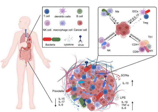
Fig. 4 The role of microbes in tumor. Modulation of inflammation and immune response induced by microbial infection in the TME which influences tumor progression by modulating immune cell and inflammatory mediator production and metabolites produced by microbes[24].
In summary, inflammation is driven by a coordinated network of pro inflammatory cytokines, chief among them IL-1β, IL-6, and TNF-α, that act through conserved NF-κB, MAPK, and JAK/STAT signaling pathways to propagate disease across metabolic, neurological, and malignant settings. Because these mediators are both biomarkers of disease activity and central nodes of immune activation, their reliable measurement is essential. The cytokine elisa assay and multiplex cytokine assay provide sensitive, quantitative detection of secreted cytokines such as IL-1β and IL-6, while intracellular cytokine staining and cytokine flow cytometry resolve the cellular sources and functional states of cytokine-producing cells. Within the tumor microenvironment, cytokine changes and cancer-associated inflammation shape immune escape and therapeutic responses, underscoring the value of inflammation-targeted strategies. Together, a deeper understanding of which cytokines drive inflammation and how they are measured will accelerate the development of precision diagnostics and therapies for inflammatory and malignant diseases.
Elabscience® Quick Overview of Popular Products:
Table 1. Research Tools for Detection
|
Cat. No. |
Product Name |
|
E-EL-H0149 |
Human IL-1β(Interleukin 1 Beta) ELISA Kit |
|
E-EL-H0109 |
Human TNF-α(Tumor Necrosis Factor Alpha) ELISA Kit |
|
E-EL-H1547 |
Human PD-L1(Programmed Cell Death Protein 1 Ligand 1) ELISA Kit |
|
E-EL-H2162 |
Human IDO(Indoleamine-2,3-Dioxygenase) ELISA Kit |
|
E-EL-H6154 |
Human IL-10(Interleukin 10)ELISA Kit |
|
E-EL-H6156 |
Human IL-6(Interleukin 6) ELISA Kit |
|
E-EL-0162 |
TGF-β1(Transforming Growth Factor Beta 1) ELISA Kit |
|
E-UNEL-H0340 |
Uncoated Human PD-1 (Programmed Cell Death Protein 1) ELISA Kit |
|
E-CK-A480 |
Human Granzyme B Activity Detection Substrate for Flow Cytometry |
|
XJM004 |
RAW 264.7 Polarized M1 Macrophage Induction and Identification Kit |
References:
[1] Feng Q, et al., Serum inflammatory cytokines in the progression of depression. Frontiers in Immunology. 2026. 17:1808989.
[2] Gebretsadik G, et al., The Role of Selected Bacteria in Breast Cancer Initiation and Development. International Journal of Molecular Sciences. 2026. 27(10):4585.
[3] Ramalingam PS, et al., Macrophage-derived long non-coding RNAs in cancer: pioneering targets for immune modulation and personalized therapy. Frontiers in Immunology. 2026. 17:1711686.
[4] González-Serrano S, et al., The immunometabolic nature of MASH: framing immune cells within metabolic dysfunction. Frontiers in Immunology. 2026. 17:1797949.
[5] Thanga Velu DPV, et al., Pro-Inflammatory Cytokines as Core Mediators of Colonic Epithelial Barrier Dysfunction: Roles of TNF-α, IFN-γ, IL-1β, and IL-6. International Journal of Molecular Sciences. 2026. 27(11):4722.
[6] Nurlybek T, et al., Immunomodulatory Mechanisms of Mesenchymal Stromal Cells: Cytokine Networks and Therapeutic Potential Across Immune-Mediated, Inflammatory, and Regenerative Disorders. Biology. 2026. 15(10):794.
[7] Mikołajczak K, et al., Neuroinflammation in Alzheimer's Disease (AD) and Glioblastoma (GBM): Shared Mechanisms and Therapeutic Insights. Cells. 2026. 15(12):1111.
[8] Zhang T, et al., Mechanisms and therapeutic prospects of DNA methylation-mucosal innate immunity crosstalk in inflammatory bowel disease. Frontiers in Immunology. 2026. 17:1877804.
[9] Huang J, et al., Luteolin and neuroinflammation: a multi-target therapeutic strategy for central nervous system disorders. Frontiers in Pharmacology. 2026. 17:1844585.
[10] Yang J, et al., Signaling pathways and targeted interventions for precancers. Signal Transduction and Targeted Therapy. 2026. 11(9).
[11] Hossain MDA, et al., Non-receptor tyrosine kinase signaling pathways and therapeutic implications. Signal Transduction and Targeted Therapy. 2026. 11(235).
[12] Wu C, et al., Inflammation in hepatocellular carcinoma progression: a "Single Wick, dual interwoven strands" regulatory framework. Frontiers in Immunology. 2026. 17:1822039.
[13] Azhar M, et al., Nanocarrier-mediated targeting of NF-κB and JAK/STAT signaling pathways in hepatocellular carcinoma: mechanisms and therapeutic strategies. Journal of Experimental & Clinical Cancer Research. 2026. 45(140).
[14] Zhu SQ, et al., Performance of Tai Chi Chuan and NF-κB-driven proinflammatory gene expression (IL-6, IL-1β, TNF-α) in adult chronic disease patients: a systematic review and meta-analysis of randomized controlled trials. Frontiers in Immunology. 2026. 17:1702830.
[15] Mukherjee S, et al., CFIm25-dependent alternative polyadenylation in AKT2 mRNA programs macrophage polarization. iScience. 2026. 29(6):115791.
[16] Stanisic T, et al., Optimization of protein extraction from skin tape strips for biomarker assessment. Scientific Reports. 2026. 16:44234.
[17] Lee KH, et al., Bioprocessed Black Rice Bran and Balloon Flower Root Cooperatively Regulate IgE, Epithelial Signaling, and Th1/Th2 Balance to Induce Therapeutic Response in a Mouse Model of Atopic Dermatitis. International Journal of Molecular Sciences. 2026. 27(6):2691.
[18] Heinemann N, et al., Plant extract mixture shows anti-inflammatory and barrier-strengthening effects and activates aryl hydrocarbon receptor in a 2D psoriasis model. Scientific Reports. 2026. 16:13638.
[19] Schulze S, et al., The RNA- and DNA-Binding Protein Y-Box Binding Protein 1 (YB-1) Regulates iNKT Cell Development. European Journal of Immunology. 2026. 56:e70201.
[20] Klinmalai C, et al., Comparison between enzyme-linked immunospot assay and intracellular cytokine flow cytometry assay of cytomegalovirus-specific T-cell response in healthy participants. PLoS One. 2026. 21(6):e0349292.
[21] Bu W, et al., Protective role of mucosa-associated invariant T cells in sepsis-related liver injury. Frontiers in Immunology. 2026. 17:1779656.
[22] Zhao M, et al., Cell-autonomous co-stimulatory function of membrane-bound CD100 promotes activation and differentiation of HBcAg-specific CD8+ T cells. Frontiers in Immunology. 2026. 17:1826036.
[23] Bhalgat A, et al., CIPHER-seq enables intracellular multimodal profiling of cytokine responses in single immune cells. Scientific Reports. 2026. 16:9693.
[24] Li Y, et al., The interaction between microbes and cytokines in cancer: unraveling the underlying effects. Journal of Translational Medicine. 2026. 24:740.
[25] Zaongo SD, et al., Bridging aging and colorectal cancer: synergistic roles of inflammaging and immunosenescence. Frontiers in Immunology. 2026. 17:1792954.
[26] Han Y, et al., Inflammation as a master regulator of immunotherapy response in head and neck squamous cell carcinoma: from malignant transformation to ecology-aware precision combinations. Frontiers in Immunology. 2026. 17:1798938.
[27] Takkar S, et al., Adipose Dysfunction Caused by Obesity and Radiation Therapy Rewires the Prostate Stroma Toward Tumor Progression. Cells. 2026. 15(15):1387.
[28] Wang Z, et al., The microbiome-inflammation-immune axis in oral squamous cell carcinoma: from mechanistic insights to therapeutic perspectives. Frontiers in Immunology. 2026. 17:1842459.

