

Antibodies that recognize their targets with exquisite specificity, coupled to enzymes that amplify a single binding event into a measurable signal, form the conceptual core of the most widely deployed diagnostic platform in modern medicine. Conceived in 1971 when the groups of Engvall and Perlmann and of Schuurs and van Weemen replaced the radioactive label of radioimmunoassay with an enzyme reporter, the enzyme-linked immunosorbent assay (ELISA) freed clinical laboratories from radiation hazards and dedicated counting facilities while preserving the sensitivity of the antigen-antibody reaction[1]. Today, fully automated instruments in medical laboratories around the world use this immunoassay principle with an enzyme as the reporter label for routine measurement of innumerable analytes in patient samples.
This review article compares ELISA technology across five practical dimensions in medical diagnostics: its core principle and assay formats, serological detection of infectious disease biomarkers, measurement of hormones and endocrine biomarkers, quantification of low-abundance analytes in clinical samples, and multiplex extensions for simultaneous biomarker profiling. Each section draws on peer-reviewed analytical and clinical studies to give actionable guidance on when to rely on classical ELISA, when to demand ultrasensitive or multiplex formats, and how to validate the chosen approach in its intended matrix.
Table of Contents
1. ELISA technology principle in medical diagnostics
2. ELISA for infectious disease biomarker detection
3. ELISA detection of hormones and endocrine biomarkers
4. How does ELISA measure low-abundance biomarkers in clinical samples?
5. Multiplex ELISA for simultaneous biomarker profiling
6. Key takeaways
7. Related resources
01 ELISA technology principle in medical diagnostics
The elisa technology grew directly out of radioimmunoassay (RIA), first described by Berson and Yalow in 1960 for measuring endogenous plasma insulin, work for which Yalow later received the Nobel Prize[1]. Concern over radiation safety, radioactive waste, and the need for expensive counting equipment motivated the search for a nonisotopic reporter, and the objection that a bulky enzyme could not be attached to an antigen or antibody without blocking the immunochemical reaction was settled only by careful coupling experiments, notably the glutaraldehyde-mediated conjugation of alkaline phosphatase (EC 3.1.3.1), glucose oxidase, and horseradish peroxidase reported in the late 1960s[1]. Engvall and Perlmann published the first ELISA in 1971, demonstrating quantitative measurement of immunoglobulin G (IgG) in rabbit serum with alkaline phosphatase as the reporter, while van Weemen and Schuurs reported quantification of human chorionic gonadotropin in urine with peroxidase labeling[1].The general procedure has changed little since: antigen in fluid phase is immobilized, usually in 96-well microtiter plates, allowed to bind a specific antibody that is itself detected by a secondary enzyme-coupled antibody, and a chromogenic or fluorogenic substrate yields a color change or fluorescence from which quantitative or qualitative measures are read[2]. Fluorogenic substrates offer higher sensitivity and accurately report antigen concentration, whereas the enzyme-mediated color reaction will proceed indefinitely, so overdevelopment can inflate the signal and produce false-positive results[2]. Three format families adapt this core to different analytical problems: indirect ELISA, in which sample antigen is adsorbed and detected through a primary plus enzyme-conjugated secondary antibody, as used to measure ovalbumin-specific immunoglobulin E (IgE) after cutaneous allergen sensitization; sandwich ELISA, in which a capture antibody selects the analyte from complex mixtures, as used to quantify keratinocyte growth factor in sera from keloid and scleroderma patients; and competitive ELISA, in which sample antigen competes with plate-bound antigen for a limited primary antibody so that absence of color indicates presence of analyte, a design applied to human immunodeficiency virus (HIV) antibody screening[2]. Ready-to-use pin-based portable versions extend the same chemistry to large-scale screening in low-resource settings[2].
02 ELISA for infectious disease biomarker detection
Serological diagnosis of infection illustrates both the power and the boundaries of elisa applications. Conventional Epstein-Barr virus (EBV) serology relies on ELISA against a limited panel of whole-protein antigens, typically Epstein-Barr nuclear antigen 1 (EBNA-1), viral capsid antigen (VCA), and early antigen-diffuse (EA-D), which classify exposure and infection stage but aggregate antibody binding across each entire antigen into a single measurement[3]. A 108-peptide microarray spanning nine EBV proteins was therefore validated across 329 specimens collected as serum, ethylenediaminetetraacetic acid (EDTA) plasma, and Streck cell-free deoxyribonucleic acid (DNA) plasma, and composite antigen scores correlated with the corresponding ELISA measurements for EBNA-1, VCA-p18, and EA-D[3]. Signals from immunoglobulin G (IgG) were stable across collection matrices, whereas immunoglobulin M (IgM) was selectively attenuated in fixative-containing Streck plasma, a pre-analytical caveat for studies reusing remnant specimens when IgM is the readout of interest[3].Veterinary serology follows the same pairing of antibody detection with nucleic acid confirmation. In 201 dam-daughter pairs from five dairy herds, bovine leukemia virus (BLV) status was assessed by ELISA for anti-BLV antibodies alongside quantitative polymerase chain reaction (qPCR) for proviral deoxyribonucleic acid (DNA), with proviral DNA detected in 33 of 200 daughters (16.5%), and maternal ELISA positivity was associated with higher odds of daughter proviral detection (odds ratio 2.74, 95% confidence interval 1.10-6.83)[4]. The durability of antibody-based testing across species and matrices does not remove its principal limitation in acute human infection, timing. A Cochrane systematic review of 57 publications covering 54 study cohorts and 15,976 samples found that pooled sensitivity of antibody tests for SARS-CoV-2 was below 30.1% during the first week after symptom onset, rising to 72.2% (95% confidence interval 63.5-79.5) in days 8-14 and 91.4% (95% confidence interval 87.0-94.4) in days 15-21, reaching 96.0% (95% confidence interval 90.6-98.3) for immunoglobulin G/immunoglobulin M (IgG/IgM) at 21-35 days, while summary specificity exceeded 98%[5]. Antibody sensitivity in the first week is therefore too low for a primary diagnostic role in coronavirus disease 2019 (COVID-19), although serology usefully complements direct detection later or documents previous infection[5].
03 ELISA detection of hormones and endocrine biomarkers
Endocrine diagnosis shows where the classical elisa assay excels and where it fails. In a prospective cohort of adults admitted to an emergency department with mild traumatic brain injury (mTBI), pituitary and thyroid hormones were measured by chemiluminescence immunoassay (CLIA) alongside neuron-specific enolase (NSE) at admission and after 3, 6, and 12 months, and higher NSE levels were associated with lower free triiodothyronine (fT3) at 3 months post injury, with no significant association at 6 or 12 months[6]. The transient coupling of a neuronal injury marker with a thyroid hormone readout supports multi-marker monitoring of post-traumatic neuroendocrine responses rather than reliance on any single analyte[6].
The harder lesson comes from testosterone, where the analyte itself exposes assay limits. Total testosterone (TT) in plasma spans three orders of magnitude with age, gender, and disease, only 1-3% circulates free of plasma proteins, structurally similar steroids interfere, and no universally recognized calibrating standard exists[7]. High-throughput direct immunoassays performed on whole serum or plasma are, for the most part, too insensitive and inaccurate to measure TT in women and children, whereas labor-intensive methods incorporating extraction and chromatography remove interfering proteins, separate cross-reacting steroids, and permit larger serum aliquots for sensitivity[7]. College of American Pathologists (CAP) proficiency data illustrate the cost of this gap: for a normal female-range sample, coefficients of variation within the same methodology across laboratories ranged from 13% to 32%, and results for the same sample varied 2- to 6-fold, severely compromising clinical usefulness at low concentrations[7]. Mass spectrometry (MS), particularly isotope-dilution gas chromatography-MS and liquid chromatography tandem MS (LC/MS-MS), both identifies and quantifies the analyte and serves as the accuracy anchor, but still requires validation for accuracy, sensitivity, and precision[7]. The Endocrine Society position statement therefore concludes that proficiency testing should assess the ability to measure samples of known concentration accurately and precisely rather than mere agreement with peers using the same method, and that normative values for total and free testosterone should be established for both genders and children once such standardization is in place[7].
04 How does ELISA measure low-abundance biomarkers in clinical samples?
Conventional immunoassays typically measure proteins above 10-12 M, yet most clinically important proteins in cancer, neurological disorders, and early infection circulate between 10-16 and 10-12 M, so the elisa test had to be reinvented at the single-molecule level[8]. In single-molecule array (Simoa) digital ELISA, sandwich immunocomplexes are formed on 2.7-micrometer paramagnetic beads, labeled with an enzyme reporter exactly as in bead-based ELISA, and then isolated in arrays of femtoliter reaction chambers holding at most one bead each, so that at low protein-to-bead ratios the fraction of labeled beads follows a Poisson distribution and fluorescence imaging counts single molecules[8]. The approach detected as few as 10-20 enzyme-labeled complexes in 100 microliters of sample (approximately 10-19 M) and routinely measured serum proteins below 10-15 M, including prostate-specific antigen (PSA) at 14 fg/mL (0.4 fM) in post-prostatectomy sera[8].
Clinical validations confirm the leap in practice. A Simoa assay for human interferon-alpha (IFN-alpha) built from high-affinity autoantibodies of autoimmune polyendocrinopathy syndrome type 1 (APS1) patients reached a limit of detection of 0.69 fg/mL with detection of all 13 IFN-alpha subtypes and no cross-reactivity, whereas commercial ELISAs for IFN-alpha subtypes stall at 1.95, 12.5, and 12.5 pg/mL, often insufficient for direct protein measurement in biological samples[9]. The same chemistry counts single immunocomplexes in 50,000 wells of 46 femtoliters each and reports average enzymes per bead (AEB), a format also applied to tumor necrosis factor-alpha (TNF-alpha) in Crohn disease, neurofilament light in brain pathologies, and human immunodeficiency virus (HIV) reservoir characterization[9]. For Amyloid-Beta 1-42 peptide (Abeta1-42) in plasma, a Simoa digital ELISA using antibodies 3D6 and 21F12 achieved a limit of detection of 0.3 pg/mL and a lower limit of quantification of 2.8 pg/mL with intra- and inter-assay coefficients of variation at or below 10%, quantified Abeta1-42 in all 84 clinical samples, and captured a rapid reduction within 1 hour of beta-site amyloid precursor protein cleaving enzyme 1 (BACE1) inhibitor treatment with a dose-dependent decrease over time[10]. Repurposed beyond biomarkers, Simoa screening of a transcription factor library detected the aristaless-related homeobox (ARX) interaction with TCF7L2 at a fold change of 2.88 (p less than 0.0001), where mass spectrometry and yeast two-hybrid screens had found nothing[11].
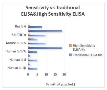
Fig. 1 Sensitivity comparison between conventional and high-sensitivity cytokine assay kits. (The data are provided by Elabscience®)
Assay Used for This Experiment:
Rat TNF-α(Tumor Necrosis Factor Alpha) ELISA Kit
Cat. No: E-EL-R2856
Sample Type: Serum
Detection Range: 15.63-1000 pg/mL
Sensitivity: 9.38 pg/mL
Detection Instrument: Microplate reader
Explore the Rat TNF-α ELISA Kit Used in 1279 Publications→
05 Multiplex ELISA for simultaneous biomarker profiling
When clinical questions involve many mediators at once, running one ELISA per target becomes impractical, and multiplex elisa formats parallelize the measurement. Discovery-phase proteomics first nominates candidates without bias, research-phase immunoassays including ELISA, electrochemiluminescence immunoassays, microfluidics, and single-molecule arrays validate them in characterized cohorts, and fully automated platforms such as Lumipulse and Elecsys carry approved cerebrospinal fluid and, more recently, blood-based Alzheimer biomarkers into clinical use, a fit-for-purpose pipeline in which each technology occupies the phase matching its analytical maturity[12]. Standardization, inter-platform variability, and demonstration of clinical utility remain the gating challenges, since only a limited number of candidates survive translation despite rapidly expanding discovery lists[12].
Whether multiplex readouts can substitute for single-analyte ELISA hinges on antibody reagents rather than on the platform itself. Multiplex bead array assays in automated 96-well format quantify many analytes in small volumes with statistical rigor from numerous beads per ligand, and comparisons show that matched monoclonal antibody clones for capture and reporting are pivotal: with reagents from the same vendor, a six-plex assay for interleukin-2 (IL-2), IL-4, IL-10, IL-12, interferon-gamma (IFN-gamma), and tumor necrosis factor-alpha (TNF-alpha) correlated with ELISA at coefficients from 0.92 (IL-2) to 1.0 (IL-10), whereas comparisons across vendors and undisclosed antibody pairs varied widely[13]. The dedicated 96-specimen validation of a nine-cytokine Luminex panel (IL-1beta, IL-4, IL-5, IL-6, IL-10, IL-12p70, IL-13, IFN-gamma, TNF-alpha) against commercial sandwich ELISA kits in culture supernatants from ten subjects found significant correlations for six of the nine analytes (all p less than 0.0001 except IL-12), with IL-13 at 0.81 and poor agreement for IL-12p70, confirming that each analyte in a multiplex panel still requires individual validation[14]. Endocrine multiplex menus already cover adrenocorticotropic hormone (ACTH), thyroid-stimulating hormone (TSH), triiodothyronine (T3), and thyroxine (T4), extending the same parallel logic to hormonal profiling[13].
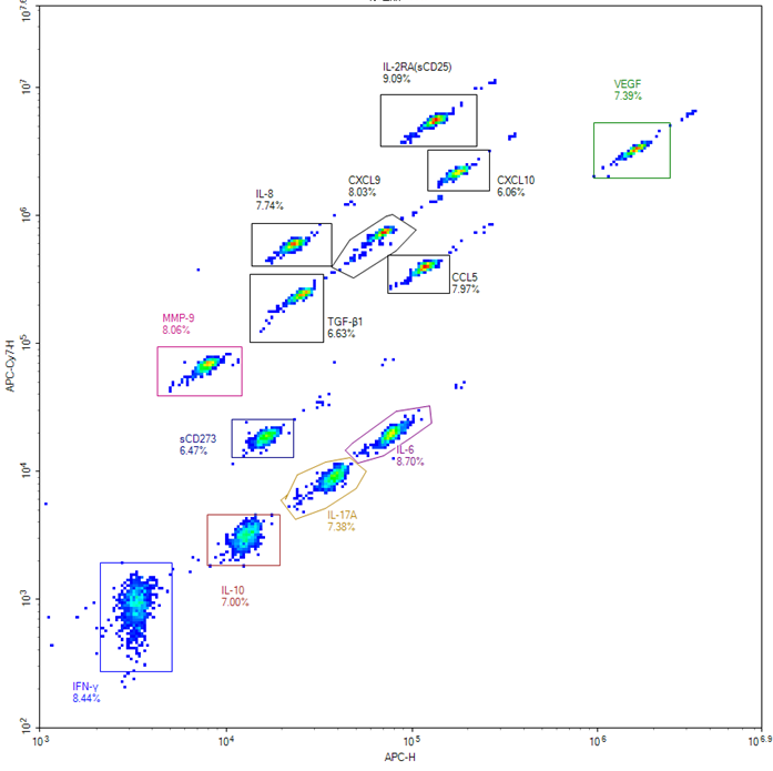
Fig. 2 Detection results of the 13 factors. (The data are provided by Elabscience®)
Assay Used for This Experiment:
Aptplex™ Human Tumor 13-Plex Panel
Cat. No: MPA010
Sample Type: Serum
Detection Instrument: Flow cytometer
Explore the Aptplex™ Human Tumor 13-Plex Panel→
In summary, ELISA remains the quantitative backbone of medical diagnostics because it converts specific antigen-antibody recognition into a robust, scalable signal, from classical 96-well formats rooted in the elisa technology of Engvall, Perlmann, Schuurs, and van Weemen to single-molecule arrays that count individual immunocomplexes. Across elisa applications, serology classifies exposure and infection stage for agents from Epstein-Barr virus to SARS-CoV-2 provided sampling respects antibody kinetics, endocrine measurement succeeds only with validated, standardized assays as the testosterone experience shows, and the modern elisa assay reaches attomolar concentrations that make plasma pharmacodynamic monitoring feasible. For laboratories choosing platforms, the elisa test in single-analyte form answers how much of one mediator is present, while multiplex elisa answers how mediator patterns covary across cohorts at lower per-analyte cost and sample volume, with the caveat that multiplex panels require analyte-by-analyte validation. Reliable quantification with a fit-for-purpose testosterone elisa kit or any validated kit underpins both biomarker detection and clinical sample analysis: validate each target in its intended matrix, standardize calibration and controls, and combine frequency, concentration, and phenotypic readouts whenever the clinical question spans humoral and cellular immunity.
Elabscience Quick Overview of Popular Products
Table 1. Research Tools for Biomarker Detection
|
Cat. No. |
Product Name |
|
E-EL-H0169 |
Human IgG(Immunoglobulin G) ELISA Kit |
|
E-EL-H6104 |
Human IgE(Immunoglobulin E) ELISA Kit |
|
E-EL-E605 |
SARS-CoV-2 Spike Protein S1 RBD ELISA Kit |
|
E-EL-0079 |
fT3(Free Triiodothyronine) ELISA Kit |
|
E-OSEL-H0007 |
QuicKey Pro Human T(Testosterone) ELISA Kit |
|
E-EL-H0091 |
Human PSA(Prostate Specific Antigen) ELISA Kit |
|
E-EL-H0109 |
Human TNF-α (Tumor Necrosis Factor Alpha) ELISA Kit |
|
E-EL-H0543 |
Human Aβ1-42(Amyloid Beta 1-42) ELISA Kit |
|
E-HSEL-H0007 |
High Sensitivity Human IFN-γ (Interferon Gamma) ELISA Kit |
|
E-EL-0100 |
T4(Thyroxine) ELISA Kit |
06 Key takeaways
Conventional ELISA and advanced immunoassay modalities should not be regarded as mutually exclusive platforms; they address distinct diagnostic objectives.
● Classical ELISA converts specific antigen–antibody recognition into a detectable enzyme-mediated chromogenic or fluorogenic signal, thereby enabling bulk quantification of soluble analytes in clinical specimens.
● Indirect, sandwich, and competitive configurations are selected according to analyte structure, sample matrix, and assay target (native antigen, specific antibody, or low-abundance cross-reacting compound).
● Conventional ELISA generally exhibits a detection limit near 10⁻¹² M, whereas single-molecule digital ELISA (Simoa) achieves attomolar sensitivity, permitting quantification of scarce cancer, neurodegenerative, and early-stage infectious biomarkers.
● Serological ELISA retrospectively characterizes prior exposure and establishes infection stage; however, its diagnostic sensitivity during acute infection is governed by the kinetics of antibody seroconversion, and it cannot substitute for direct nucleic-acid detection within the first week of symptom onset.
● Endocrine and steroid measurements mandate methodologically validated and standardized assays; high-throughput direct immunoassays may lack requisite accuracy at low concentrations, whereas extraction and chromatography coupled to mass spectrometry serve as the reference accuracy benchmark.
● Multiplex bead arrays enable simultaneous quantification of numerous mediators with reduced sample consumption and lower per-analyte cost, yet capture/detection antibody performance is analyte-dependent, and each target requires independent validation against single-analyte ELISA in the intended biological matrix.
● Clinical interpretation necessitates fit-for-purpose assay selection, matrix-specific validation, standardized calibration and quality controls, and orthogonal confirmation whenever diagnostic conclusions carry therapeutic or prognostic implications.
The recommended workflow is therefore: Define the clinical question → Select the primary assay modality → Incorporate a complementary or orthogonal reference method → Interpret measurements within the appropriate specimen matrix → Conduct cross-method validation and mechanistic confirmation as warranted
07 Related resources
● Explore ELISA principles, protocols, and applications to support reliable immunoassay design and data analysis.
Download Your Free ELISA Technology Guide →
● Explore the principles, workflow, and applications of ELISpot technology for immune cell analysis and cytokine detection.
Read A Window into the World of Immunity: Detailed Explanation of ELISpot Technology →
● Understand the key differences between ELISpot and ELISA in cytokine detection, including their principles, sensitivity, and research applications.
Read ELISpot vs ELISA for Sensitive Cytokine Detection and Immune Response Analysis →
References:
[1] Lequin RM, et al. Enzyme immunoassay (EIA)/enzyme-linked immunosorbent assay (ELISA). Clin Chem. 2005;51(12):2415-2418.
[2] Gan SD, et al. Enzyme immunoassay and enzyme-linked immunosorbent assay. J Invest Dermatol. 2013;133(9):e12.
[3] Peterson M, et al. Analytical validation of a multiplexed peptide microarray for multi-antigen Epstein-Barr virus serological profiling across diverse clinical specimens. Front Immunol. 2026;17:1919983.
[4] Sokacz ME, et al. Maternal BLV infection markers and BoLA-DRB3 polymorphism are associated with BLV outcomes in dairy youngstock. Int J Mol Sci. 2026;27(19):8627.
[5] Deeks JJ, et al. Antibody tests for identification of current and past infection with SARS-CoV-2. Cochrane Database Syst Rev. 2020;6:CD013652.
[6] Kalas M, et al. Transient association between elevated NSE and reduced fT3 levels following mild traumatic brain injury: a 12-month prospective pilot study. J Clin Med. 2026;15(14):5676.
[7] Rosner W, et al. Utility, limitations, and pitfalls in measuring testosterone: an Endocrine Society position statement. J Clin Endocrinol Metab. 2007;92(2):405-413.
[8] Rissin DM, et al. Single-molecule enzyme-linked immunosorbent assay detects serum proteins at subfemtomolar concentrations. Nat Biotechnol. 2010;28(6):595-599.
[9] Llibre A, et al. Development and validation of an ultrasensitive single molecule array digital enzyme-linked immunosorbent assay for human interferon-alpha. J Vis Exp. 2018;(136):e57421.
[10] Song L, et al. A digital enzyme-linked immunosorbent assay for ultrasensitive measurement of amyloid-beta 1-42 peptide in human plasma with utility for studies of Alzheimer disease therapeutics. Alzheimers Res Ther. 2016;8:58.
[11] Lim Y, et al. Single-molecule array (Simoa) technology as a highly sensitive screening tool for protein-protein interactions. ACS Sens. 2025;10(11):8256-8261.
[12] Agnello L, et al. Analytical methods for fluid biomarkers in Alzheimer disease from discovery to clinical implementation. Int J Mol Sci. 2026;27:4518.
[13] Elshal MF, et al. Multiplex bead array assays: performance evaluation and comparison of sensitivity to ELISA. Methods. 2006;38(4):317-323.
[14] duPont NC, et al. Validation and comparison of Luminex multiplex cytokine analysis kits with ELISA: determinations of a panel of nine cytokines in clinical sample culture supernatants. J Reprod Immunol. 2005;66(2):175-191.

