

Chimeric antigen receptor (CAR) T cell therapy has reshaped the oncology landscape, with six CD19-directed products now approved by the U.S. Food and Drug Administration. As the field expands from ex vivo manufactured products toward in vivo CAR-T strategies, the demand for robust, reproducible characterization tools grows in parallel. Flow cytometry antibodies sit at the center of this effort: they enable detection of CAR surface expression, resolve memory and exhaustion phenotypes, quantify cytotoxic function, and provide the quality control framework that underpins both product release and in vivo pharmacokinetic monitoring.
Table of Contents
1. Detecting CAR expression using Anti-Idiotype and Protein L flow cytometry antibodies
2. Phenotyping memory CAR-T cells with CD45RA, CCR7, and CD62L markers
3. Monitoring exhaustion markers in CAR-T cells during chronic antigen exposure
4. Tracking in vivo CAR-T generation following LNP-mediated gene delivery
5. Assessing CAR-T cell cytotoxicity through degranulation marker CD107a
6. Quality control strategies for in vivo CAR-T programs using flow cytometry antibodies
01 Detecting CAR expression using Anti-Idiotype and Protein L flow cytometry antibodies
Accurate CAR expression detection is the first critical step in CAR-T development, directly affecting manufacturing efficiency and product release. Anti-idiotype antibodies targeting the scFv unique epitope achieve a median background of 0.02%, outperforming Protein L (0.65 to 0.80%)[1]. Protein L binds the immunoglobulin kappa light chain and provides a construct-independent detection method suitable for novel construct screening[2]. Anti-linker monoclonal antibodies E7O2V (targeting G4S linker and E3U7Q (targeting Whitlow 218 linker, present in approximately 90% of clinical constructs) enable universal, construct-agnostic CAR detection[3]. The standard flow cytometry antibodies staining protocol involves incubating transduced T cells with anti-idiotype antibody or biotinylated Protein L, followed by fluorophore-conjugated streptavidin. Isotype control flow cytometry and untransduced T cells serve as negative references. Selecting the appropriate isotype control matched by host species, immunoglobulin subclass, fluorophore, and concentration is essential for distinguishing specific CAR staining from nonspecific Fc receptor binding, which is elevated in activated CAR-T cells.
Table 1. Comparison of common CAR detection reagents by flow cytometry
|
Detection reagent |
Target |
Background (median) |
Construct specificity |
Best application |
|
Anti-idiotype antibody |
scFv unique epitope |
~0.02% |
Construct-specific |
Clinical pharmacokinetic monitoring |
|
Protein L |
Ig kappa light chain |
~0.73% |
Broad (kappa-containing scFv) |
Novel construct screening |
|
Anti-FMC63 |
FMC63 scFv (CD19 CAR) |
<0.1% |
FMC63-derived constructs |
Novel construct screening CD19 CAR-T tracking |
|
Anti-G4S linker |
(GGGGS)4 scFv linker |
<0.5% |
Broad (G4S-linked scFv) |
Universal CAR detection, multispecific programs |
|
Anti-Whitlow 218 linker |
Whitlow 218 peptide |
<0.5% |
Broad (Whitlow-linked scFv, ~90% of clinical constructs) |
Universal CAR detection, translational studies |
Table 2. Recommended Antibodies for CAR Detection
|
Detection reagent |
Clone |
Cat. No. |
Notes |
|
Anti-FMC63 (CD19 CAR) |
DA001 |
AN010000P |
FMC63-derived CAR constructs |
|
Anti-G4S linker |
DA010 |
AN011300P |
Universal G4S-linked CAR detection |
|
Anti-Whitlow 218 linker |
DA011 |
AN009990P |
~90% of clinical CAR constructs |
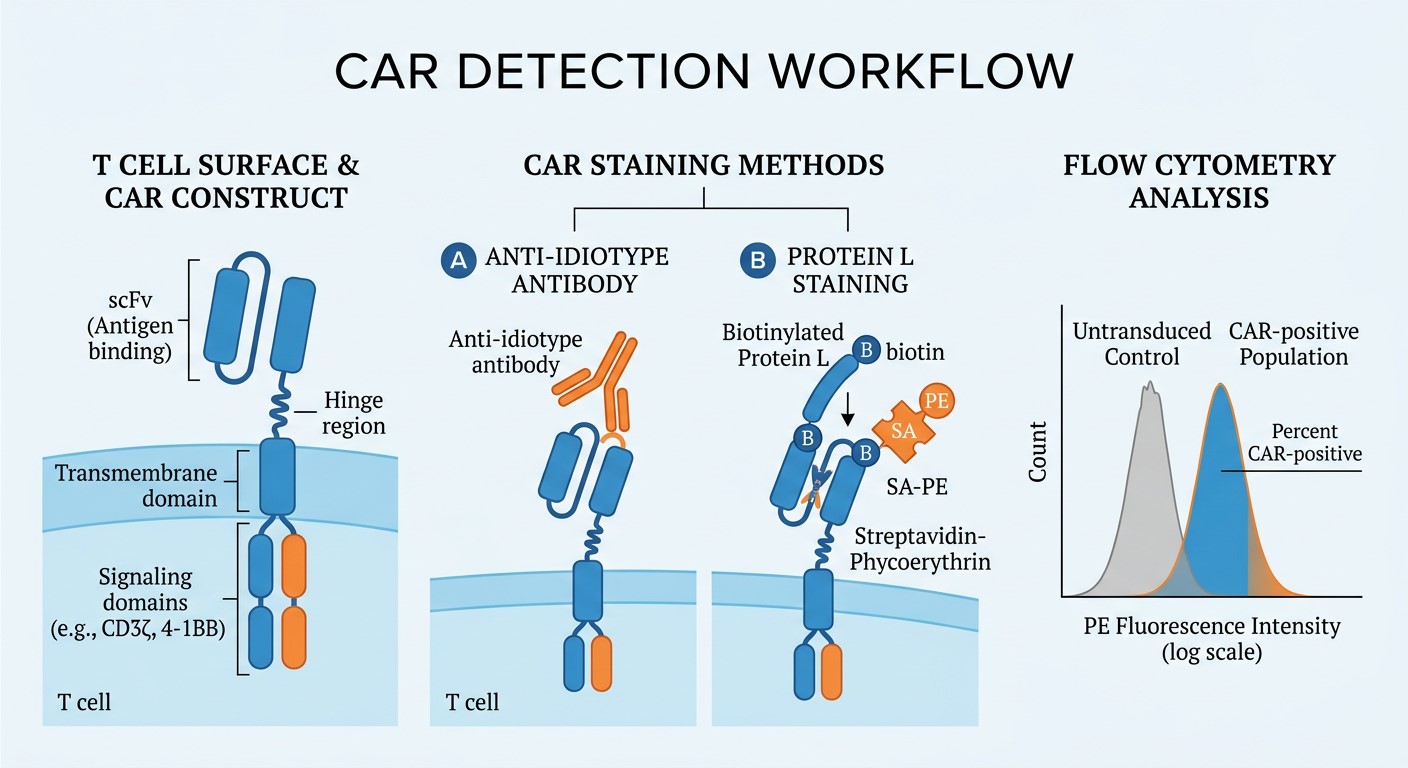
Fig. 1 Schematic of the CAR detection workflow by flow cytometry. Transduced T cells are stained with anti-idiotype antibody or biotinylated Protein L followed by fluorophore-conjugated streptavidin. Isotype controls and untransduced T cells serve as negative references.
02 Phenotyping memory CAR-T cells with CD45RA, CCR7, and CD62L markers
T cell phenotyping through flow cytometry antibodies has become indispensable for predicting CAR-T product potency. CD45RA, CCR7, CD62L and CD44 together resolve the full spectrum of T cell differentiation states. The CD44 CD62l axis is particularly informative: naive T cells (CD44lowCD62Lhigh), central memory (TCM, CD44highCD62Lhigh), effector memory (TEM, CD44highCD62Llow), and CD45RA-positive effector memory (TEMRA, CD44lowCD62Llow) subsets each carry distinct implications for in vivo persistence. Enrichment of TSCM and TCM populations correlates with long-term antitumor activity, and multiple CAR-T developers now optimize memory-enriched products based on this gating strategy. A 20-color full-spectrum flow cytometry panel incorporating CD45RA, CCR7, CD62L, CD95, CD25, and CD127 resolves all major T cell subsets in a single tube for standardized multicenter trials[4,5]. This approach allows researchers to assess product fitness before infusion and to track differentiation drift during manufacturing. The FACS staining protocol for memory phenotyping requires Fc receptor blocking and viability dye inclusion to ensure accurate subset resolution in activated CAR-T samples.
_.png)
Fig. 2 Purified Anti-Human CD62L Antibody (Cat. No.E-AB-F1051M, Clone DREG56).
%20Antibody%20(Cat_%20No_E-AB-F1159M,%20Clone%20G043H7)_.png)
Fig. 3 Purified Anti-Human CCR7 (CD197) Antibody (Cat. No.E-AB-F1159M, Clone G043H7).
Table 3. Recommended Antibodies for Memory CAR-T Cell Phenotyping
|
Marker |
Clone |
Cat. No. |
Purpose |
|
CD45RA |
HI100 |
E-AB-F1052A |
Naive/TEMRA identification |
|
CCR7 |
G043H7 |
E-AB-F11590 |
Lymph node homing, TCM gating |
|
CD62L |
DREG-56 |
E-AB-F10510 |
Lymph node homing, memory gating |
|
CD44 |
IM7 |
E-AB-F11000 |
Activation and memory marker |
|
CD3 |
UCHT1 |
E-AB-F12300 |
T cell lineage gating |
|
CD4 |
RPA-T4 |
E-AB-F11090 |
Helper T subset |
|
CD8 |
SK1 |
AN003370P |
Cytotoxic T subset |
03 Monitoring exhaustion markers in CAR-T cells during chronic antigen exposure
CAR-T cell exhaustion remains one of the most actively investigated barriers to durable remission. The anti PD-1 antibody (clone EH12.2H7) serves as the sentinel tool for tracking T cell exhaustion in CAR-T research. PD-1, TIM-3, and LAG-3 upregulation correlates inversely with cytotoxicity and cytokine secretion[6]. In a NALM6-Luc xenograft model, PD-1 and LAG-3 rose continuously while CD25 and 4-1BB peaked at day 30 then declined, marking progressive exhaustion coinciding with tumor outgrowth[7]. A multiplexed iQue assay confirmed persistently high PD-1, LAG-3, and TIM-3 with markedly lower IFN-γ and TNF secretion in exhausted T cells[8].The standard CAR-T cell exhaustion markers panel requires longitudinal sampling at pre-infusion baseline, early expansion (day 7 to 14), peak proliferation (day 21 to 30), and late persistence (day 60+). Rising PD-1 at late time points, particularly accompanied by TIM-3 and LAG-3 co-expression, signals progressive functional exhaustion. A proper FACS staining protocol with matched isotype control flow cytometry for each exhaustion marker ensures reproducible gating across serial time points.
Table 4. Purified Antibodies for CAR-T Cell Exhaustion Markers
|
Marker |
Clone |
Cat. No. |
Purpose |
|
PD-1 |
EH12.2H7 |
E-AB-F12290 |
Inhibitory receptor, exhaustion hallmark |
|
TIM-3 |
F38-2E2 |
AN009710 |
Inhibitory receptor, late exhaustion |
|
LAG-3 |
11C3C65 |
AN010470P |
Inhibitory receptor, exhaustion hallmark |
04 Tracking in vivo CAR-T generation following LNP-mediated gene delivery
In vivo CAR-T strategies deliver CAR-encoding mRNA to T cells via targeted lipid nanoparticles (LNPs), bypassing the complex and costly ex vivo manufacturing process[9]. In humanized mouse models (NSG, NOG, BRGS), chimerism is confirmed by co-staining hCD45 (HI30) and mCD45 (30-F11) with at least 25% hCD45-positive cells as the enrollment criterion. CD3-targeted LNPs achieve T cell transfection with surface CAR detectable within 24 hours[10], and anti-CD5-targeted LNPs yield 11.6% CAR-positive T cells in lymphoid organs[11]. Flow cytometry antibodies are essential for this tracking workflow: panels must include CD3, CD4, CD8, a CAR detection reagent or epitope tag antibody, and a viability dye. The FACS staining protocol for in vivo CAR-T monitoring requires careful panel design to accommodate the limited blood volumes available from lymphodepleted animals, with viability and lineage markers prioritized at every time point. Tracking CAR-positive T cell frequency and phenotype over time provides critical data on in vivo transfection efficiency, CAR-T expansion kinetics, and target cell depletion.
Table 5. Recommended Antibodies for In Vivo CAR-T Tracking
|
Marker |
Clone |
Cat. No. |
Purpose |
|
hCD45 |
HI30 |
E-AB-F1137A |
Human hematopoietic chimerism |
|
mCD45 |
30-F11 |
E-AB-F1136A |
Mouse hematopoietic reference |
|
CD3 |
UCHT1 |
E-AB-F12300 |
Human T cell lineage gating |
|
CD4 |
RPA-T4 |
E-AB-F11090 |
Helper T subset |
|
CD8 |
SK1 |
AN003370P |
Cytotoxic T subset |
|
CD19 |
HIB19 |
AN008610 |
B cell depletion readout |
05 Assessing CAR-T cell cytotoxicity through degranulation marker CD107a
The CD107a marker (LAMP-1) provides a direct readout of cytotoxic granule exocytosis and has become a standard method for evaluating CAR-T killing function[12]. The assay involves co-culturing CAR-T cells with antigen-positive target cells in the presence of anti-CD107a antibody and monensin for 4 to 6 hours, then quantifying CD107a-positive events within the CAR-positive CD8-positive T cell population. Combined CD107a and annexin V staining enables simultaneous readout of degranulation and target cell apoptosis[13]. Standard effector-to-target ratios are 1:1, 5:1, and 10:1, with antigen-negative target cells serving as specificity controls. The gating strategy proceeds through lymphocytes, singlets, live cells, CD3-positive, CD8-positive, and CAR-positive populations before assessing CD107a positivity. This approach is experimentally accessible and provides functional data that complements surface phenotype characterization, making it suitable for routine batch release testing of CAR-T products.
Table 6. Purified Antibodies for CAR-T Cell Cytotoxicity
|
Marker |
Clone |
Cat. No. |
Purpose |
|
CD107a |
H4A3 |
E-AB-F1149A |
Degranulation readout |
06 Quality control strategies for in vivo CAR-T programs using flow cytometry antibodies
In vivo CAR-T QC relies on post-administration peripheral blood monitoring rather than final-product release testing. Cai et al. established a cryopreserved CAR-T cell bank as flow cytometry QC standards: transduction efficiency and vector identity markers remained stable over one year, with post-thaw viability exceeding 50%[14], enabling inter-operator and inter-instrument comparability. Blache et al. demonstrated standardized 13-color panels using pre-formulated dry antibody formats for characterizing CAR-T persistence, activation, and cytotoxic molecule expression[5]. The UNITC consortium has emphasized standardizing QC procedures across academic production sites[15]. A practical QC panel includes viability dye, CD45/CD3/CD4/CD8, CAR detection reagent, CD45RA/CCR7/CD62L, and PD-1/TIM-3/LAG-3. For humanized mouse models, hCD45 and mCD45 staining should track lot-to-lot with a spiked PBMC:splenocyte reference standard (e.g., 50:50)
Table 7. Recommended flow cytometry QC panel for in vivo CAR-T monitoring
|
Category |
Markers |
Purpose |
Frequency |
|
Viability and lineage |
Live/dead dye, CD45, CD3, CD4, CD8 |
Population gating and viability |
Every time point |
|
CAR detection |
Anti-idiotype or Protein L |
Transduction efficiency |
Every time point |
|
Human chimerism |
hCD45 (HI30), mCD45 (30-F11) |
Engraftment validation (humanized models) |
Every sampling time point |
|
Memory phenotyping |
CD45RA, CCR7, CD62L, CD44 |
Differentiation state |
Pre-infusion and key milestones |
|
Exhaustion Isotype and FMO controls |
PD-1, TIM-3, LAG-3 Matched isotype antibodies |
Functional competence Gating reference |
Serial time points Per batch |
References:
[1] Schanda, N., Sauer, T., Kunz, A., Hückelhoven-Krauss, A., Neuber, B., Wang, L., Schmitt, M., & Schmitt, A. (2021). Sensitivity and Specificity of CD19.CAR-T Cell Detection by Flow Cytometry and PCR. Cells, 10(11), 3208.
[2] Zheng, Z., Chinnasamy, N., & Morgan, R. A. (2012). Protein L: a novel reagent for the detection of Chimeric Antigen Receptor (CAR) expression by flow cytometry. Journal of Translational Medicine, 10(1), 29.
[3] Singh, A., L'Heureux, S., Sinapius, R., Kvorjak, M., Wade, E., Lohmueller, J., Fisher, J., & Prioli, R. (2023). Generation and validation of anti-linker monoclonal antibodies for the detection of surface expressed scFv-based CARs (Abstract No. 898). Cancer Research, 83(7_Supplement).
[4] Vaughan, K., Muthusamy, S., Zhao, C., Carbajal, A., Fu, C., Dakappagari, N., & Alfonso, Z. (2024). A patient centric full spectrum flow cytometry assay for comprehensive phenotyping of CAR T cells in clinical trials (Abstract No. 6424). Cancer Research, 84(6_Supplement).
[5] Blache, U., Weiss, R., Boldt, A., Kapinsky, M., Blaudszun, A.-R., Quaiser, A., Pohl, A., Miloud, T., & Koehl, U. (2021). Advanced flow cytometry assays for immune monitoring of CAR-T cell applications. Frontiers in Immunology, 12.
[6] Zhang, Z., Liu, S., Zhang, B., Qiao, L., Zhang, Y., & Zhang, Y. (2020). T cell dysfunction and exhaustion in cancer. Frontiers in Cell and Developmental Biology, 8.
[7] D. W. Draper, D. Germain, S. Roys, O. Nelsonand S. Wise, “Preclinical assessment of chimeric antigen receptor (CAR) T persistence and functionality in the disseminated NALM6-Luc human B cell acute lymphoblastic leukemia (ALL) model”, Cancer Research, vol. 83, no. 7\_Supplement, p. 2749, 2023, doi: 10.1158/1538-7445.am2023-2749.
[8] Carter, M., Chauhan, S., O'Rourke, J., & Senutovitch, N. (2020). Development of a cells and bead-based, advanced flow cytometry assay for exhausted T cell quantitation. The Journal of Immunology, 204(1_Supplement), 86.29.
[9] V. K. Yadav, P. Yadav, S. Mallappaand P. Neeli, “In vivo mRNA-lipid nanoparticle CAR-T cell engineering: advances, challenges, and clinical translation”, Biomedicines, vol. 14, no. 6, p. 1276, 2026.
[10] M. M. Billingsley et al., “In vivo mRNA CAR T cell engineering via targeted ionizable lipid nanoparticles with extrahepatic tropism”, Small, vol. 20, no. 9, p. 2304378, 2023.
[11] N. Zhang et al., “In situ CAR T cell engineering using lipid nanoparticles for B cell lymphoma therapy”, Cancer Research, vol. 84, no. 6\_Supplement, p. 35, 2024.
[12] M. R. Betts et al., “Sensitive and viable identification of antigen-specific CD8+ T cells by a flow cytometric assay for degranulation”, Journal of Immunological Methods, vol. 281, no. 1–2, pp. 65–78, 2003.
[13] M. W. Burkett, K. A. Shafer-Weaver, S. Strobl, M. Baselerand A. Malyguine, “A novel flow cytometric assay for evaluating cell-mediated cytotoxicity”, Journal of Immunotherapy, vol. 28, no. 4, pp. 396–402, 2005.
[14] Cai, Y., Prochazkova, M., Jiang, C., Song, H. W., Jin, J., Moses, L., Gkitsas, N., Somerville, R. P., Highfill, S. L., Panch, S., Stroncek, D. F., & Jin, P. (2021). Establishment and validation of in-house cryopreserved CAR/TCR-T cell flow cytometry quality control. Journal of Translational Medicine, 19(1), 523.
[15] Marton, C., Clémenceau, B., Dachy, G., Demerle, C., Derenne, S., Ferrand, C., Giverne, C., Latouche, J. B., Lemée, L., Martinet, J., Bonig, H., Bensoussan, D., Chabannon, C., Köhl, U., Deschamps, M., De Vos, J., Diana, J. S., Dougé, A., Forcade, E., Galaine, J., Thiant, S., Galy, A., Larghero, J., Reppel, L., Viel, S., Boyer, O., & Yakoub-Agha, I. (2025). Harmonisation of quality control tests for academic production of CAR-T cells: a position paper from the WP-bioproduction of the UNITC consortium. Bone Marrow Transplantation, 60(9), 1209–1217.

