

Induced pluripotent stem cells have reshaped regenerative medicine by providing a scalable, ethically unencumbered source of therapeutically relevant cell types. Yet the journey from a somatic cell to a clinical-grade iPSC-derived product is long: reprogramming must be confirmed, pluripotency validated, differentiation efficiency quantified, safety risks mitigated, and functional potency verified. At every step, flow cytometry antibodies provide the analytical foundation. Elabscience® supplies a comprehensive portfolio of validated reagents purpose-built for the iPSC workflow, from pluripotency characterization through clinical release testing.
Table of Contents
1. Confirming iPSC pluripotency using OCT4, SOX2, TRA-1-60, and SSEA-4 markers
2. Detecting residual undifferentiated iPSCs to improve cell therapy safety
3. Monitoring immune checkpoint expression in iPSC-derived immune cells
4. Identifying senescence markers during long-term iPSC culture
5. Profiling surface markers during organoid differentiation from iPSCs
6. Biomarker-based release testing for iPSC-derived therapeutic cells
01 Confirming iPSC Pluripotency Using OCT4, SOX2, TRA-1-60, and SSEA-4 Markers
Pluripotency validation is the founding quality gate of iPSC research. A standard characterization panel pairs intracellular transcription factors with surface antigens: OCT4 (POU5F1), SOX2, and NANOG inside the nucleus, and SSEA-4, TRA-1-60, and TRA-1-81 on the membrane. Multiparametric stem cell flow cytometry resolves this full set of iPSC pluripotency markers simultaneously at single-cell resolution, delivering both the percentage of marker-positive cells and the uniformity of expression across the population. A homogeneous, >90% positive readout for multiple markers confirms successful reprogramming, while heterogeneous or dim expression flags clones that require further scrutiny before entering the iPSC differentiation pipeline[1].
Elabscience® provides validated intracellular-staining reagents that deliver sharp, unambiguous separation from isotype controls. The anti SOX2 antibody (clone O30-678, Cat. No. AN011220P) and anti OCT4 antibody (clone 40/Oct-3, Cat. No. AN010010P) have been validated in human iPSC and NCCIT teratocarcinoma models, with nearly 100% positive populations in both systems. Surface pluripotency antibodies targeting SSEA-4 (clone MC813-70), TRA-1-60 (clone TRA-1-60, Cat. No. AN011240P), and TRA-1-81 round out the panel, with multiple fluorochrome conjugates available for flexible panel design.
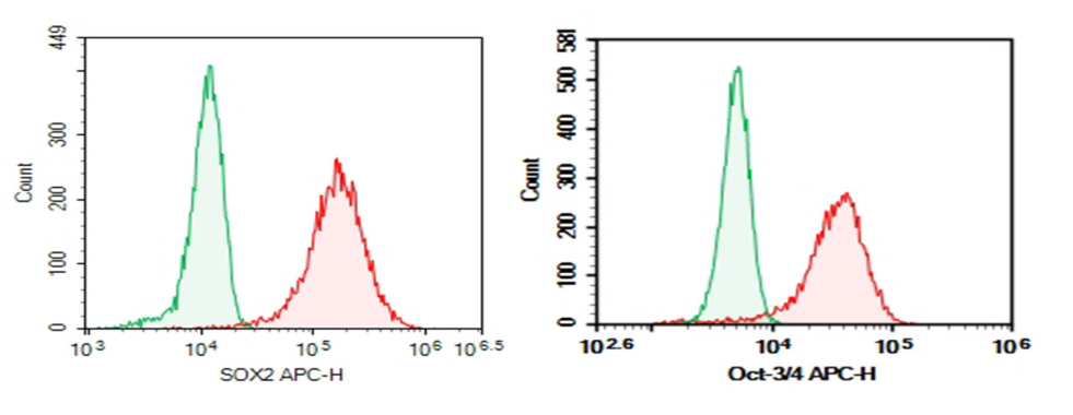
Fig. 1 Intracellular flow cytometry validation of Elabscience® anti-SOX2 and anti-OCT4 antibodies in human teratocarcinoma NCCIT cells. Specific antibody signal (red) shows clear separation from isotype control (blue), with nearly 100% positive populations.
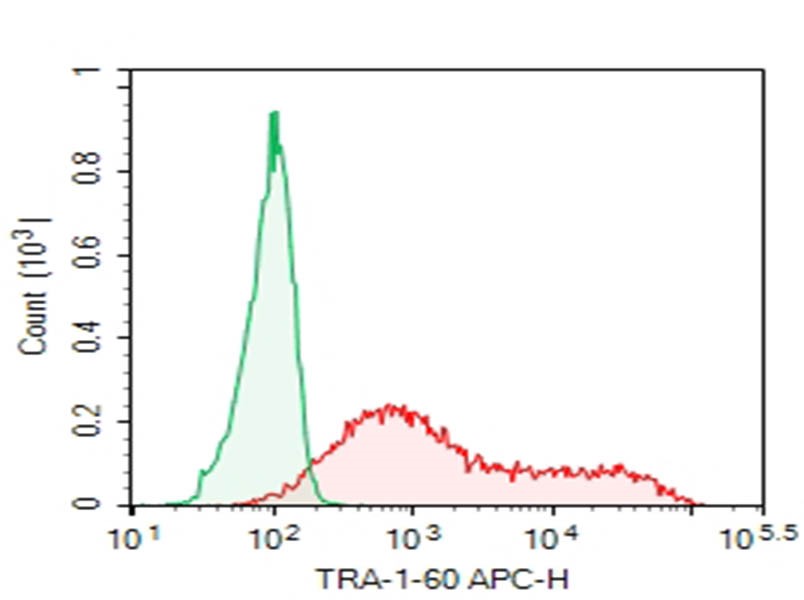
Fig. 2 Surface staining of human NCCIT cells with Elabscience® Purified Anti-Human TRA-1-60 Antibody (clone TRA-1-60). The specific antibody (right) shows strong positive signal versus mouse IgM isotype control (left).
02 Detecting Residual Undifferentiated iPSCs to Improve Cell Therapy Safety
Tumorigenicity arising from residual undifferentiated iPSCs is the paramount safety concern in iPSC-derived cell therapy. Landmark studies have established that as few as a few hundred undifferentiated cells can seed teratomas in immunodeficient hosts, making sensitive detection a regulatory prerequisite for clinical translation[2]. Flow cytometry using pluripotency surface markers provides a core in-process and release safety assay that is quantitative, reproducible, and amenable to GMP qualification. The anti-TRA-1-60 antibody has demonstrated detection sensitivity of 0.1% residual undifferentiated iPSCs spiked into differentiated populations such as cardiomyocytes and retinal pigment epithelial cells, and anti-SSEA-4 and anti-TRA-1-81 offer complementary specificity when used in combination. Co-staining with lineage-specific markers of the target cell type provides orthogonal, cross-validated purity data.
Elabscience® antibodies targeting TRA-1-60 (clone TRA-1-60), SSEA-4 (clone MC813-70), and TRA-1-81 maintain consistent staining performance across differentiation models, helping researchers and manufacturers ensure that final cell products remain below acceptable residual iPSC thresholds.
03 Monitoring Immune Checkpoint Expression in iPSC-Derived Immune Cells
One of the most active frontiers in iPSC research is the generation of off-the-shelf immune effector cells (T cells and NK cells) for adoptive cancer immunotherapy. However, iPSC-derived immune cells are not immune to exhaustion[3]. Sustained antigenic stimulation in the tumor microenvironment triggers a sequential checkpoint cascade: PD-1 rises on early activated cells, while TIGIT and TIM-3 accumulate as exhaustion deepens toward a terminally dysfunctional state. Each wave of checkpoint upregulation further erodes proliferative capacity and cytotoxic output, directly compromising therapeutic potency[4].
Quantitative checkpoint profiling is therefore integral to the functional characterization and batch-to-batch consistency assessment of iPSC-derived immune cell products. Elabscience® provides a validated panel covering the full exhaustion spectrum: an anti PD-1 antibody (clone EH12.2H7, Cat. No. E-AB-F1229D) for early exhaustion detection, together with anti-TIGIT (clone A15153G, Cat. No. AN009730P) and anti-TIM-3 (clone F38-2E2, Cat. No. E-AB-F1192A) for advanced dysfunction. These reagents have been validated across multiple cell models and are available in PE, APC, and FITC conjugates to integrate seamlessly into multi-color panels alongside functional markers such as IFN-γ, granzyme B, and perforin.
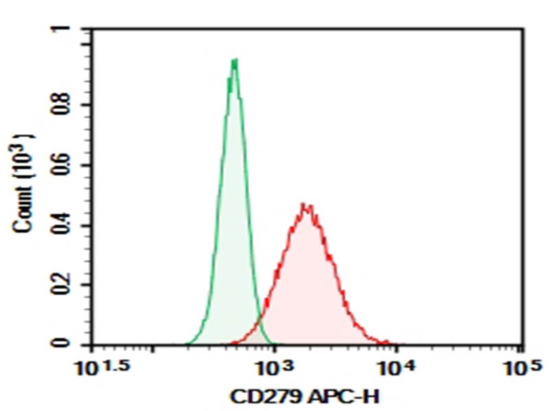
Fig. 3 Molt-4 cells activated with MIX for 4 hours, stained with Elabscience® Purified Anti-Human CD279/PD-1 Antibody (clone EH12.2H7, right) and mouse IgG1 isotype control (left), followed by APC-conjugated secondary antibody.
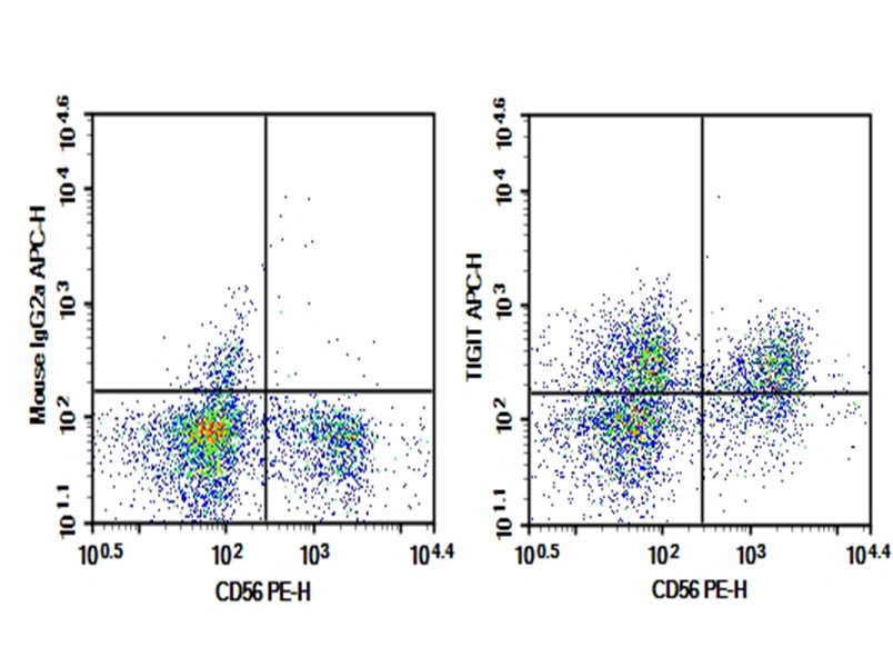
Fig. 4 Human peripheral blood lymphocytes stained with Elabscience® Purified Anti-Human TIGIT Antibody (clone A15153G, right) and mouse IgG2a isotype control (left), co-stained with anti-human CD56 PE.
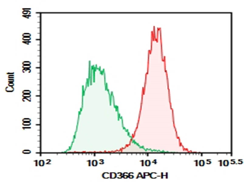
Fig. 5 Daudi cells stained with Elabscience® Purified Anti-Human CD366/TIM-3 Antibody (clone F38-2E2, right) and mouse IgG1 isotype control (left), followed by APC-conjugated secondary antibody.
04 Identifying Senescence Markers During Long-Term iPSC Culture
Long-term iPSC culture drives progressive replicative senescence characterized by irreversible cell cycle arrest, enlarged and flattened morphology, and the emergence of a senescence-associated secretory phenotype. Senescent iPSCs exhibit reduced differentiation efficiency and accumulate genomic abnormalities, making routine senescence surveillance essential for master and working cell bank maintenanc[5]. Traditional detection relies on cytochemical staining for senescence-associated β-galactosidase (SA-β-gal) activity at pH 6.0, a method that is labor-intensive, subjective, and poorly suited to quantitative batch release. Flow cytometry offers a superior alternative. Multiparameter panels simultaneously quantify SA-β-gal activity using fluorogenic substrates such as C12FDG, cell cycle inhibitors p16INK4a and p21Cip1, DNA damage marker γH2AX, and proliferation indicators Ki-67 and EdU, all at single-cell resolution and in a single workflow.
Elabscience® supports senescence monitoring with validated intracellular antibodies against p16, p21, p53, and γH2AX, plus fluorophore-conjugated Ki-67 for proliferation tracking. For live-cell identification and sorting of senescent populations, C12FDG-based SA-β-gal detection reagents are also available.
05 Profiling Surface Markers During Organoid Differentiation from iPSCs
Organoid differentiation pushes iPSC differentiation into three dimensions, producing self-organizing structures that recapitulate tissue architecture and harbor multiple interacting cell lineages. This complexity demands rigorous surface marker profiling to confirm lineage composition and maturity at each developmental stage. Across common organoid models, well-defined surface markers correspond to key commitment points: CXCR4 (CD184) and CD117 (c-Kit) mark definitive endoderm; CD271 and CD133 identify neural progenitor populations; and tissue-specific markers such as aquaporin-5 or surfactant proteins characterize mature pulmonary organoids. Multiparametric stem cell flow cytometry enables quantitative, stage-resolved surface marker tracking through the full organoid differentiation timeline, providing an objective readout of protocol reproducibility and batch quality[6].
Elabscience® provides a broad portfolio of surface marker antibodies applicable across organoid lineages, including anti-CXCR4 (CD184), anti-CD117 (c-Kit), anti-CD271, and anti-CD133, all validated for flow cytometry and compatible with multi-color organoid panels.
06 Biomarker-Based Release Testing for iPSC-Derived Therapeutic Cells
As iPSC-derived cell therapies advance through clinical development, regulatory frameworks from the FDA and EMA mandate comprehensive release testing covering identity, purity, potency, and safety. Flow cytometry is central to this testing matrix because it delivers quantitative, multiparametric data on defined cell populations in a format that translates directly from discovery-phase characterization to GMP-validated release assays. For iPSC master cell banks, consensus release criteria require ≥70% positive expression for a panel of iPSC pluripotency markers (SSEA-4, Oct-3/4, TRA-1-60, and TRA-1-81) and <5% expression of differentiation-associated markers. Terminal differentiated products demand lineage-identity confirmation (e.g., CD3 for T cells, CD56 for NK cells), quantification of residual undifferentiated iPSCs, and viability assessment at ≥70%. The same antibody clones and panel designs employed during process development can carry forward into qualified release methods, creating a seamless analytical continuum from bench to bedside.
Elabscience® flow cytometry antibodies support the full release testing workflow with rigorously validated clones, broad fluorochrome selection spanning FITC, PE, APC, PerCP/Cyanine5.5, and the Elab Fluor® series (488, 647, Violet 450, Red 780), together with free panel design and spectrum analysis services. Products are cited in over 33,000 peer-reviewed publications.
Table 1.Elabscience® iPSC Flow Cytometry Antibody Portfolio
|
Target |
Clone |
Cat. No. |
|
Oct-3/4 |
40/Oct-3 |
AN010010P |
|
SOX2 |
O30-678 |
AN011220P |
|
NANOG |
23D2-3C6 |
AN010430P |
|
SSEA-4 |
MC813-70 |
AN012800P |
|
TRA-1-60 |
TRA-1-60 |
AN011240P |
|
PD-1 (CD279) |
EH12.2H7 |
E-AB-F1229D |
|
TIGIT |
A15153G |
AN009730P |
|
TIM-3 (CD366) |
F38-2E2 |
E-AB-F1192A |
For iPSC culture and iPSC differentiation support, Elabscience® also supplies recombinant cytokines (IL-2, IL-7, IL-15, SCF) and functional antibodies for immune cell activation and expansion. View the complete flow cytometry antibodies at www.elabscience.com/products/flow-cytometry-antibodies or contact Elabscience® scientific support team for custom panel design services.
References:
[1] Chan, E. M., et al. (2009). Live cell imaging distinguishes bona fide human iPS cells from partially reprogrammed cells. Nature Biotechnology,27 (11),1033–1037.
[2] Kuroda, T., et al. (2025). Flow cytometry detection limit for residual undifferentiated human iPSCs in retinal pigment epithelial cell products. Regenerative Medicine, 20 (4), 489–501.
[3] Cichocki, F., et al. (2020). iPSC-derived NK cells combined with PD-1 checkpoint blockade eliminate solid tumor xenografts. Cell Reports Medicine, 1 (8), 100145.
[4] Anderson, A. C., & Long, A. H. (2023). T cell exhaustion markers PD-1, TIGIT and TIM-3 in iPSC-derived tumor-infiltrating lymphocytes. Journal of Immunotherapy of Cancer, 11 (2), e006892.
[5] Liu, X., et al. (2024). Senescence profiling of long-term cultured human iPSCs via multiparameter flow cytometry. Stem Cell Reports, 19 (5), 781–794.
[6] Müller, S., et al. (2022). Multiparametric flow cytometry for lineage tracking in human iPSC-derived organoids. Organoids, 1 (1), 23–37.

