

Cytotoxic T lymphocytes (CTLs), predominantly CD8-positive (CD8+) T cells, are essential effectors of adaptive immunity that eliminate virus-infected and malignant cells through directed cytolysis. Their activation is tightly regulated by antigen recognition, co-stimulatory signals, and metabolic reprogramming, and its accurate measurement underpins progress in vaccine development, adoptive cell therapy, and immune monitoring. Dysregulated cytotoxic T cell activation contributes to immunopathology or tumor evasion, while robust, antigen-specific responses correlate with protective immunity. Consequently, understanding the molecular basis of CTL activation and applying reproducible detection strategies are central to both basic immunology and translational research.
This review article systematically examines how cytotoxic T cells are activated and how this process can be quantified. The first sections dissect T cell receptor (TCR) signaling that initiates activation and the contribution of CD8 co-stimulation, particularly CD28-mediated signals. We then describe how activated CTLs execute target-cell killing via the perforin-granzyme and Fas-Fas ligand (FasL) pathways, how cellular metabolism shifts to support effector function, and finally how multiparameter flow cytometry enables quantitative assessment of activation status using defined marker panels.
Table of Contents
1. How does TCR signaling initiate cytotoxic T cell activation?
2. What is the role of CD8 co-stimulation in cytotoxic T cell activation?
3. How do activated cytotoxic T cells kill target cells?
4. How does T cell metabolism change during cytotoxic T cell activation?
5. How can flow cytometry quantify cytotoxic T cell activation?
01 How does TCR signaling initiate cytotoxic T cell activation?
Cytotoxic T lymphocyte (CTL) activation is initiated when the T cell receptor (TCR) recognizes a peptide antigen presented by major histocompatibility complex class I (pMHC-I) on the surface of an antigen-presenting or target cell. This engagement triggers a highly orchestrated intracellular signaling cascade that ultimately drives clonal expansion, cytokine secretion, and targeted cytolysis[1].
Step 1: CD8 co-receptor-mediated Lck recruitment. The CD8 co-receptor binds to the conserved α3 domain of MHC-I, stabilizing the TCR–pMHC interaction and simultaneously delivering the Src-family tyrosine kinase Lck (lymphocyte-specific protein tyrosine kinase) into proximity of the TCR–CD3 complex[2]. Lck is the critical initiating kinase: its association with CD8 is essential for phosphorylation of the immunoreceptor tyrosine-based activation motifs (ITAMs) within the cytoplasmic tails of the CD3 chains (CD3ε, CD3γ, CD3δ, and CD3ζ/CD247)[3]. Notably, CD8-bound Lck is particularly important for T cell responses to low-affinity antigens, whereas free Lck (not bound to CD8) can still support anti-viral and anti-tumor responses in vivo[4].
Step 2: ZAP70 recruitment and activation. Phosphorylated ITAMs on CD3ζ serve as docking sites for the tandem SH2 domains of ζ-chain-associated protein kinase 70 (ZAP70). Upon recruitment, ZAP70 is itself phosphorylated and activated by Lck[5]. Activated ZAP70 then phosphorylates the transmembrane adaptor protein LAT (linker for activation of T cells), which nucleates a multi-protein signaling complex known as the LAT signalosome[6]. LAT phosphorylation is facilitated by a molecular bridging mechanism in which Lck simultaneously binds phospho-ZAP70 (via its SH2 domain) and a proline-rich motif in LAT (via its SH3 domain), thereby colocalizing the kinase with its substrate[7].
Step 3: Three downstream signaling branches. The LAT signalosome coordinates activation of three major downstream pathways: PLCγ1–Ca²⁺–NFAT pathway: Phospholipase C-γ1 (PLCγ1) is recruited to LAT and hydrolyzes phosphatidylinositol-4,5-bisphosphate (PIP₂) to generate inositol-1,4,5-trisphosphate (IP₃) and diacylglycerol (DAG). IP₃ triggers Ca²⁺ release from the endoplasmic reticulum, followed by store-operated Ca²⁺ entry through CRAC channels. Sustained Ca²⁺ elevation activates the phosphatase calcineurin, which dephosphorylates NFAT (nuclear factor of activated T cells), enabling its nuclear translocation and transcription of effector genes such as IL2, IFNG, and TNF[8]. Ras–MAPK pathway: DAG and the adaptor protein GRB2–SOS activate Ras, leading to a kinase cascade (Raf → MEK → ERK). ERK phosphorylates transcription factors including Elk-1 and c-Fos, promoting cell proliferation and differentiation[9]. PKCθ–NF-κB pathway: DAG also recruits protein kinase C-θ (PKCθ), which, together with the CARMA1–BCL10–MALT1 complex, activates the IKK signalosome, leading to nuclear translocation of NF-κB and transcription of pro-survival and pro-inflammatory genes[10].
Step 4 : Effector function execution. The integration of these transcriptional programs drives CTL effector functions: polarized release of cytotoxic granules containing perforin and granzyme B, de novo synthesis of cytokines (IFN-γ, TNF, IL-2), and upregulation of activation markers (CD69, CD25, HLA-DR)[11].
02 What is the role of CD8 co-stimulation in cytotoxic T cell activation?
Productive cytotoxic t cell activation requires a second, co-stimulatory signal delivered principally through CD28, which synergizes with TCR ligation to lower the activation threshold and sustain clonal expansion. CD8 co-receptor binding to conserved regions of pMHC-I stabilizes the TCR-pMHC interaction and recruits LCK to the immunological synapse, amplifying ITAM phosphorylation and downstream LAT signalosome assembly. CD28 engagement by CD80 (B7-1) or CD86 (B7-2) on antigen-presenting cells activates phosphoinositide 3-kinase (PI3K)-AKT and nuclear factor-kappa B (NF-kappa B, NF-kB) cascades, drives interleukin 2 (IL-2) transcription, upregulates cell-cycle regulators, and antagonizes activation-induced cell death. Direct ex vivo evidence demonstrates that CD28-driven culture can generate stem-like memory CD8+ T cells even when cluster of differentiation 3 (CD3)/TCR signaling is bypassed, underscoring that co-stimulation independently programs survival and memory fate[12]. Inflammaging research reveals that age-related erosion of CD8 co-stimulatory circuits, particularly reduced CD28 expression on senescent CD8+ T cells, contributes to diminished cytotoxic t cell activation and impaired vaccine responsiveness in older adults[13]. In practice, comparing stimulation with anti-CD3 alone versus anti-CD3 plus anti-CD28 cleanly distinguishes abortive TCR-only activation from fully licensed effector differentiation, as judged by CD25, CD137, and interferon gamma (IFN-gamma) upregulation.
03 How do activated cytotoxic T cells kill target cells?
Activated cytotoxic T lymphocytes (CTLs) eliminate targets through two complementary, contact-dependent programs: the perforin-granzyme pathway and the Fas-Fas ligand (FasL) pathway, both tightly gated by prior TCR-induced activation. Upon immune synapse formation, polarization of lytic granules delivers perforin, which forms calcium-dependent pores in the target membrane, permitting entry of granzyme B (GZMB) and granzyme A that activate caspase-dependent and caspase-independent apoptosis. Parallel upregulation of FasL on CTLs engages Fas (CD95) on targets to trigger extrinsic caspase 8 activation and mitochondrial amplification. Decidual NK cell studies demonstrate that activation can be uncoupled from killing: they secrete cytokines rather than kill trophoblasts due to failed perforin polarization, yet efficiently degranulate and kill virus‑infected stromal cells when KIR2DS1‑HLA‑C2 licensing is satisfied[14]. Broader tumor immunity work shows that cytotoxic CD4+ T cells, which acquire perforin-granzyme machinery in chronic antigen settings, employ the same pore-forming mechanism to lyse class II-positive targets, supporting the generalizability of this pathway beyond CD8 lineages[15]. Engineering approaches that model CD8 response design confirm that moderate activation thresholds balance rapid infection clearance against toxicity, with granule-mediated killing as the dominant effector output[16]. For quantification, t cell activation flow cytometry combined with co-culture readouts provides direct functional evidence: surface CD107a (lysosomal-associated membrane protein 1, LAMP-1) externalization marks degranulation, while annexin V, TUNEL, or lactate dehydrogenase (LDH) release assays score target apoptosis.
04 How does T cell metabolism change during cytotoxic T cell activation?
TCR engagement with co-stimulation drives cytotoxic CD8+ T cells out of an oxidative-phosphorylation (OXPHOS)-dependent quiescent state and into aerobic glycolysis, and this glycolytic switch is fate instructive rather than merely permissive: upregulation of glucose transporter 1 (GLUT1), hexokinase 2 (HK2), and lactate dehydrogenase A (LDHA) raises glucose uptake and lactate output, and the resulting glycolytic flux is specifically required for effector differentiation, clonal expansion, and cytokine production, whereas cells that dial glycolysis back and retain OXPHOS capacity are the ones that survive as long-lived memory[17, 18].
Mitochondrial metabolism independently shapes memory T cell formation: memory, but not effector, CD8+ T cells maintain substantial spare respiratory capacity, undergo IL-15-driven mitochondrial biogenesis, and ramp up fatty-acid oxidation through carnitine palmitoyltransferase 1a, while mitochondria also feed TCA-cycle intermediates into the anabolic program of dividing effectors[19, 20].
The mTOR/AMPK nutrient-sensing axis reads out this bioenergetic state. mTORC1 integrates TCR, cytokine, and amino-acid signals to lock activated cells into the glycolytic, effector state; AMPK, acting upstream of mTORC1, senses glucose/ATP stress and favors the memory program, so rapamycin-mediated mTORC1 inhibition biases CD8+ T cells toward memory[21].
Experimentally, the activation-induced changes are tracked with the fluorescent glucose analog 2-NBDG (glucose uptake), extracellular acidification rate (ECAR) or medium lactate (glycolytic flux), oxygen consumption rate (OCR) or ATP assays (mitochondrial ATP generation), and TMRM or JC-1 staining (mitochondrial membrane potential); reading these alongside CD44, CD62L, and granzyme B ties metabolic state to the correct effector/memory stage[17, 20].
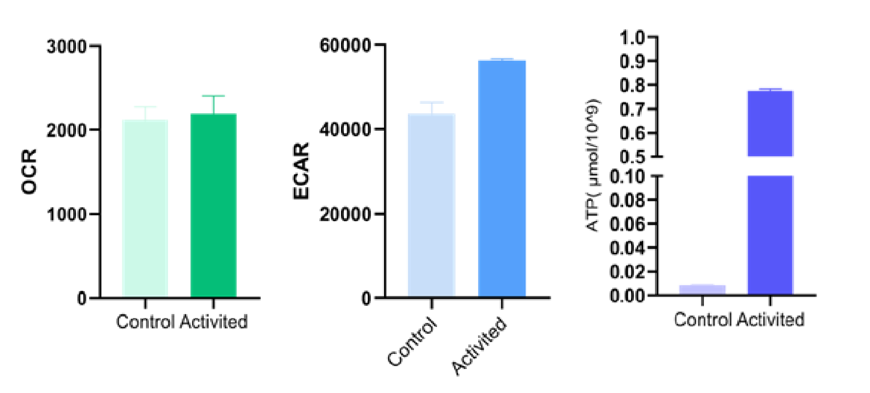
Fig. 1 The activation status of mouse naive CD8⁺ T cells was assessed following 72‑hour stimulation with Mouse CD3/CD28 T cell activation beads.
05 How can flow cytometry quantify cytotoxic T cell activation?
Multiparameter flow cytometry remains the gold standard to quantify cytotoxic T cell activation because it resolves identity, activation, and effector function at single-cell resolution. A robust T cell flow cytometry panel first gates lymphocytes by forward and side scatter, excludes doublets and dead cells, and identifies CD3+CD8+ cytotoxic T cells before overlaying activation readouts. Canonical CD8+ T cell markers include CD3 and CD8 as lineage anchors, while activation is scored with CD69 (early), CD25 (interleukin 2 receptor alpha, IL-2Ra), CD137 (4-1BB), and human leukocyte antigen-DR (HLA-DR), and effector capacity with intracellular interferon gamma (IFN-gamma), tumor necrosis factor alpha (TNF-alpha), perforin, and granzyme B after brief restimulation. Using CD3/CD28 T cell activation beads to deliver signal 1 plus co-stimulation provides a standardized, bead-to-cell ratio-controlled stimulus that uniformly upregulates these readouts across donors, enabling comparison of activation thresholds. Complementary chimeric antigen receptor (CAR)-T studies demonstrate that immunometabolic determinants, including elevated oxidative phosphorylation and mitochondrial fitness in infusion products, predict long-term persistence and can be co-monitored by flow-based mitochondrial dyes[22]. Conversely, pharmacologic inhibition of LCK and PI3K with ponatinib skews CD8+ T cells toward stem cell memory, while soluble uric acid drives exhaustion via KSR1-mediated MAPK hyperactivation, both detectable as altered CD62L, CCR7, CD45RA, and PD-1 profiles that illustrate how flow cytometry captures activation versus exhaustion bifurcations[23, 24]. Practically, investigators should include fluorescence-minus-one (FMO) and single-stained controls for spectral unmixing, use fixable viability dyes, and combine surface staining with intracellular cytokine staining to link phenotype to function, thereby converting frequencies into quantitative measures of cytotoxic t cell activation.
Table 1. Markers of T-cell activation status
|
Markers |
Clone |
Function |
|
CD69 |
FN50 |
CD69 is one of the earliest markers upregulated upon T cell activation, and its expression becomes diluted as cells proliferate. |
|
CD38 |
HIT2 |
ADP ribosyl cyclase mediates cell adhesion and signal transduction. It is constitutively expressed on naive T cells, downregulated on resting memory T cells, and then upregulated again on activated cells. |
|
CD25 |
CHI621 |
The α chain of the IL 2 receptor (CD25) is expressed on regulatory and resting memory T cells, and is upregulated within 24 hours after stimulation of the TCR/CD3 complex. |
|
HLA-DR |
L243 |
It is constitutively expressed on APCs, and its expression is upregulated on T cells upon stimulation. |
|
CD30 |
Ki-4 |
Expressed on activated lymphocytes, it regulates cell growth, proliferation, and apoptosis via multiple signaling pathways. |
|
CD134 |
Ber-ACT35 |
A member of the TNF receptor superfamily, it inhibits apoptosis. |
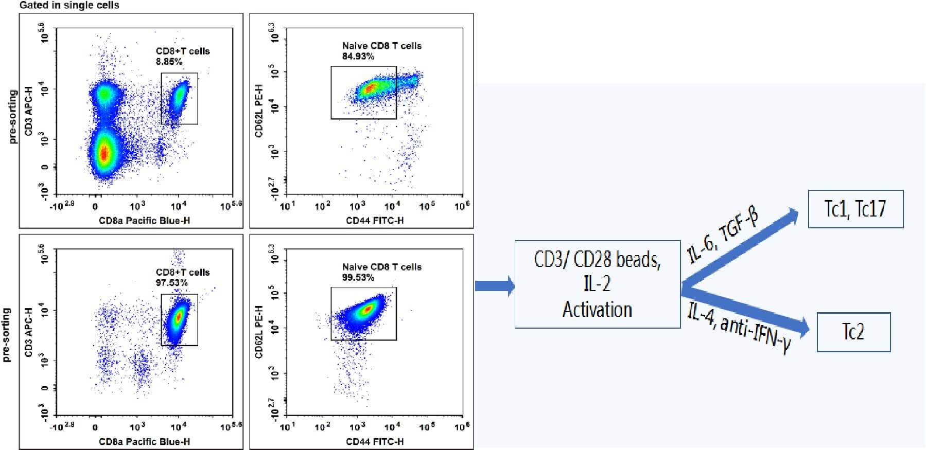
Fig. 2 The purity of naive CD8⁺ T cells from C57 mouse splenocytes was evaluated before (upper panel) and after (lower panel) cell sorting using APC‑CD3, EV450‑CD8a, FITC‑CD44, and PE‑CD62L. The sorted cells were then subjected to directed differentiation using activation beads, cytokines, and antibodies.
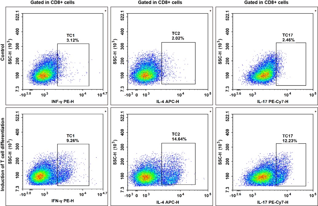
Fig. 3 Evaluation of in vitro directed differentiation of mouse naive CD8⁺ T cells.
In summary, cytotoxic T cell activation is orchestrated by TCR-initiated phosphorylation of CD3, ZAP70, and LAT, amplified by CD28 co-stimulation, translated into perforin-granzyme and Fas-FasL-mediated killing, and sustained by a glycolytic and oxidative metabolic switch that can be monitored alongside canonical activation signatures. Understanding these integrated layers is essential for designing reproducible immune assays and for optimizing adoptive immunotherapies. For researchers quantifying these processes, the t cell activation assay provides a rapid readout of proximal signaling and CD69 upregulation, while cytotoxic t cell activation assessed by CD107a degranulation and cytokine release captures functional competence. Coupling t cell activation flow cytometry with a validated T cell flow cytometry panel that includes CD8+ T cell markers and cytotoxic t cell markers such as CD44, CD62L, granzyme B, and CD137 improves subset resolution and allows correlation of phenotype with killing activity. Standardized stimulation with CD3/CD28 T cell activation beads ensures consistent signal 1 and signal 2 delivery across experiments, and metabolic fitness can be confirmed with a glycolysis assay that measures glucose uptake, extracellular acidification, or lactate production. Together, these complementary approaches enable robust, comparable assessment of CTL activation in health, infection, and cancer.
Elabscience® Quick Overview of Popular Products:
Table 2. Research Tools for Biomarker Detection
|
Cat. No. |
Product Name |
|
MIM001A |
Mouse CD3/CD28 T Cell Activation Beads |
|
E-EL-H0108 |
Human IFN-γ (Interferon Gamma) ELISA Kit |
|
E-EL-H0109 |
Human TNF-α (Tumor Necrosis Factor Alpha) ELISA Kit |
|
ESP-H0002 |
Human IFN-γ (Interferon Gamma) ELISPOT Kit |
|
E-CK-A480 |
Human Granzyme B Activity Detection Substrate for Flow Cytometry |
|
E-EL-H1617 |
Human GzmB(Granzyme B) ELISA Kit |
|
E-EL-M0594 |
Mouse GzmB(Granzyme B) ELISA Kit |
|
E-EL-H6215 |
Human CD30(Cluster of Differentiation 30) ELISA Kit |
|
E-AB-F1149D |
PE Anti-Human CD107a/LAMP-1 Antibody[H4A3] |
|
E-AB-F1254C |
FITC Anti-Mouse CD107a/LAMP-1 Antibody[1D4B] |
|
E-AB-F1187D |
PE Anti-Mouse CD69 Antibody[H1.2F3] |
|
E-AB-F1058E |
APC Anti-Human CD38 Antibody[HIT2] |
|
E-AB-F1102J |
PerCP/Cyanine5.5 Anti-Mouse CD25 Antibody[PC-61.5.3] |
|
E-AB-F1111G |
PE/Cyanine5 Anti-Human/Monkey HLA-DR Antibody[L243] |
|
E-AB-F1153C |
FITC Anti-Human CD134/OX40 Antibody[Ber-ACT35] |
References:
[1] SHAH K, AL-HAIDARI A, SUN J, KAZI J U. T cell receptor (TCR) signaling in health and disease [J]. Signal Transduction and Targeted Therapy, 2021, 6(1): 412.
[2] LI Y, MARIUZZA R. Structural and Biophysical Insights into the Role of CD4 and CD8 in T Cell Activation [J]. Frontiers in Immunology, 2013, Volume 4 - 2013.
[3] HORKOVA V, DROBEK A, PAPRCKOVA D, et al. Unique roles of co-receptor-bound LCK in helper and cytotoxic T cells [J]. Nature Immunology, 2023, 24(1): 174-85.
[4] LIANG Y, YE L. Bound to be perfect: Lck and T cell co-receptors [J]. Nature Immunology, 2023, 24(1): 5-7.
[5] FERNáNDEZ-AGUILAR L M, VICO-BARRANCO I, ARBULO-ECHEVARRIA M M, AGUADO E. A Story of Kinases and Adaptors: The Role of Lck, ZAP-70 and LAT in Switch Panel Governing T-Cell Development and Activation [J]. Biology, 2023, 12(9):1163.
[6] WANGE R L. LAT, the Linker for Activation of T Cells: A Bridge Between T Cell-Specific and General Signaling Pathways [J]. Science's STKE, 2000, 2000(63): re1-re.
[7] LO W L, SHAH N A-O, AHSAN N, et al. Lck promotes Zap70-dependent LAT phosphorylation by bridging Zap70 to LAT [J]. Nat Immunol. 2018, 19(7):733-741.
[8] KASCHEK L, ZöPHEL S, KNöRCK A, HOTH M. A calcium optimum for cytotoxic T lymphocyte and natural killer cell cytotoxicity [J]. Seminars in Cell & Developmental Biology, 2021, 115: 10-8.
[9] HWANG J-R, BYEON Y, KIM D, PARK S-G. Recent insights of T cell receptor-mediated signaling pathways for T cell activation and development [J]. Experimental & Molecular Medicine, 2020, 52(5): 750-61.
[10] HUANG W, LIN W, CHEN B, et al. NFAT and NF-κB dynamically co-regulate TCR and CAR signaling responses in human T cells [J]. Cell Reports, 2023, 42(7).
[11] OJANEN M J T, CARO F M, AITTOMäKI S, et al. FURIN regulates cytotoxic T-lymphocyte effector function and memory cell transition in mice [J]. European Journal of Immunology, 2023, 53(6): 2250246.
[12] IHARA F, OHASHI Y, ZHENG E Y F, et al. CD28-driven ex vivo generation of stem-like memory CD8+ T cells bypassing CD3/TCR signaling [J]. Proceedings of the National Academy of Sciences, 2026, 123(17): e2524626123.
[13] STURMLECHNER I, LEBRA SSEUR N K, WEYAND C M, GORONZY J J. The immunology behind inflammaging—causes, sources, and mechanisms [J]. Journal of Allergy and Clinical Immunology, 2026, 158(3): 629-41.
[14] TILBURGS T. Decidual immune activation by extravillous trophoblasts drives maternal-fetal tolerance and immunity [J]. Trends in Immunology, 2026, 47(3): 180-3.
[15] LAI L, RAN S, LI Y, et al. Cytotoxic CD4+ T cells: origin, biological functions, diseases and therapeutic targets [J]. Signal Transduction and Targeted Therapy, 2026, 11(1): 85.
[16] UKOGU O A, MONTAGUE Z, ALTAN-BONNET G, NOURMOHAMMAD A. Design principles of the cytotoxic CD8+ T cell response [J]. Proceedings of the National Academy of Sciences, 2026, 123(17): e2528202123.
[17] MACIVER N J, MICHALEK RD FAU - RATHMELL J C, RATHMELL J C. Metabolic regulation of T lymphocytes [J]. Annu Rev Immunol. 2013;31:259–283.
[18] CHANG C-H, CURTIS JONATHAN D, MAGGI LEONARD B, JR., et al. Posttranscriptional Control of T Cell Effector Function by Aerobic Glycolysis [J]. Cell, 2013, 153(6): 1239-51.
[19] VAN DER WINDT G J, EVERTS B FAU - CHANG C-H, CHANG CH FAU - CURTIS J D, et al. Mitochondrial respiratory capacity is a critical regulator of CD8+ T cell memory development [J]. Immunity. 2012;36(5):687–695.
[20] STEINERT E M, VASAN K, CHANDEL N S. Mitochondrial Metabolism Regulation of T Cell–Mediated Immunity [J]. Annual Review of Immunology, 2021, 39(1): 395-416.
[21] ARAKI K, AHMED R. AMPK: a metabolic switch for CD8+ T-cell memory [J].Eur J Immunol. 2013;43(4):897–899.
[22] GOLDBERG L, HAAS E R, WU J, et al. Immunometabolic determinants of long-term response in leukemia patients receiving CD19 CAR T cell therapy [J]. Nature Communications, 2026, 17(1): 2967.
[23] OKUHIRO Y, ITO S, WATANABE K, et al. Ponatinib inhibits LCK and PI3K signaling and promotes CD8+ T stem cell memory cell development [J]. Nature Communications, 2026, 17(1): 4934.
[24] LIU A, ZUO F, LI M, et al. Soluble Uric Acid Drives CD8+ T-cell Exhaustion by Inducing KSR1-Mediated MAPK Hyperactivation [J]. Cancer Research, 2026, 86(14): 3459-79.

