

On October 5, the 2026 Nobel Prize in Physiology or Medicine was announced and awarded to Karl Deisseroth, Peter Hegemann, and Georg Nagel for their discovery of light-gated ion channels and the founding of optogenetics. The technology arose from basic research into the light-sensing mechanism of the green alga Chlamydomonas reinhardtii: Hegemann and Nagel identified channelrhodopsins and demonstrated that light can gate ion channels; Deisseroth subsequently introduced them into neuroscience, enabling specific neurons to be precisely activated or inhibited under illumination and permitting neural-circuit manipulation in living animals. Neuroscience thereby advanced from observing correlation toward manipulating causation, and research on memory, emotion, reward, sleep, and the circuits underlying neuropsychiatric disorders has been profoundly reshaped. The award recognizes a foundational discovery and once again illustrates how tool innovation reshapes the life sciences.
This article systematically reviews five aspects: (1) the discovery timeline from bacteriorhodopsin in 1971 to light-controlled neurons in 2005; (2) the technical principle of ChR2: photoreception, gating, conduction, and signal generation, revealing how light is converted into a neural signal; (3) the paradigm shift that moves neuroscience from observing correlation to verifying causation; (4) key nodes in downstream research, including flow-cytometric phenotypic validation, ELISA-based molecular detection, and cellular metabolic monitoring; and (5) applications of optogenetics in disease intervention. Looking ahead, optogenetics will overcome the bottlenecks of light penetration and manipulation, advancing toward more refined, safer, and more intelligent closed-loop neuromodulation.
Table of Contents
1. Retrospective of the award-winning work: A relay passed between three scientists
2. Technical principle: How a beam of light becomes a neural signal
3. Impact of optogenetics on the neuroscience research paradigm
4. Key technical nodes in downstream optogenetics research
5. Optogenetics and disease intervention
6. Related resources
01 Retrospective of the award-winning work: A relay passed between three scientists
On October 5, 2026, the Karolinska Institute awarded the Nobel Prize in Physiology or Medicine to Karl Deisseroth (Stanford University / Howard Hughes Medical Institute, USA), Peter Hegemann (Humboldt University of Berlin, Germany), and Georg Nagel (University of Würzburg, Germany) "for their discoveries concerning light-gated ion channels and optogenetics."
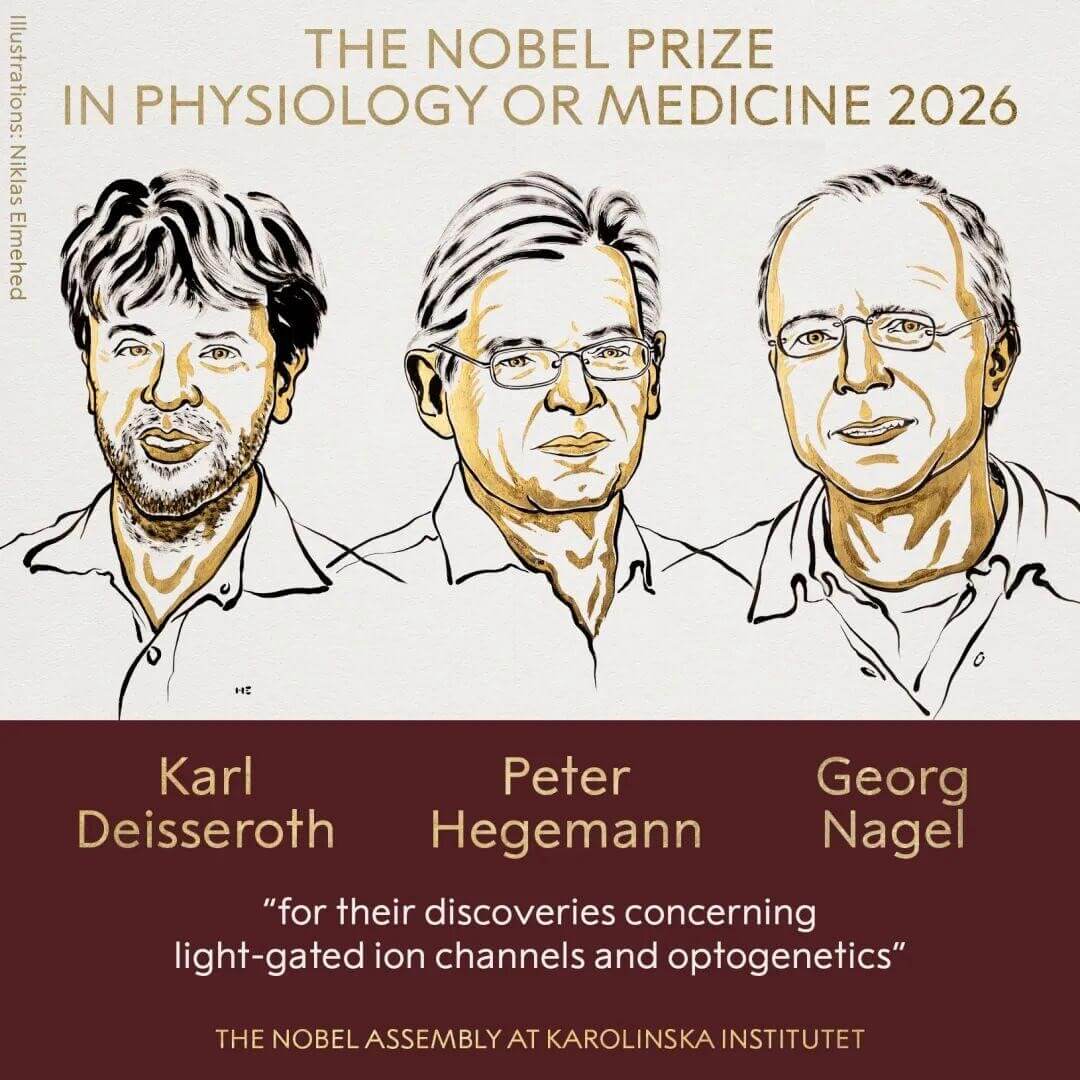
Fig. 1 The Nobel Prize in physiology or medicine 2026.
1.1 1971–1999: Microbes supply the "parts," crick proposes the dream
In 1971, Oesterhelt and Stoeckenius discovered bacteriorhodopsin in the purple membrane of halophilic microorganisms, first revealing that light can drive transmembrane proton movement, this is the first cornerstone of optogenetics. In 1979 and 1999, Francis Crick, co-discoverer of the DNA double helix, proposed that if specific neurons could be made light-sensitive, one could precisely switch a given class of nerve cells on or off. Seemingly science fiction at the time, this idea planted the central question for the technology decades later.
1.2 2002–2003: Isolating the key molecule from green algae
Hegemann had long asked how the unicellular green alga Chlamydomonas reinhardtii "chases light." In 2002, together with Nagel, Bamberg, and colleagues, he reported channelrhodopsin-1 (ChR1), and in 2003 demonstrated that channelrhodopsin-2 (ChR2) is itself a cation channel that can be opened directly by blue light; being simultaneously a photoreceptor and an ion channel, it was precisely the "switch" that neuroscience had long sought[1,2]. Meanwhile, from 2002 onward Miesenböck had already used genetics to specify neurons and light to exert control, providing the conceptual proof that "genes determine whom to control, while light determines when to control."
1.3 2004–2005: Blue light first moves a neuron
In August 2004, Boyden at Stanford introduced the ChR2 gene into mammalian neurons, where a flash of blue light triggered action potentials. In 2005, Boyden, Zhang, Bamberg, Nagel, and Deisseroth published their results, achieving millisecond-scale, cell-type-specific optical control of neuronal firing and marking the formal birth of optogenetics[3]. Zhang's key contribution was establishing a viral-vector system for delivering ChR2 into neurons[3].
1.4 2006–2007: Toward the living brain
In 2006, Deisseroth and colleagues systematically described the technical roadmap of optogenetics[4]. In 2007, their team targeted ChR2 to hypocretin neurons in the mouse hypothalamus; illumination through an implanted optical fiber awoke sleeping mice, establishing for the first time in the living brain a causal relationship between the activity of specific neurons and behavior[5].
1.5 2007 to the present: Global impact and clinical exploration
Subsequently, inhibitory tools (such as halorhodopsin) emerged, allowing neurons to be both "switched on" and "switched off" by light. In 2012, Tonegawa and colleagues used the technology to reactivate the neural "engram" of a fear memory. Today, laboratories worldwide use it to dissect the circuits underlying pain, social behavior, reward, and circadian rhythm, and are attempting to restore partial vision in patients with retinitis pigmentosa. Optogenetics has fundamentally changed the way humans understand the brain.
02 Technical principle: How a beam of light becomes a neural signal
The core of optogenetics is to introduce the gene encoding a naturally occurring light-gated ion channel, channelrhodopsin-2 (ChR2), into specific neurons, endowing the cells with the ability to "fire upon seeing light." Its signal transduction can be decomposed into four steps.
2.1 Photoreception: Photoisomerization of retinal
ChR2 is the phototactic receptor of the green alga Chlamydomonas reinhardtii. Seven transmembrane helices enclose a covalently bound retinal chromophore, linked via a protonated Schiff base to K257. In the dark, retinal is in the all-trans configuration and the channel is closed. Upon absorption of blue light at ~470 nm, the photon energy isomerizes the C13=C14 double bond to the 13-cis configuration within ~3 ps, generating a thermally relaxing K intermediate (P1500)[6,7]. This step is the "light-to-chemical-energy" conversion.
2.2 Gating: Three-gate gating and proton transfer
The isomerization strain is propagated downstream through reorganization of the charge cluster. The Schiff base transfers a proton to D253 (~10 µs), forming the M-like pre-open state P2390[6,7]; water molecules progressively enter, forming a transmembrane "water pore" that sequentially opens the extracellular gate (ECG), the central gate (CG), and the intracellular gate (ICG)[7]. After ~180–225 µs, the channel becomes fully open, constituting the conducting P520 state.
2.3 Conduction: Cation influx
ChR2 is a directly light-gated, non-selective cation channel permeable to Na⁺, K⁺, Ca²⁺, and H⁺. Based on expression-density estimates, its single-channel conductance is approximately 50 fS (dominated by protons under physiological conditions)[2]. The channel opens within about 0.2 ms of illumination[2], and cations flow inward down the electrochemical gradient.
2.4 Signal generation: Depolarization and action potentials
Cation influx depolarizes the membrane potential; upon reaching threshold, voltage-gated Na⁺ channels activate and generate an action potential that propagates along the axon to synapses, ultimately converting the light signal into a transmissible neural signal. After light offset, the channel closes within about 10 ms and the neuron returns to its resting state. This millisecond-scale switching is precisely the physical basis for the ability of optogenetics to control neural activity with high precision[3].
The elegance of optogenetics lies in a "single protein serving two roles": ChR2 is itself both the photoreceptor and the ion channel and requires no intracellular second-messenger cascade, thereby compressing the "light-to-electricity" delay to the sub-millisecond level and achieving spatiotemporal precision in the control of neural activity.
03 Impact of optogenetics on the neuroscience research paradigm
The most profound contribution of optogenetics is not the provision of a new tool, but the shift of systems neuroscience from an "observational discipline" to one grounded in causal intervention. Its paradigm impact can be summarized in four points.
3.1 From "correlation" to "causation": A fundamental change in how questions are posed
For a long time, electrophysiology, lesion, pharmacological, and imaging methods excelled at establishing correlations between neural activity and perception, movement, or emotion, yet lacked the ability to precisely manipulate a specific cell type within an intact circuit while matching neural computation on a millisecond timescale[8]. Traditionally, if a group of neurons was active during fear or foraging, researchers could only say that they "were related to" the behavior. Optogenetics changed the experimental logic: switch it on and the behavior appears; switch it off and the behavior disappears. This "manipulate-and-read out" logic for the first time allowed researchers to directly verify the causal link between neurons and behavior in the living brain[3,8].
3.2 Cell-type specificity: Solving the mixed-circuit problem
Excitatory, inhibitory, and modulatory neurons in the brain often coexist intermingled, and neighboring cells may even drive opposite functions. Optogenetics uses genetic means to "specify whom to control," enabling researchers to manipulate only a molecularly defined cell type or its projection pathway rather than an entire brain region[3]. As researchers have put it, the cerebral cortex was nearly a "black box" before; only now can it be truly disassembled.
3.3 Memory engram: A paradigm example of causal research
Memory engrams long lacked causal proof because the specific cells altered during learning could not be selectively accessed. Using activity-dependent genetic labeling, the Tonegawa laboratory expressed ChR2 in dentate gyrus neurons of the hippocampus that were active during fear learning; optically stimulating these cells in a neutral environment elicited freezing (fear recall), proving that activating a sparse population of engram cells is sufficient to evoke a specific memory[9]. This was the first direct demonstration that certain cells are both necessary and sufficient for a specific memory.
3.4 Universal causal testing from sleep to behavior, and the rise of a "toolbox" mentality
In 2007, optical stimulation of hypothalamic hypocretin neurons awoke sleeping mice, establishing the causal relationship by which the activity of specific cells drives arousal[5]. Thereafter, optogenetics expanded from a single "activation switch" into a platform of dozens of tools, including inhibition, red-shifted (two-color) control, and axonal-terminal manipulation, whose architecture of "genetic targeting + molecular actuator + optical control + physiological readout" can be transferred to multiple systems such as sleep, reward, pain, social behavior, the heart, and the gut[10].
04 Key technical nodes in downstream optogenetics research
Optogenetics achieves millisecond-scale manipulation of specific neurons through "genetic targeting + light-controlled switching." However, manipulation itself is only the starting point; truly answering "what did this group of switched-on/off cells do" still requires three types of downstream assays, corresponding to readouts at the phenotypic, molecular, and metabolic levels.
4.1 Phenotypic validation after cell-type-specific manipulation: The role of flow cytometry
Following optogenetic manipulation, researchers typically need to verify the transduction/expression efficiency of the target population (e.g., the positivity rate of ChR2 fused to a fluorescent protein), cell viability, and changes in subset proportions and functional phenotype, which is particularly critical in experiments involving neuro–immune interactions. Flow cytometry is the standard method for identifying and sorting immune-cell subsets, and its data quality depends heavily on the design of the antibody panel: from the choice of fluorophore-conjugated antibodies to the gating strategy for multicolor combinations, all affect the accuracy of subset discrimination[11]. Sample-processing strategy matters equally: in highly heterogeneous samples such as brain tissue, the target population is often first enriched with immunomagnetic beads (MACS) and then its purity and viability are verified by flow cytometry. For example, positive selection from a mouse brain single-cell suspension using CD11b beads rapidly yields a myeloid-enriched population. In ex vivo or in vitro models (primary cultures, co-cultures, organoids), the above workflow can provide a sufficient starting population with a well-defined state; in in vivo optogenetic models, tissue should be harvested after manipulation and phenotypic confirmation performed using the same gating strategy.
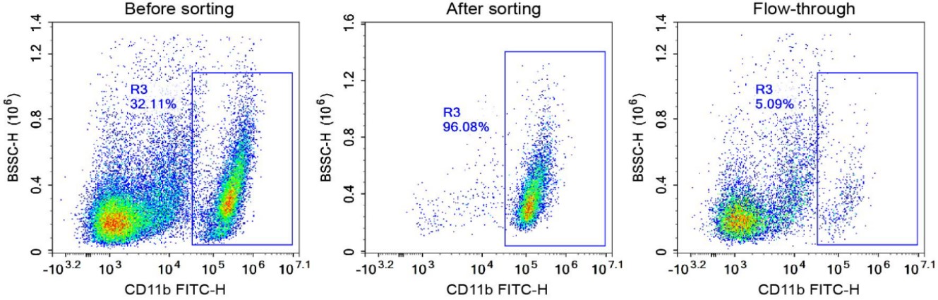
Fig. 2 Comparison of cell purity before and after CD11b-positive magnetic-bead sorting of C57 mouse brain tissue. Before sorting, the purity of CD11b-positive cells was 32.11%; after sorting, the purity of CD11b+ cells reached 96.08% (The data are provided by Elabscience®).
4.2 Detection of molecular effects after neural-circuit modulation: ELISA and protein quantification
After optogenetic activation or inhibition of specific neurons, downstream changes in neurotransmitter release, inflammatory-cytokine secretion, and signaling-pathway phosphorylation are core outputs of mechanistic research. ELISA, Western blot, and immunofluorescence are routine methods for quantifying these molecular events. For example, after optical stimulation of basal forebrain cholinergic neurons, ELISA detection of the changes in brain TNF-α, IL-6, and IL-1β revealed that they were significantly suppressed, thereby revealing the role of the cholinergic anti-inflammatory pathway[12]. Such high-sensitivity, high-specificity ELISA kits are routine tools for dissecting neuro–immune interaction mechanisms.
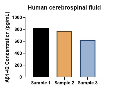
Fig. 3 The detection concentration of Aβ1-42 in human cerebrospinal fluid. The test was conducted using the Human Aβ1-42 (Amyloid Beta 1-42) ELISA Kit (Cat. No. E-EL-H0543) (The data are provided by Elabscience®).
Assay Used for This Experiment:
Human Aβ1-42(Amyloid Beta 1-42) ELISA Kit
Cat. No.: E-EL-H0543
Detection Range: 15.63-1000 pg/mL
Sensitivity: 9.38 pg/mL
Explore the Human Aβ1-42 ELISA Kit Used in 117 Publications →
4.3 Neuronal energy metabolism and cell state monitoring
Neurons are among the most energy-demanding cells in the brain, and synaptic transmission accounts for the bulk of brain energy consumption[13]. The calcium signals and firing activity elicited by optogenetic manipulation are directly coupled to changes in cellular metabolic state: action potentials and glutamatergic postsynaptic effects consume most of the signaling-related energy[13], and mitochondria at active synapses enlarge and increase cristae density to simultaneously meet ATP supply, Ca²⁺ buffering, and ROS regulation[14]. Therefore, dynamically monitoring mitochondrial function, ROS levels, ATP production, and glycolytic activity helps elucidate the coupling between neural activity and energy metabolism. Kits for ATP detection, ROS detection, mitochondrial membrane potential detection, and glycolysis-rate detection provide convenient quantitative means to assess the metabolic impact of optogenetic manipulation.
The three nodes form a progressive "phenotype–molecule–metabolism" research chain: flow cytometry answers "whom did we manipulate, and did they survive"; ELISA/protein quantification answers "what signaling molecules were released"; and metabolic assays answer "how did the cellular state change." Only by combining the three can the causal manipulation of optogenetics be translated into interpretable mechanistic conclusions.
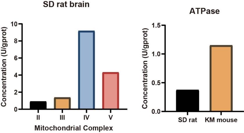
Fig. 4 The results of mitochondrial complex and ATPase detection. Left figure: Using the mitochondrial complex detection kit (Cat. No. E-BC-K150-M、E-BC-K151-M、E-BC-K152-M、E-BC-K153-M), the mitochondrial complexes II, III, IV, and V in the brain tissue homogenate of SD rats were detected. Right figure: The brain tissue homogenates of SD rats and Kunming mice were tested using the ATPase detection kit (Cat. No. E-BC-K826-M) respectively (The data are provided by Elabscience®).
05 Optogenetics and disease intervention
With millisecond-scale temporal precision and cell-type specificity, optogenetics can activate or inhibit neurons and is gradually moving from a research tool for dissecting neural circuits toward disease intervention, following a translational logic of "mechanism dissection, target identification, and therapeutic attempt." To date, optogenetics has found some application in the intervention of Parkinson's disease, epilepsy, retinal degeneration, mood disorders, and chronic pain.
In Parkinson's disease research, deep brain stimulation (DBS) of the subthalamic nucleus still benefits only a limited population. Valverde et al. found that DBS corrects pyramidal-cell hyperactivity by activating somatostatin (SST)-positive interneurons and inhibiting parvalbumin (PV)-positive interneurons in the motor cortex; in a mouse model, optogenetic activation of these SST-positive interneurons alone alleviated motor symptoms, suggesting a less invasive alternative strategy[15]. Ledri et al. noted that optogenetics can selectively modulate specific interneurons, helping to clarify their role in the initiation of seizures and to reveal interactions between remote circuits and hippocampal hyperexcitable networks; a closed-loop system combined with real-time monitoring can suppress abnormal discharges on demand before seizure onset[16]. Regarding retinal degeneration, exploiting the fact that inner retinal neurons survive while photoreceptors are lost, Sahel et al. injected a viral vector encoding ChrimsonR into the eye of a blind patient and, in combination with light-stimulating goggles, activated retinal ganglion cells; the patient could perceive and count objects, achieving partial restoration of visual function for the first time[17]. Russo and Nestler reviewed the role of the reward circuitry (the VTA–NAc pathway) in depression, showing that optogenetics can directly dissect the opposing effects of D1- and D2-type medium spiny neurons, providing a framework for circuit-targeted intervention in treatment-resistant depression[18]. Moreover, Guo et al. concluded that optogenetics can modulate specific neurons at multiple levels, inculding peripheral, spinal, and cerebral, revealing the roles of spinal dorsal horn interneurons, descending anterior cingulate cortex pathways, and glial cells in pain facilitation[19].
Overall, the application of optogenetics in disease remains constrained by factors such as light delivery, long-term expression, and immunogenicity; with the development of red-shifted opsins and closed-loop systems, its path from a mechanistic research tool toward a disease therapy is gradually becoming clearer.
From the photoreceptor protein of green algae to the rewriting of the neuroscience paradigm, optogenetics turns light into a key for controlling cells, allowing researchers for the first time to "switch" neural activity on and off with millisecond precision and cell-type specificity, pushing neuroscience from "observing correlation" to "verifying causation." Today it has extended to the intervention of multiple neurological diseases, bringing benefits to human health. In the next two decades, optogenetics will go beyond controlling neurons toward the spatiotemporal programming of life processes. When the ability to manipulate is no longer the barrier, how to validate its consequences with high quality cell-subset identification, factor quantification, and metabolic monitoring will become the key to unleashing its full potential. Imagination is becoming the only limit.
Elabscience® Quick Overview of Popular Products:
Table 1. Flow Cytometry Products for Neuroimmunology Research
|
Cat. No. |
MTT Assay |
|
E-AB-F1136US |
Elab Fluor® Red 780 Anti-Mouse CD45 Antibody[30-F11] |
|
E-AB-F1013C |
FITC Anti-Mouse CD3 Antibody[17A2] |
|
E-AB-F1353D |
PE Anti-Mouse CD4 Antibody[RM4-5] |
|
AN00408Q |
Elab Fluor® Violet 450 Anti-Mouse CD8a Antibody[YTS169.4] |
|
E-AB-F1081J |
PerCP/Cyanine5.5 Anti-Mouse/Human/Monkey CD11b Antibody[M1/70] |
|
AN01022C |
FITC Anti-Mouse CX3CR1 Antibody[SA011F11] |
|
E-AB-F1133H |
PE/Cyanine7 Anti-Human CD274/PD-L1 Antibody[29E.2A3] |
|
MIM010N |
EasySort™ Mouse Monocyte Isolation Kit |
Table 2. ELISA Kits for Neurosciences Research
|
Cat. No. |
MTT Assay |
|
E-EL-M3111 |
Mouse GFAP (glial fibrillary acidic protein) ELISA Kit |
|
E-EL-M3010 |
Mouse Aβ1-42(Amyloid Beta 1-42) ELISA Kit |
|
E-EL-M3009 |
Mouse Aβ1-40(Amyloid Beta 1-40) ELISA Kit |
|
E-EL-H0543 |
Human Aβ1-42(Amyloid Beta 1-42) ELISA Kit |
|
E-EL-M3136 |
Mouse BDNF(Brain Derived Neurotrophic Factor) ELISA Kit |
|
E-EL-M1289 |
Mouse pMAPT /pTAU(phosphorylated microtubule-associated protein tau) ELISA Kit |
|
E-EL-H0109 |
Human TNF-α(Tumor Necrosis Factor Alpha) ELISA Kit |
|
E-HSEL-M0003 |
High Sensitivity Mouse IL-6 (Interleukin 6) ELISA Kit |
|
CQM003 |
CellaQuant™ Mouse IL-1β (Interleukin 1 Beta) ELISA Kit |
Table 3. Cell Metabolism Kits for Neurosciences Research
|
Cat. No. |
MTT Assay |
|
E-BC-K149-M |
Mitochondrial Complex I (NADH-CoQ Reductase) Activity Assay Kit |
|
E-BC-K150-M |
Mitochondrial Complex II Activity Assay Kit |
|
E-BC-K151-M |
Mitochondrial Complex Ⅲ (Coenzyme Q-Cytochrome C Reductase) Activity Assay Kit |
|
E-BC-K152-M |
Mitochondrial Complex IV (Cytochrome C Oxidase ) Activity Assay Kit |
|
E-BC-K153-M |
Mitochondrial Complex Ⅴ(F₀F₁-ATPase/ATP Synthase) Activity Assay Kit |
|
E-BC-K826-M |
ATPase/GTPase Activity Colorimetric Assay kit |
|
E-BC-K212-S |
Ca2+-ATPase Activity Assay Kit |
|
E-BC-K774-M |
ATP Colorimetric Assay Kit (Enzyme Method) |
|
E-BC-K138-F |
Reactive Oxygen Species (ROS) Fluorometric Assay Kit (Green) |
|
E-BC-F069 |
Extracellular Acidification Rate (ECAR) Fluorometric Assay Kit |
|
E-BC-F070 |
Enhanced Oxygen Consumption Rate (OCR)Fluorometric Assay Kit |
06 Related resources
● Optogenetics determines "what is activated", and flow cytometry answers "what happens in the cell", the downstream verification of neuro-immune interactions begins here.
Download Your Free Application of Flow Cytometry Guide →
● Tracking the metabolic remodeling of neurons through cellular metabolic analysis takes the next step in downstream research of optogenetics.
Download Your Free Cellular Metabolism Integration Guide →
References:
[1] Nagel G, Ollig D, Fuhrmann M, Kateriya S, Musti AM, Bamberg E, Hegemann P. Channelrhodopsin-1: a light-gated proton channel in green algae. Science. 2002;296(5577):2395-2398. PMID: 12089443.
[2] Nagel G, Szellas T, Huhn W, Kateriya S, Adeishvili N, Berthold P, Ollig D, Hegemann P, Bamberg E. Channelrhodopsin-2, a directly light-gated cation-selective membrane channel. Proc Natl Acad Sci U S A. 2003;100(24):13940-13945. PMID: 14615590.
[3] Boyden ES, Zhang F, Bamberg E, Nagel G, Deisseroth K. Millisecond-timescale, genetically targeted optical control of neural activity. Nat Neurosci. 2005;8(9):1263-1268. PMID: 16116447.
[4] Deisseroth K, Feng G, Majewska AK, Miesenböck G, Ting A, Schnitzer MJ. Next-generation optical technologies for illuminating genetically targeted brain circuits. J Neurosci. 2006;26(41):10380-10386. PMID: 17035522.
[5] Adamantidis AR, Zhang F, Aravanis AM, Deisseroth K, de Lecea L. Neural substrates of awakening probed with optogenetic control of hypocretin neurons. Nature. 2007;450(7168):420-424. PMID: 17943086.
[6] Ardevol A, Hummer G. Retinal isomerization and water-pore formation in channelrhodopsin-2. Proc Natl Acad Sci U S A. 2018;115(14):3550-3555. PMID: 29555736.
[7] Kuhne J, Vierock J, Tennigkeit SA, et al. Unifying photocycle model for light adaptation and temporal evolution of cation conductance in channelrhodopsin-2. Proc Natl Acad Sci U S A. 2019;116(19):9380-9389. PMID: 31004059.
[8] Deisseroth K. Optogenetics: 10 years of microbial opsins in neuroscience. Nat Neurosci. 2015;18(9):1213-1225. PMID: 26308982.
[9] Liu X, Ramirez S, Pang PT, Puryear CB, Govindarajan A, Deisseroth K, Tonegawa S. Optogenetic stimulation of a hippocampal engram activates fear memory recall. Nature. 2012;484(7394):381-385. PMID: 22441246.
[10] Josselyn SA, Tonegawa S. Memory engrams: Recalling the past and imagining the future. Science. 2020;367(6473):eaaw4325. PMID: 31896692.
[11] Matevossian A, Akbarian S. Neuronal nuclei isolation from human postmortem brain tissue. J Vis Exp. 2008;(20):914. PMID: 19078943.
[12] Yun Y, Wang X, Xu J, Chen J, Wang X, Yang P, Qin L. Optogenetic stimulation of basal forebrain cholinergic neurons prevents neuroinflammation and neuropsychiatric manifestations in pristane-induced lupus mice. Behav Brain Funct. 2023;19(1):11. PMID: 37322485.
[13] Attwell D, Laughlin SB. An energy budget for signaling in the grey matter of the brain. J Cereb Blood Flow Metab. 2001;21(10):1133-1145. PMID: 11598490.
[14] Faria-Pereira A, Morais VA. Synapses: The Brain's Energy-Demanding Sites. Int J Mol Sci. 2022;23(7):3627. PMID: 35408993.
[15] Valverde S, Vandecasteele M, Piette C, Derousseaux W, Gangarossa G, Aristieta Arbelaiz A, Touboul J, Degos B, Venance L. Deep brain stimulation-guided optogenetic rescue of parkinsonian symptoms. Nat Commun. 2020;11(1):2388. PMID: 32404907.
[16] Ledri M, Andersson M, Wickham J, Kokaia M. Optogenetics for controlling seizure circuits for translational approaches. Neurobiol Dis. 2023;184:106234. PMID: 37479090.
[17] Sahel JA, Boulanger-Scemama E, Pagot C, Arleo A, Galluppi F, Martel JN, et al. Partial recovery of visual function in a blind patient after optogenetic therapy. Nat Med. 2021;27(7):1223-1229. PMID: 34031601.
[18] Russo SJ, Nestler EJ. The brain reward circuitry in mood disorders. Nat Rev Neurosci. 2013;14(9):609-625. PMID: 23942470.
[19] Guo F, Du Y, Qu FH, Lin SD, Chen Z, Zhang SH. Dissecting the Neural Circuitry for Pain Modulation and Chronic Pain: Insights from Optogenetics. Neurosci Bull. 2022;38(4):440-452. PMID: 35249185.

