

On April 7, 2026, the team of Yuan Yexian at Northwest A&F University published a research paper online in Cell Metabolism titled “Microbial-derived 3-phenylpropionic acid orchestrates immune–progenitor cell crosstalk to promote beige adipogenesis and energy expenditure”. The study found that cold exposure increases the abundance of the gut bacterium Prevotella copri (P. copri) and elevates the level of its metabolite 3-phenylpropionic acid (3-PPA). Rather than acting directly on adipocytes, 3-PPA sequentially activates FFAR1 on M2-like macrophages in adipose tissue, induces CXCL13 secretion, recruits T follicular helper (Tfh) cells, and ultimately promotes the differentiation of SMA⁺ adipocyte progenitor cells into thermogenic beige adipocytes via IL-4/STAT6 signaling. This work proposes a novel mechanism by which “gut microbiota–immune cells–adipocyte progenitors” coordinately regulate thermogenesis and energy expenditure.
DOI:10.1016/j.cmet.2026.01.017
01 Background: Why Does “Beige Fat” Deserve Attention?
An important approach to obesity treatment is not just reducing intake but also increasing expenditure. The fat in the human body and in mammals is not all the same:
White adipose tissue (WAT): primarily responsible for energy storage;
Brown adipose tissue (BAT): rich in UCP1, capable of expending energy through non-shivering thermogenesis;
Beige adipocytes: can be induced in white adipose tissue by stimuli such as cold exposure and exercise, and also possess strong thermogenic potential.
Among these, the browning or beiging of inguinal white adipose tissue (iWAT) is considered to help increase energy expenditure, improve glucose and lipid metabolism, and potentially counteract obesity. The question is: how exactly does external cold exposure transmit signals through the gut microbiota to adipose tissue and initiate beige adipogenesis?
Previous studies have observed that cold exposure can remodel the gut microbiota, but which specific bacteria are involved, what metabolites they produce, and through which host cells and molecular pathways they act, remain unclear.
02 Core finding: Cold induces a “gut–immune–adipose progenitor cell” thermogenic pathway
The mechanistic chain presented in this study can be summarized as:
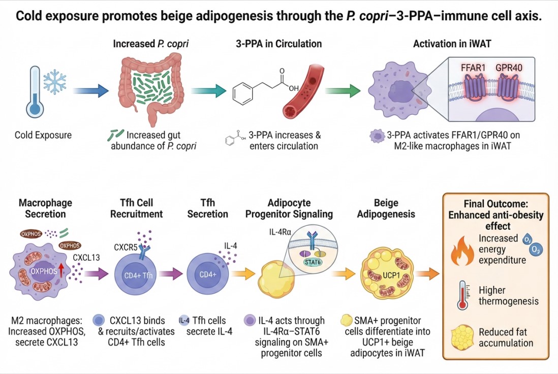
Fig. 1 Cold exposure promotes beige adipogenesis through the P. copri–3-PPA–immune cell axis.
The importance of this pathway lies in its connection of a gut microbiota-derived small-molecule metabolite to immune cells, chemokines, T cells, and adipose progenitor cells, forming a multicellular synergistic metabolic regulatory network.
03 How was the research carried out step by step?
3.1 Cold exposure altered the gut microbiota, and P. copri emerged as a key candidate bacterium.
The researchers housed mice at room temperature (23°C) or in a cold environment (6°C) for 14 days, and then performed 16S rRNA sequencing and metabolomic analysis on colonic contents. The results showed that cold exposure not only increased multilocular adipocytes and enhanced UCP1 expression in iWAT, but also significantly altered the composition of the gut microbiota; among these changes, the genus Prevotella, particularly P. copri, was markedly increased.
To verify its function, the authors first depleted the gut microbiota with antibiotics, and then colonized the mice with PBS, E. coli, or P. copri, respectively. Under cold exposure conditions, compared with the control group, mice colonized with P. copri exhibited:
(1) better cold tolerance and survival;
(2) higher core body temperature;
(3) higher UCP1 expression in iWAT;
(4) more multilocular beige adipocytes with typical characteristics.
It is noteworthy that the effects of P. copri were primarily concentrated on iWAT beiging, without significantly enhancing UCP1 expression in classical BAT, suggesting that it does not broadly and non-specifically activate all thermogenic adipose tissue, but rather preferentially regulates the beiging of white adipose tissue.
3.2 Identifying the key metabolite: 3-PPA serves as the messenger between P. copri and thermogenesis
Metabolomic analysis revealed that cold exposure significantly reshaped phenylalanine metabolism in the colon. Among the metabolites, 3-PPA was elevated in both the colon and serum. Further evidence demonstrated that:
(1) P. copri produced more 3-PPA when supplied with phenylalanine as a substrate;
(2) antibiotic depletion of the microbiota markedly reduced serum 3-PPA levels;
(3) recolonization with P. copri restored 3-PPA levels;
(4) mono-colonization of germ-free mice with P. copri increased serum 3-PPA, and cold exposure further enhanced this effect.
Additionally, strains such as Clostridium sporogenes and Peptostreptococcus anaerobius also produced 3-PPA and promoted UCP1 expression in iWAT. This suggests that 3-PPA itself may be closer to the true functional effector molecule than any single bacterial strain.
3.3 Can 3-PPA supplementation truly improve energy metabolism?
The authors added 0.5% 3-PPA to the drinking water of cold-exposed mice for 5 weeks. The results showed that 3-PPA, without altering food or water intake:
(1) reduced body weight and fat mass;
(2) decreased the weight of the liver, iWAT, and epididymal adipose tissue;
(3) elevated core body temperature;
(4) increased oxygen consumption and total energy expenditure;
(5) enhanced the expression of thermogenic genes such as UCP1 and Cidea in iWAT;
(6) promoted the formation of multilocular beige adipocytes.
However, in BAT, tissue weight, UCP1 expression, histology, and thermogenic genes were not significantly altered. In other words, the primary effect of 3-PPA is to drive beiging of white adipose tissue.
A particularly noteworthy finding is that supplementation with 3-PPA at room temperature did not independently induce iWAT beiging. This suggests that 3-PPA is not a single agent that switches on thermogenesis, but rather acts more like an amplifier that enhances the thermogenic response in a cold context.
04 Mechanistic Breakthrough: 3-PPA Does Not Directly “Burn Fat” but First Remodels the Immune Microenvironment
4.1 The Receptor for 3-PPA Is FFAR1 (a Gq-Coupled Receptor), Not Aryl Hydrocarbon Receptor (AhR)
3-PPA has previously been reported to participate in processes such as intestinal barrier function via AhR. However, in this study, knockdown of AhR did not affect 3-PPA-promoted UCP1 expression, indicating that AhR is not the primary mediator in the context of adipose beiging.
The researchers subsequently found that:
(1) Isotope labeling experiments suggested that 3-PPA does not freely enter cells to a significant extent;
(2) After 3-PPA treatment, IP3/DAG signaling increased in iWAT, while cAMP showed no significant change, consistent with activation of a Gq-coupled receptor;
(3) In HEK293T cells overexpressing FFAR1, 3-PPA induced significant Ca²⁺ flux;
(4) Molecular docking and CETSA experiments jointly supported binding between 3-PPA and FFAR1 as well as evidence of target stabilization;
(5) The FFAR1 antagonist GW1100, Ffar1 knockdown, or adipose tissue-specific Ffar1 deletion all significantly attenuated or blocked 3-PPA-induced beiging.
Therefore, this study identifies FFAR1/GPR40 as the key receptor mediating 3-PPA-induced beige adipogenesis.
4.2 The true key target cells: M2-like macrophages
FFAR1 is not primarily expressed in mature adipocytes or Pdgfrα⁺ adipose progenitor cells. Single-cell transcriptomics, protein detection, and conditional knockout experiments have revealed that the core action of 3-PPA lies in M2-like macrophages within adipose tissue. After 3-PPA treatment, the following changes occurred in mouse iWAT:
(1) M1-like macrophage-associated markers decreased;
(2) M2-like macrophage markers, such as Arg1, Il10, and CD206, increased;
(3) The proportion of CD45⁺F4/80⁺CD206⁺ M2-like macrophages increased;
(4) Macrophage-specific knockout of Ffar1 significantly attenuated both basal beiging and 3-PPA-induced beiging.
This indicates that 3-PPA does not directly instruct adipocytes to become beige, but rather first reshapes the adipose tissue immune microenvironment into a state more conducive to thermogenic remodeling.
4.3 CXCL13: A Key Chemokine Linking Macrophages and Tfh Cells
After transcriptomic analysis of M2 macrophages treated with 3-PPA, the authors found that multiple cytokines were altered, among which CXCL13 showed the most significant upregulation. 3-PPA enhanced the mitochondrial oxidative phosphorylation (OXPHOS)-related program in macrophages and promoted CXCL13 secretion via NF-κB activation; inhibiting mitochondrial ATP synthesis or NF-κB signaling both reduced CXCL13 secretion.
Table 1. Functional validation of CXCL13
|
Intervention |
Observation |
Explanation |
|
Recombinant CXCL13 treatment of SVF cells |
Increased UCP1 and thermogenesis-related markers |
CXCL13 promotes beige adipocyte differentiation |
|
Mice injected with recombinant CXCL13 |
Increased UCP1 in iWAT and increased multilocular adipocytes |
CXCL13 promotes beiging in vivo |
|
CXCL13 neutralizing antibody |
Attenuated 3-PPA-induced UCP1 upregulation |
CXCL13 is a key downstream effector of 3-PPA |
|
Macrophage Ffar1 knockdown |
3-PPA fails to effectively induce CXCL13 |
CXCL13 production depends on FFAR1 |
05 Tfh cells and SMA⁺ progenitor cells: Who is truly manufacturing beige fat?
The classical receptor for CXCL13 is CXCR5. Studies have found that CXCL13 can increase local CXCR5 expression in iWAT and promote the accumulation of CD4⁺CXCR5⁺PD-1⁺ Tfh cells. Knockdown of Cxcr5 blocks CXCL13-induced upregulation of UCP1.
Further co-culture experiments showed that:
(1) In an SVF culture system lacking Tfh cells, CXCL13 cannot effectively promote beiging;
(2) Only in the presence of Tfh cells can CXCL13 enhance beige adipogenesis;
(3) CXCL13 promotes the secretion of IL-4, but not IL-13, by Tfh cells;
(4) After blocking IL-4Rα or STAT6 phosphorylation, the pro-beiging effects of 3-PPA/IL-4 are significantly diminished.
Finally, through lineage tracing, the authors confirmed that 3-PPA-induced nascent beige adipocytes mainly originate from SMA⁺ adipocyte progenitor cells, while the contribution of Pdgfrα⁺ and Pdgfrβ⁺ progenitor cells is relatively limited.
This implies that what 3-PPA drives is not simply the "transdifferentiation" of mature white adipocytes, but rather the activation of specific progenitor cell populations through immune signaling, leading to the differentiation of nascent beige adipocytes.
06 Anti-obesity effects and translational value: promise exists, but clinical application remains distant
In mice fed a high-fat diet combined with cold exposure, 3-PPA supplementation reduced body weight gain, fasting blood glucose, fat mass, and the weights of the liver and white adipose tissue, while increasing core body temperature, oxygen consumption, and energy expenditure. These anti-obesity and pro-thermogenic effects were no longer significant in Ffar1 knockout mice, further demonstrating that FFAR1 is an indispensable node for this effect.
The research was further extended to human-derived cells and porcine models:
(1) In human adipose SVF cells cultured at 31°C, 10 μM 3-PPA increased the expression of UCP1 and Perilipin 1;
(2) In porcine adipose SVF cells, 3-PPA increased the expression of thermogenesis-related genes such as UCP3, CIDEA, and DIO2;
(3) Cold-tolerant Tibetan pigs had higher colonic and serum 3-PPA levels than cold-sensitive Bama pigs, and exhibited a stronger thermogenesis-related phenotype.
07 Main Innovations of This Study
7.1 Advancing from Overall Microbiota Changes to a Causal Chain at the Strain–Metabolite–Receptor Level
This study did not stop at the associative description that cold exposure alters microbiota composition. Instead, through experiments involving microbiota depletion, P. copri colonization, mono-colonization in germ-free mice, metabolite supplementation, and receptor knockout, it progressively constructed a relatively complete causal framework:
P. copri → 3-PPA → FFAR1 → CXCL13 → Tfh → IL-4/STAT6 → SMA⁺ progenitor cells.
7.2 Discovery of a Novel Role for FFAR1 in Adipose Thermogenesis
FFAR1 has long been primarily recognized for its role in fatty acid sensing and insulin secretion in pancreatic β-cells. This study extends its function to the adipose tissue immune microenvironment, proposing that macrophage FFAR1 serves as a key receptor linking microbial metabolites to beige adipogenesis.
7.3 Incorporating Tfh Cells into the Adipose Thermogenesis Regulatory Network
Previous research on adipose thermogenesis immunity has largely focused on macrophages, eosinophils, ILC2s, and other cell types. This study further demonstrates that Tfh cells can enter adipose tissue via the CXCL13/CXCR5 axis and promote progenitor cell beiging through IL-4/STAT6, thereby expanding the cellular atlas of adipose tissue immune.
08 Limitations and Next Steps
This mechanistic framework is rigorous and the chain of evidence is relatively complete, yet its translation still faces several key issues.
Table 2. Limitations
|
Key limitations |
Why it matters |
|
The effect is highly dependent on cold exposure |
3-PPA failed to independently induce beiging at room temperature, suggesting it is more likely a thermogenic enhancer rather than a weight-loss agent that can act independently of environmental stimuli. |
|
Lack of human in vivo evidence |
To date, only human SVF cells have been shown to respond to 3-PPA in vitro; there is no causal evidence linking human plasma 3-PPA, P. copri abundance, cold adaptation, and metabolic benefits. |
|
Long-term safety unknown |
The authors explicitly state that the long-term effects of chronically elevated 3-PPA on systemic inflammation, glucose homeostasis, and metabolic flexibility still require evaluation. |
|
Dosage and exposure route need optimization |
Mice received 3-PPA via 0.5% drinking water; how to convert this to an acceptable human dose, frequency, and formulation remains an open question. |
|
P. copri cannot simply be equated with a probiotic |
Microbial effects are typically strain-specific, host-dependent, and diet-dependent; even if 3-PPA has metabolic potential, this does not justify directly recommending P. copri supplementation. |
These limitations do not diminish the mechanistic value of the paper; rather, they clarify the direction of next-stage translational research: population-based association studies, short-term cold exposure interventions, pharmacokinetic/safety evaluation of 3-PPA, and pharmacological validation targeting the FFAR1 or CXCL13 pathways.
09 Summary: A Noteworthy New Pathway of Postbiotic-Promoted Thermogenesis
The most valuable aspect of this study is not merely the discovery that 3-PPA can promote beige adipogenesis, but rather its elucidation of the underlying mechanism.
Cold exposure does not only directly activate adipose tissue through the sympathetic nervous system; it may also first remodel the gut microbiota. After gut microbes produce 3-PPA, it acts on adipose tissue macrophages via blood-borne or local metabolic signals. Subsequently, macrophages, Tfh cells, and adipocyte progenitor cells form a cascade, ultimately converting environmental cold signals into beige adipogenesis and enhanced energy expenditure.
From a translational medicine perspective, what deserves future exploration is not simply “probiotic supplementation” or “metabolite supplementation”, but rather the establishment of more precise intervention strategies centered on the following nodes: Controllable 3-PPA postbiotic intervention, targeted activation of macrophage FFAR1, regulation of CXCL13–CXCR5 immune recruitment, and restoration of the thermogenic differentiation capacity of SMA⁺ adipocyte progenitor cells.
Final conclusion: 3-PPA is not yet a mature anti-obesity therapeutic regimen, but this study provides a highly instructive mechanistic paradigm for the therapeutic approach of reshaping the adipose immune microenvironment and enhancing energy expenditure through gut microbial metabolites.
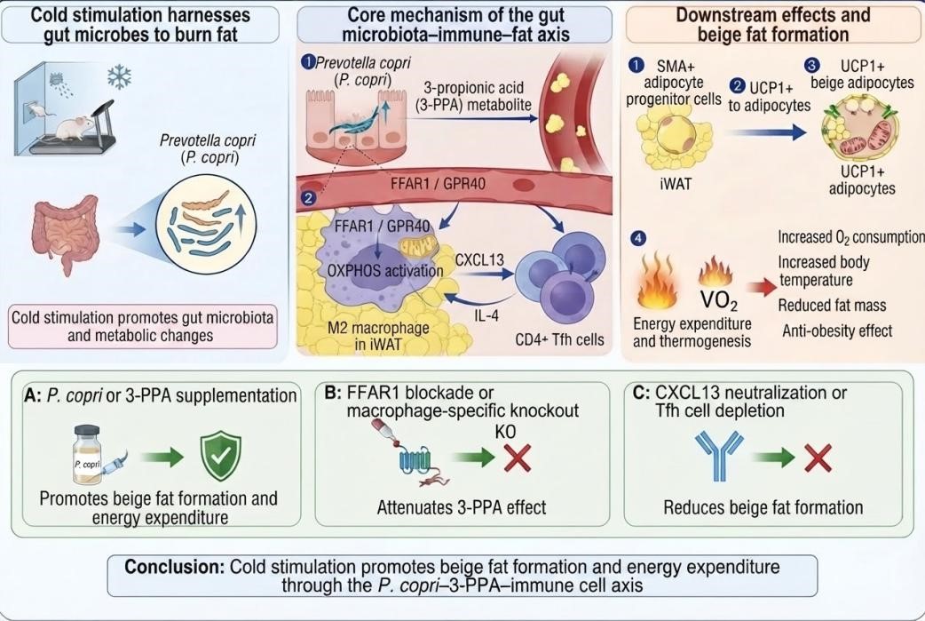
Fig. 2 Cold stimulation promotes beige fat formation and energy expenditure through the P. copri–3-PPA–immune cell axis.
● Key experimental techniques used in this study
16S rRNA sequencing, metabolomics sequencing, transcriptomics sequencing, LC-MS, microbiota colonization and germ-free mice, flow cytometry, ELISA, RNA-seq, Western Blot, SVF/BMDM culture, [Ca2+]i assay, molecular docking, cellular thermal shift assay (CETSA), conditional knockout, lineage tracing, metabolic cages, and histological staining.
● Elabscience® products used in this study
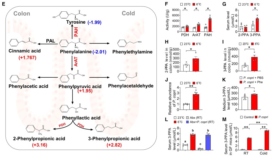
Fig. 3 Cold induces 3-PPA, which is predominantly derived from P. copri.
Caption: In Fig. 3F, the authors used Elabscience® Mouse PDH Activity Assay Kit (Cat. No.: E-BC-K650-M) to detect phenylpyruvate dehydrogenase (PDH) activity in the colon, providing important evidence for the discovery of the potential microbiota-derived metabolite 3-PPA.
Explore related Elabscience® products for energy metabolism research and enjoy 15% OFF from Sep 1 to Dec 18, 2026. → View Promotion
Table 3. Product recommendations that Elabscience® can provide for related studies
|
Cat. No. |
Product Name |
|
E-AB-F1136F |
PerCP Anti-Mouse CD45 Antibody[30-F11] |
|
E-AB-F0995C |
FITC Anti-Mouse F4/80 Antibody[CI:A3-1] |
|
E-AB-F1135D |
PE Anti-Mouse CD206/MMR Antibody[C068C2] |
|
E-AB-F1097C |
FITC Anti-Mouse CD4 Antibody[GK1.5] |
|
E-AB-F1131E |
APC Anti-Mouse CD279/PD-1 Antibody[29F.1A12] |
|
MIM010N |
EasySort™ Mouse Monocyte Isolation Kit |
|
E-EL-M0043 |
Mouse IL-4(Interleukin 4) ELISA Kit |
|
E-EL-M0727 |
Mouse IL-13(Interleukin 13) ELISA Kit |
|
E-EL-0056 |
cAMP(Cyclic adenosine monophosphate) ELISA Kit |
|
E-EL-0059 |
IP3(Inositol Triphosphate) ELISA Kit |
|
E-EL-0047 |
NA/NE(Noradrenaline/Norepinephrine) ELISA Kit |
|
E-BC-K650-M |
Pyruvate Dehydrogenase (PDH) Activity Assay Kit |
Copy the catalog number and search it on Elabscience.com for more product details and related publications.
Related Resources
• Download Your Free Energy Metabolism Guide
• Watch Experimental Videos:
(1) Oxygen Consumption Rate (OCR) Fluorometric Assay Kit Operation Guide Video
(2) Extracellular Acidification Rate (ECAR) Fluorometric Assay Kit Operation Guide Video

