

Apoptosis is an active, controllable form of programmed cell death, and serves as a core physiological mechanism for maintaining organismal homeostasis, eliminating abnormal cells, regulating tissue development, and suppressing tumor proliferation. Within the complete signaling pathway of apoptosis, Caspase 3/7 are the absolute core execution targets. Regardless of whether the apoptotic signal is intrinsic or extrinsic, all signals ultimately converge to activate Caspase 3/7, initiating the irreversible apoptotic process. Today, Elabscience® will systematically dissect the apoptosis inducing pathways, introduce the experimental principles of combined Caspase 3/7 detection strategies, and provide result interpretation.
Table of Contents
1. Overview of apoptosis
2. Intrinsic and extrinsic apoptotic pathways
3. Combined detection of Caspase 3/7 for apoptosis analysis
4. Related resources
01 Overview of apoptosis
Apoptosis is extensively involved in the entire physiological and pathological processes of the organism. Under normal physiological conditions, apoptosis precisely eliminates senescent, superfluous, and damaged cells. Under pathological conditions, excessive apoptosis can induce neurological diseases and autoimmune disorders, whereas suppressed apoptosis allows mutated cells to escape surveillance and undergo unlimited proliferation, driving tumorigenesis, metastasis, and the development of drug resistance.
Apoptosis exhibits typical morphological features throughout its course: cell shrinkage, chromatin condensation, nuclear fragmentation, membrane blebbing, and apoptotic body formation. During the early and intermediate stages, apoptotic cells maintain membrane integrity; in the late stage, they form apoptotic bodies. This process typically does not cause massive leakage of intracellular contents or elicit an obvious inflammatory response, which is one of the important distinctions between apoptosis and necrosis.
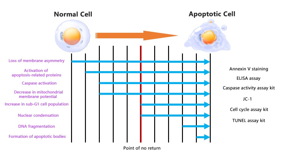
Fig. 1 Apoptosis progression diagram.
02 Intrinsic and extrinsic apoptotic pathways
Apoptotic inducing signals are mainly divided into the extrinsic death receptor pathway and the intrinsic mitochondrial pathway. These two pathways initiate independently, complement each other, and ultimately both target and activate Caspase 3/7 to complete the apoptotic program.
2.1 Extrinsic apoptotic pathway and death receptor signaling
This pathway is initiated by death receptors on the cell membrane surface and primarily responds to extracellular apoptotic signals. Typical stimuli include FasL, TNF-α, and TRAIL. When extracellular apoptotic ligands specifically bind to cell membrane death receptors, they recruit intracellular adaptor proteins and activate the initiator protease Caspase-8. Activated Caspase-8 further cleaves and activates downstream Caspase 3/7, rapidly initiating apoptosis. This pathway is mainly involved in immune clearance and the killing of cells infected by viruses.
2.2 Intrinsic apoptotic pathway and mitochondrial signaling
This pathway is triggered by intracellular stress signals, including DNA damage, oxidative stress, drug toxicity, nutrient deprivation, and radiation damage. Stress signals dysregulate the balance of Bcl-2 family proteins, promoting increased mitochondrial membrane permeability and the release of cytochrome C, which binds Apaf-1 to form the apoptosome, activating the initiator Caspase-9. This further activates Caspase 3/7 in a cascade manner. It is a major pathway for apoptosis caused by drugs in tumor cells and apoptosis caused by cell damage.
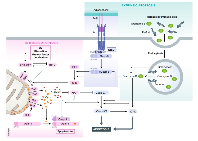
Fig. 2 Intrinsic and extrinsic apoptotic pathways[1].
03 Combined detection of Caspase 3/7 for apoptosis analysis
3.1 Combined detection of Caspase 3/7 and Annexin V
Detection principle: Annexin V specifically binds to phosphatidylserine (PS) that is externalized to the membrane surface during apoptosis, labeling apoptotic cells undergoing PS externalization. Caspase 3/7 fluorescent substrates label the activation of intracellular Caspase 3/7. Combined staining of the two allows the distinction of live cells (negative for both), early apoptotic cells with PS externalization but without significant Caspase activation yet (positive for Annexin V only), early apoptotic cells in which Caspase activation precedes PS externalization (positive for Caspase 3/7 only), and early to mid to late apoptotic cells with relatively high Caspase activity (positive for both), thereby reducing the risk of misinterpretation associated with assessment using a single indicator.
Result presentation:
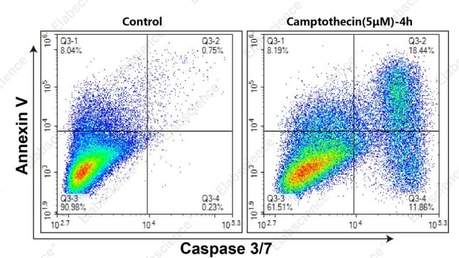
Fig. 3 Molt-4 cells induced with 5 μM camptothecin and stained with both Caspase 3/7 and Annexin V.
Result interpretation: Molt-4 cells were treated with 5 μM camptothecin (right) and without drug (left) for 4 h, stained using the Caspase 3/7 and Annexin V Double Staining Apoptosis Kit (E-CK-A831), and analyzed by flow cytometry. Cells positive for Annexin V only (Q3-1) are early apoptotic cells undergoing PS externalization; cells positive for both Annexin V and Caspase 3/7 (Q3-2) are early to mid to late apoptotic cells with relatively high Caspase 3/7 activity; cells negative for both Annexin V and Caspase 3/7 (Q3-3) are live cells that have not undergone apoptosis; cells positive for Caspase 3/7 only (Q3-4) are early apoptotic cells in which Caspase 3/7 enzymes have already been activated.
3.2 Combined detection of Caspase 3/7 and PI or DAPI
Detection principle: This addresses the experimental challenge of distinguishing apoptosis from necrosis. Through the dual indicators of "molecular apoptotic signal + cell membrane integrity," it precisely distinguishes early apoptosis, late apoptosis, necrosis, or cells with disrupted membranes, avoiding interference from necrotic cells in the statistical results of apoptosis experiments.
Result presentation:
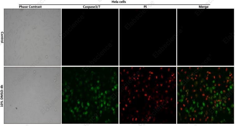
Fig. 4 HeLa cells treated with 10% DMSO for 4 h and stained with both Caspase 3/7 and PI.
Result interpretation: HeLa cells treated with 10% DMSO for 4 h were stained using the Caspase 3/7 and PI Double Staining Kit (E-CK-A832) and photographed under a fluorescence microscope. Cells stained only with Caspase 3/7 are early apoptotic cells, as Caspase 3/7 has been activated, but the cell membrane remains intact; cells stained with both Caspase 3/7 and PI are late apoptotic cells, as Caspase 3/7 is activated and cell membrane permeability is increased; cells stained only with PI are necrotic cells or cells with disrupted membranes, indicating that the cell membrane is disrupted but apoptosis dependent on Caspase 3/7 has not occurred.
Apoptosis is regulated by both the extrinsic death receptor pathway and the intrinsic mitochondrial pathway. As the core execution molecules where the two pathways converge, Caspase 3/7 are key markers for determining whether cells have undergone programmed apoptosis. A single detection indicator is prone to false positives or false negatives. By combining Caspase 3/7 with Annexin V or PI/DAPI, respectively, for flow cytometry or fluorescence imaging detection, live cells, apoptotic cells at different stages, and necrotic cells can be distinguished across multiple dimensions, including molecular activation, cell membrane integrity, and cell morphology, making apoptosis experimental results more rigorous and reliable.
Elabscience® Products related to Caspase activity assays
Table 1. Elabscience® products for caspase activity detection and apoptosis analysis
04 Related resources
● Explore the mechanisms of caspase-mediated apoptosis and learn how to detect Caspase-3/7/8/9 activation using complementary staining methods.
References:
[1] Galluzzi L, et al. Cell death pathways: intricate connections and disease implications[J]. The EMBO Journal, 2021, 40(10): e106934.

