

Sandwich enzyme-linked immunosorbent assay (ELISA) remains the workhorse for specific protein quantification in research and diagnostics, because two antibodies that grip the same antigen at different epitopes deliver a selectivity that single-binder formats cannot match. From picogram-level cytokine monitoring in inflammation to antigen quantification in vaccine quality control, the format underpins decisions in immunology, infectious disease, and oncology. Yet the gap between a kit insert protocol and a truly optimized assay is wide: mismatched antibody pairs, under-blocked plates, rushed incubations, and uncorrected matrix effects quietly erode sensitivity, reproducibility, and quantitative credibility, and troubleshooting usually starts only after precious samples are spent.
This review article walks through the six decisions that close that gap for sandwich ELISA developers and users. It explains which components must be used as a matched pair and why non-competing epitopes are non-negotiable, how to select capture and detection antibodies for potency and variant coverage, how to optimize antibody coating and blocking conditions for a clean background, how to tune incubation and washing steps for each specific pair, how to push detection down to low-abundance cytokines with amplified readouts, and how to handle sample dilution and matrix effects in serum, plasma, and culture supernatants.
Table of Contents
1. Which components of a sandwich ELISA must be used as a matched pair?
2. How to select capture and detection antibodies for sandwich ELISA
3. Optimizing antibody coating and blocking conditions
4. Optimizing incubation and washing steps in sandwich ELISA
5. Detecting low-abundance cytokines with sandwich ELISA
6. Sample dilution and matrix effects in sandwich ELISA
7. Key takeaways
8. Related resources
01 Which components of a sandwich ELISA must be used as a matched pair?
A sandwich ELISA works only when its two antibodies are used as a matched pair: a capture antibody immobilized on the plate and a detection antibody that recognizes two distinct, non-overlapping epitopes on the same antigen at the same time[1]. If the epitopes overlap or the antibodies compete, sandwich formation collapses and sensitivity is lost, so systematic pairwise screening rather than single-antibody affinity alone determines assay success. Rossotti et al. formalized this into a standardized sandwich elisa protocol for pair selection using camelid single-domain antibodies (nanobodies)[1]. By biotinylating 96 clones in vivo and saturating avidin-coated wells to normalize capture density, they ranked all clones by reactivity with labeled soluble epoxide hydrolase (sEH), grouped them into four epitope families, and tested a restricted panel of five capture antibodies against the full clone set. This epitope-binning strategy yielded a sandwich ELISA for sEH with a detection limit of 63 pg/mL, a dynamic range of about four logs (SC50 3.97 plus-or-minus 0.15 ng/mL), and excellent recovery in different tissue extracts[1]. That pairs must additionally be validated in real matrices, not just with recombinant protein, was shown by Watzlawik et al., who screened 15 anti-PRKN antibodies and compared every capture/detection combination in human cells and mouse brain[2]. Only pairs #1/#11 and #2/#11 performed best on human samples (limit of quantification below 10 pg/mL, i.e. below 194 fM; linearity 0 to 100 and 0 to 50 ng/mL; coefficient of variation below 5%), whereas #3/#13 and #4/#11 were superior on mouse samples, and knockout (KO)-verified testing across wild-type, PRKN KO, and PINK1 KO genotypes confirmed specificity and even gene-dosage resolution[2]. The same screen-then-validate logic underpins diagnostic development: Liu et al. generated six monoclonal antibodies against monkeypox virus (MPXV) A29L protein and, from the characterized panel, built a sandwich ELISA with a detection limit of 31.25 pg/mL, a linear range of 31.25 to 2,000 pg/mL, and 95% sensitivity with 100% specificity on plasma samples[3]. Together, these studies define the matched-pair rule: two non-competing antibodies, selected by combinatorial screening and confirmed in the intended sample matrix.
02 How to select capture and detection antibodies for sandwich ELISA
Selecting the capture and detection antibodies is the single most consequential step of any elisa assay protocol, and three criteria decide whether a candidate pair will survive contact with real samples. First, each antibody needs proven specificity for the target plus an epitope that stays accessible in the sample matrix. Cai et al. illustrate disciplined selection: from broad-spectrum neutralizing antibodies against rabies virus they characterized three human-mouse chimeric monoclonal antibodies (05-2F10, 06-2A12, and 07-4D8-1C1), ranked them by in vitro pseudovirus neutralization potency and breadth, and only then assembled a double-antibody sandwich ELISA to quantify glycoprotein (G) antigen in human rabies vaccines[4]. The resulting assay tracked the traditional in vivo National Institutes of Health (NIH) mouse challenge potency data, confirming that functionally potent antibodies make quantitatively reliable assay reagents[4]. Second, affinity alone is not enough when the target is polymorphic. Lopez-Borrego et al. compared five alleles of major histocompatibility complex (MHC) class I chain-related gene A (MICA), the ligand of the activating immune receptor NKG2D (Natural Killer group 2 member D), and found that a dimorphism at position 129 governs binding: MICA002 and MICA011 carry methionine (Met129) and bind NKG2D with higher affinity, while MICA008, MICA009, and MICA019 carry valine (Val129)[5]. Allele-specific structural dynamics further changed recognition by certain anti-MICA antibodies commonly used for immunoassays, so an antibody pair validated against only one variant can under-detect others[5]. Third, antibodies must tolerate the intended matrix: EV-associated versus soluble antigen forms behaved differently in the same study, reminding developers to test pairs in the actual sample type rather than buffer alone[5]. In practice, then, a defensible selection workflow screens for specificity and non-overlapping epitopes, ranks candidates by functional potency, verifies detection across known variants, and confirms the final pair against a reference method before locking the assay.
03 Optimizing antibody coating and blocking conditions
Coating and blocking set the ceiling for every sandwich elisa assay: too little capture antibody starves the signal, while incomplete blocking lets sample proteins stick to the plastic and inflate the background. Cai et al. show what a locked-down sandwich protocol looks like: two non-competing capture monoclonal antibodies (07-4D8-1C1 and 06-2A12) blended in equal ratio in carbonate buffer and coated onto microtiter plates with overnight incubation at 4 degrees Celsius to build a sandwich ELISA for rabies virus glycoprotein (G) antigen[4]. Plates were coated at 100 microliters per well, washed three times with PBS containing Tween (PBST), and blocked with 200 microliters of blocking buffer at 37 degrees Celsius for 2 to 4 hours before drying overnight[4]. The acceptance logic is instructive: a linear range of 0.003125 to 0.05 international units per mL with curve fits above R squared 0.999, intra- and inter-day precision within the 15% coefficient of variation (CV) acceptance criterion, and specificity confirmed by flat baselines for non-target vaccines and serum matrices[4]. That a well-blocked surface lets even demanding formats work is illustrated by Zheng et al., whose epitope-mapped monoclonal antibodies against Echinococcus multilocularis thioredoxin peroxidase (EmTPx), targeting the surface-exposed linear epitopes 174HPGSK178 and 189SFMSS193, supported a sandwich ELISA detecting parasite antigen directly in feces with 85% positivity at early infection, a linear range of 0 to 250 ng/mL, a 0.1724 cutoff, and intra- and inter-assay precision below 10%[6]. The practical rule is therefore to titrate coating to saturation, block with the validated buffer and timing for each pair, and re-verify blank, cutoff, and precision whenever either changes.
04 Optimizing incubation and washing steps in sandwich ELISA
Incubation and washing are where elisa antibody pairs either prove themselves or fall apart, because every one of the 16 possible capture/detection combinations can behave differently once time, temperature, and stringency enter the picture. Zhang et al. make this concrete: screening 16 capture/detection combinations for feline panleukopenia virus (FPV) VP2, they found that a single antibody, 4D10, gave the highest signal-to-noise ratio and could serve as both capture and detection reagent in a homologous double-antibody sandwich ELISA (DAS-ELISA)[7]. The lesson is structural: 4D10 was raised against recombinant VP2 expressed in Escherichia coli, biasing selection toward linear, denaturation-resistant epitopes that survive plate coating and sample handling, whereas antibodies against native conformational epitopes often lose reactivity in the same workflow[7]. The resulting assay detected 10^1.67 TCID50 per 0.1 mL and agreed completely with conventional polymerase chain reaction (PCR) across clinical samples, showing that epitope-type choice directly controls field performance[7]. Ye et al. illustrate the complementary variables on the detection side with a shark-derived variable new antigen receptor (VNAR) nanobody against avian influenza H9N2: their workflow fixes blocking at 3% to 5% bovine serum albumin (BSA), stringent Tris-buffered saline-Tween (TBST) washes between every step, and defined serum and secondary-antibody incubations (room temperature to 37 degrees Celsius, up to 2 hours), because residual unbound reagent is the dominant background source when amplification is pushed[8]. In practice, optimization therefore means titrating capture coating against incubation time and temperature for each pair, washing until blank wells stay flat, adding repeat Tween washes whenever backgrounds creep up, and preferring antibodies whose epitopes tolerate the denaturing realities of coated plates and clinical matrices.
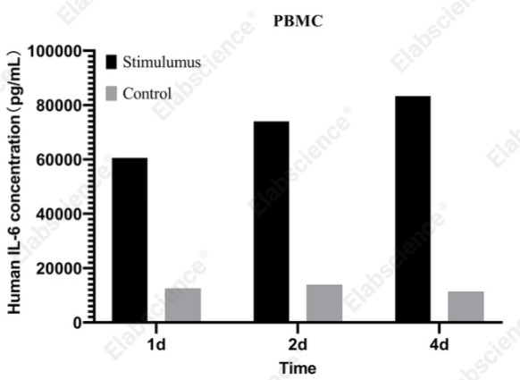
Fig. 1 An IL-6 detection kit for measuring IL-6 released from cell samples. (The data are provided by Elabscience®)
Assay Used in This Experiment:
CellaQuant™ Human IL-6(Interleukin 6) ELISA Kit
Cat. No: CQH001
Sample Type: cell culture supernatant, cell lysates or other biological fluids
Detection Range: 4.69-300 pg/mL
Sensitivity: 2.81 pg/mL
Detection Instrument: Microplate reader
Explore the CellaQuant™ Human IL-6 ELISA Kit Used in 200 Publications →
05 Detecting low-abundance cytokines with sandwich ELISA
Detecting low-abundance cytokines with a sandwich ELISA means fighting on two fronts at once: the capture antibody must pull vanishingly few molecules out of a complex fluid, and the readout must then make those few events visible. Zhang et al. attack the readout side with radical-mediated in situ fluorescence dye deposition (RIFD) on barcode beads, where free radicals trigger local, ultrafast (within 5 minutes) dye trapping specifically on target-positive beads through amide condensation on lysine residues, covalent biphenyl formation on tyrosine residues, or radical-dye co-deposition[9]. The RIFD immunoassay reaches a limit of detection of 12 fg/mL for interleukin 10 (IL-10), a 100-fold improvement over the conventional suspension-chip method, pushing protein detection into the sub-picogram-per-milliliter regime that early diagnosis demands[9]. Elkhiyari et al. take a multiplexing route with amplified enzymatic silver metallization on a plastic substrate, detecting picomolar levels of multiple cytokines including interferon gamma (IFN-gamma), the key readout for latent tuberculosis infection where only trace cytokine is released[10]. When even enzymatic amplification falls short, Han et al. fuse immunoassay to nucleic-acid amplification: their phage display-mediated immuno-PCR (PD-iPCR) uses high-affinity nanobodies and tandem NanoTags composed of VHH05 and 127D01 to quantify the insulin-binding protein ImpL2 in less than 100 nanoliters of Drosophila hemolymph, tracking its rise during starvation and gut tumorigenesis[11]. Finally, Lehmann et al. show that automation consolidates such gains into clinical-grade precision: their fully automated anti-IA2 ELISA reports intra- and inter-lot imprecision at or below 3.2% coefficient of variation (CV), with 76.0% sensitivity in new-onset type 1 diabetes and up to 98.4% specificity, outperforming the predecessor radioimmunoassay[12]. The common thread is quantitative rigor: the limit of blank (LoB), limit of detection (LoD), and limit of quantification (LoQ) determined per standardized guidelines; without which a low-abundance claim cannot be trusted.
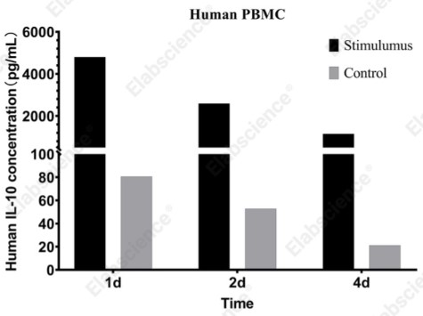
Fig. 2 An IL-10 detection kit for measuring IL-10 released from cell samples. (The data are provided by Elabscience®)
Assay Used in This Experiment:
CellaQuant™ Human IL-10 (Interleukin 10) ELISA Kit
Cat. No: CQH002
Sample Type: cell culture supernatant, cell lysates or other biological fluids
Detection Range: 1.56-100 pg/mL
Sensitivity: 0.94 pg/mL
Detection Instrument: Microplate reader
Explore the CellaQuant™ Human IL-10 ELISA Kit Used in 49 Publications →
06 Sample dilution and matrix effects in sandwich ELISA
Serum, plasma, and culture supernatants can suppress, mimic, or distort the antigen-antibody reaction, so sample dilution and matrix effects must be measured rather than assumed. Zhao et al. provide a textbook matrix-effect workup for a total human fibroblast growth factor 21 (FGF21) sandwich ELISA built on the rabbit monoclonal pair 5F6/20C3-Biotin, selected from 87 single B-cell clones and reaching a lower limit of quantification of 1.526 pg/mL, the highest sensitivity reported for this analyte[13]. Because human plasma components interfered with quantification, the team compared undiluted, 2-fold, and 3-fold diluted plasma: the 3-fold dilution stabilized the coefficient of variation (CV) across samples and brought quality-control recoveries into the 80 to 120% acceptance band at high, medium, and low control levels (576.0, 80.00, and 15.00 pg/mL), effectively diluting out the matrix effect while keeping the analyte on the standard curve (640.0 down to 1.250 pg/mL in 0.5% BSA-PBST)[13]. Validated across 22 human plasma samples, the assay even resolved the morning rise of circulating FGF21 diurnal rhythm, proving that a diluted matrix can still carry fine physiological detail[13]. The general principle extends to quality-control matrices: Cai et al. quantified glycoprotein antigen directly in formulated rabies vaccines with a double-antibody sandwich ELISA whose readout correlated with the in vivo National Institutes of Health potency test, showing that matrix-matched calibration holds even in adjuvanted formulations[4]. Practically, every new sample type deserves a dilution-linearity and spike-recovery check with the final detection antibody format, and any persistent non-linearity calls for a larger minimum required dilution or a matrix-matched standard curve rather than wishful reading of the numbers.
In summary, a sensitive and reproducible sandwich ELISA is engineered rather than assembled: a capture and detection antibody pair proven to bind non-overlapping epitopes, selected for functional potency and validated across antigen variants, coated to saturation and shielded by a matrix-appropriate blocker, incubated and washed under conditions tuned for each specific pair, and calibrated with dilution-linearity and spike-recovery checks in the real sample matrix. For routine low-abundance work, a ready-made cytokine elisa kit built on these principles saves development time, while custom targets reward the full optimization path. A concrete starting point is an IL-6 elisa, the classic low-picogram cytokine assay where pair quality, blocking, and matrix dilution visibly move the standard curve. Whatever the analyte, the developer who quantifies LoB, LoD, and LoQ instead of assuming them will hold an assay that performs in the publication exactly as it did on the bench.
Elabscience® Quick Overview of Popular Products:
Table 1. Research Tools for Biomarker Detection
|
Cat. No. |
Product Name |
|
E-EL-H0169 |
Human IgG(Immunoglobulin G) ELISA Kit |
|
E-EL-H0108 |
Human IFN-γ(Interferon Gamma) ELISA Kit |
|
E-EL-H6156 |
Human IL-6(Interleukin 6) ELISA Kit |
|
E-EL-R0015 |
Rat IL-6(Interleukin 6) ELISA Kit |
|
E-EL-M0044 |
Mouse IL-6(Interleukin 6) ELISA Kit |
|
E-EL-R0016 |
Rat IL-10(Interleukin 10) ELISA Kit |
|
E-EL-H6154 |
Human IL-10(Interleukin 10)ELISA Kit |
|
E-EL-M0046 |
Mouse IL-10(Interleukin 10) ELISA Kit |
|
CQH002 |
CellaQuant™ Human IL-10 (Interleukin 10) ELISA Kit |
|
CQM004 |
CellaQuant™ Mouse IL-10 (Interleukin 10) ELISA Kit |
|
CQR004 |
CellaQuant™ Rat IL-10 (Interleukin 10) ELISA Kit |
07 Key takeaways
Optimizing a sandwich ELISA is a chain of six linked decisions, and the assay is only as strong as its weakest link.
● Use two antibodies as a matched pair only after combinatorial screening proves non-competing epitopes, as shown by nanobody epitope binning reaching 63 pg/mL and knockout-verified PRKN pairs quantifying below 10 pg/mL.
● Select capture and detection antibodies for functional potency and variant coverage, since single-variant validation can miss alleles that escape detection.
● Coat capture antibody to saturation and block with the validated buffer and timing for each pair, holding precision within the 15% CV acceptance criterion.
● Tune incubation time and temperature plus wash stringency per antibody pair, preferring denaturation-resistant linear epitopes when plates and clinical matrices are harsh.
● Detect low-abundance cytokines with amplified readouts, from 12 fg/mL dye deposition to immuno-PCR in sub-100-nanoliter volumes, consolidated by automation.
● Dilute out matrix effects with demonstrated dilution linearity and 80 to 120% recovery, as shown for plasma FGF21 at 1.526 pg/mL.
The recommended workflow is therefore: Define the analyte and matrix → Select a matched pair by combinatorial screening → Lock coating, blocking, incubation, and wash conditions → Verify dilution linearity and spike recovery in the real matrix → Choose amplified readout when LoQ demands it.
08 Related resources
● Explore ELISA principles, protocols, and applications to support reliable immunoassay design and data analysis
Download Your Free ELISA Technology Guide →
● Learn the key steps for operating uncoated ELISA kits and performing reliable antibody-based detection.
Watch the Uncoated ELISA Kit Operation Video →
● Learn how to fit ELISA standard curves and accurately interpolate sample concentrations for quantitative analysis.
Use ELISA Curve Fitting Calculation Tool →
References:
[1] Rossotti MA, et al., A Method for Sorting and Pairwise Selection of Nanobodies for the Development of Highly Sensitive Sandwich Immunoassays. Analytical Chemistry, 2015. 87(23): 11907-11914.
[2] Watzlawik JO, et al., Robust and sensitive ELISA detection of total and activated PRKN. Autophagy, 2026. 22(11): 3014-3029.
[3] Liu J, et al., Development and preliminary clinical validation of A29L mAb-based ELISA and lateral flow immunoassays for monkeypox virus detection. Microbiology Spectrum, 2026. 14(7): e00167-26.
[4] Cai M, et al., A novel double-mAb sandwich ELISA utilizing broad-spectrum neutralizing antibodies as an in vitro alternative to the rabies vaccine NIH potency assay. Frontiers in Immunology, 2026. 17: 1960986.
[5] Lopez-Borrego S, et al., MICA Allelic Variation Modulates Secretion and Protein Flexibility, Impacting on NKG2D-Mediated Immune Responses and Antibody Detection. European Journal of Immunology, 2026. 56(10): e70304.
[6] Zheng L, et al., Conserved surface-exposed linear epitopes of Echinococcus multilocularis thioredoxin peroxidase enable non-invasive diagnosis of alveolar echinococcosis in intermediate host. PLOS Neglected Tropical Diseases, 2026. 20(9): e0014755.
[7] Zhang SJ, et al., Homologous Double-Antibody Sandwich ELISA Targeting a Linear Epitope of FPV VP2 and Demonstration of Microtubule-Dependent Intracellular Trafficking. Microorganisms, 2026. 14(10): 2412.
[8] Ye H, et al., Development and application of a VNAR-based detection nanobody for avian influenza virus H9N2. Frontiers in Immunology, 2026. 17: 1735255.
[9] Zhang J, et al., Radical-Mediated In Situ Fluorescence Dye Deposition: A Simple Interfacial Signal Amplification Reaction for Ultrasensitive Immunoassay on Barcode Beads. Advanced Science, 2026. 13(57): e76670.
[10] Elkhiyari K, et al., Inexpensive High-Throughput Multiplexed Cytokine Detection for Tuberculosis Diagnostics Using Amplified Enzymatic Metallization. ACS Sensors, 2026. DOI: 10.1021/acssensors.5c00160.
[11] Han M, et al., Phage display-mediated immuno-PCR to detect low-abundance secreted proteins in Drosophila. PNAS, 2026. 123(15): e2531777123.
[12] Lehmann M, et al., Performance assessment of a novel fully automated ELISA for anti-IA2 autoantibody detection in type 1 diabetes. Clinica Chimica Acta, 2026. 594: 122866.
[13] Zhao S, et al., Screening and validation of rabbit monoclonal antibody pair against human FGF21 for human plasma FGF21 high sensitivity bioanalysis. iScience, 2026. 29(6): 116260.

