

Ferroptosis has emerged as a key mechanism linking tumor cell death with antitumor immunity. Increasing evidence shows that ferroptosis regulates tumor immune microenvironment remodeling, CD8⁺ T cell function, and the efficacy of immune checkpoint blockade. Targeting ferroptosis-related pathways can overcome immune evasion, enhance immune cell infiltration, and improve responses to anti-PD-1/PD-L1 therapy across multiple tumor types.
Research Application Overview
This page summarizes representative peer-reviewed studies demonstrating how Elabscience® reagents have been applied in ferroptosis-related cancer immunotherapy research, covering iron metabolism, lipid peroxidation, GPX4 activity, immune activation, cytokine profiling, and immune checkpoint regulation.
Research Scope at a Glance
• Disease / Field: Cancer Immunotherapy
• Key Research Focus: Ferroptosis and tumor immune microenvironment, CD8⁺ T cell activation and immune evasion, PD-1/PD-L1 immunotherapy, Iron metabolism, ROS, and GPX4 signaling
• Experimental Models: Orthotopic and syngeneic mouse tumor models, genetically engineered mouse models, tumor cell lines, patient-derived tumor tissues, and CD8⁺ T cell-specific knockout models
• Common Assays: Iron quantification, GPX4 activity, ROS / lipid peroxidation assays, ELISA cytokine analysis, flow cytometry, multiplex immunofluorescence, and single-cell RNA sequencing
Literature-Based Experimental Application Matrix
The table below summarizes how Elabscience® reagents have been used across peer-reviewed studies in this research area.
Table 1. Literature-Based Application Matrix of Elabscience® Reagents in Ferroptosis–Immunotherapy Research
|
Research Focus |
Model |
Assay/Method |
Key Outcome |
Reference |
|
|
Tumor microenvironment remodeling via ferroptosis |
Orthotopic LUAD mouse model |
Iron metabolism assays, flow cytometry, mIF |
Ferrous Iron Colorimetric Assay Kit (E-BC-K773-M), Cell Total Iron Assay Kit (E-BC-K880-M), Total Iron Assay Kit (E-BC-K772-M) |
↑ TIMELESS expression → ↓ TF mRNA stability → ↓ iron uptake and ferroptosis sensitivity; ↓ 4-HNE; ↑ M2 macrophage infiltration, ↓CD4⁺/CD8⁺ T cells and M1 macrophages. TIMELESS promotes ferroptosis resistance and immune evasion, enhancing LUAD progression and PD-1 therapy resistance. |
[1] Hu et al., Cancer Communications (2026) |
|
Ferroptosis resistance and immune infiltration |
HCC mouse model |
ROS, lipid peroxidation, metabolic enzyme assays |
(E-BC-K099-M), (E-BC-K025-M), (E-BC-K097-M) |
↑ FADS expression → ↓ lipid peroxidation (↓ MDA) → ↑ ferroptosis resistance; ↓ CD8⁺ T cell infiltration; FADS knockdown → ↑ ROS, ↑ MDA, ↑ CD8⁺ T cell infiltration. FADS-driven VB2 metabolism suppresses ferroptosis and antitumor immunity in HCC. |
[2] Chao et al., Nature Communications (2025) |
|
CD8⁺ T cell ferroptosis and activation |
PCIF1 knockout mouse model |
EdU flow cytometry, activation assays |
E-Click EdU Cell Proliferation Flow Cytometry Assay Kit (Green,Elab Fluor® 488) (E-CK-A371) |
PCIF1 knockout → ↑ CD8⁺ T cell infiltration, ↑ CD69 expression → ↑ T cell activation; ↑ Fth1, Slc3a2 (ferroptosis resistance genes); ↓ tumor growth. PCIF1 limits CD8⁺ T cell antitumor activity by regulating ferroptosis-related gene networks and T cell activation. |
[3] Xiang et al., Nature Immunology (2025) |
|
Ferroptosis induction and T cell exhaustion reversal |
ESCC mouse model |
Lipid metabolism + immune profiling |
AA (Arachidonic Acid) ELISA Kit (E-EL-0051) |
c-Myc–CARM1 inhibition → ↑ arachidonic acid → ↑ lipid ROS → ↑ ferroptosis; ↑ CD8⁺ T cell infiltration; ↓ T cell exhaustion. Targeting the c-Myc complex enhances ferroptosis and restores antitumor immunity in ESCC. |
[4] Wang et al., International Journal of Biological Sciences (2026) |
|
Immune evasion through ferroptosis suppression |
LUAD cells + mouse tumor model |
Iron metabolism + cytokine assays |
Human GzmB ELISA Kit (E-MSEL-H0019), High Sensitivity Mouse IFN-γ ELISA Kit (E-HSEL-M0007), High Sensitivity Human IL-2 ELISA Kit (E-HSEL-H0002), MDA Fluorometric Assay Kit (E-BC-F007), Cell Ferrous Iron Colorimetric Assay Kit (E-BC-K881-M) |
↑ UCHL1 expression → enhanced FHL2 deubiquitination → ↓ ferroptosis; ↓ MDA and ↓ Fe²⁺ release; ↓ CD8⁺ T cell cytotoxicity. UCHL1 knockdown reverses these effects. UCHL1 suppresses ferroptosis to enable immune escape in LUAD. |
[5] Chen et al., Immunology (2025) |
|
ROS-driven ferroptosis and immunotherapy synergy |
TP53-mutant bladder cancer cells + syngeneic and orthotopic bladder tumor models |
ROS, cytokine profiling |
Human IP-10/CXCL10 ELISA Kit (E-EL-H0050), Mouse IP-10/CXCL10 ELISA Kit (E-EL-M0021), Penicillin-Streptomycin Solution, 100 × (PB180120) |
APR-246 → ↑ ROS → ↑ ferroptosis and apoptosis; ↑ CCL5/CXCL10 → ↑ CD8⁺/CD4⁺ T cells and NK cells; enhanced anti-PD-1 response. ROS-induced ferroptosis reprograms the TME and synergizes with PD-1 blockade in bladder cancer. |
[6] Zhang et al., Scientific Reports (2026) |
|
PD-1/PD-L1 signaling and ferroptosis-mediated immunity |
CRPC cell lines + xenograft mouse model |
PD-L1 + immune killing assays |
High Sensitivity Human IL-2 ELISA Kit (E-HSEL-H0002), High Sensitivity Human IFN-γ ELISA Kit (E-HSEL-H0007), Human PD-L1 ELISA Kit (E-EL-H1547) |
↑ HnRNP L → ↑ PD-L1; ↓ ferroptosis sensitivity → ↓ CD8⁺ T cell infiltration. Knockdown reverses phenotype. HnRNP L promotes immune evasion by inhibiting ferroptosis and upregulating PD-L1 in CRPC. |
[7] Zhou et al., Acta Pharmaceutica Sinica B (2022) |
|
GPX4-mediated ferroptosis resistance |
TNBC mouse model |
Enzyme activity + hydroxylation analysis |
GPX4 Activity Assay Kit (E-BC-K883-M) |
↑ PSAT1 phosphorylation→ ↑ GPX4 stability → ↓ lipid ROS → ↓ ferroptosis; PSAT1 inhibition reverses all above phenotypes. PSAT1–GPX4 axis blocks ferroptosis and reduces immunotherapy efficacy in TNBC. |
[8] Zheng et al., Nature Chemical Biology (2025) |
Data summarized from peer-reviewed publications. Experimental details may vary between studies.
Representative Use Cases
Below are selected examples illustrating how our Elabscience® were applied in real experimental workflows.
Use Case 1: TIMELESS promotes tumor immune escape by suppressing transferrin-mediated ferroptosis
Source: [1] Hu et al., Cancer Communications (2026)
Research Question
How does the RNA-binding protein TIMELESS regulate ferroptosis and remodel the tumor immune microenvironment to impair anti-PD-1 immunotherapy?
Experimental Workflow
LUAD cell lines + orthotopic mouse model
↓
TIMELESS knockdown
↓
Erastin treatment ± anti-PD-1 antibody
↓
Iron metabolism assays + Flow cytometry + Multiplex IF + RNA sequencing
↓
Ferroptosis, immune infiltration, tumor growth, treatment response
Assay Strategy
• Integrated ferroptosis analysis with tumor immunotherapy evaluation
• Combined iron metabolism assays with immune cell profiling
• Validated molecular mechanisms using RNA sequencing and protein interaction analyses
• Assessed therapeutic synergy between ferroptosis induction and PD-1 blockade
Key Findings
• TIMELESS accelerated transferrin mRNA degradation and suppressed ferroptosis.
• TIMELESS depletion significantly increased intracellular iron accumulation and ferroptotic cell death.
• Combination of TIMELESS depletion, erastin, and anti-PD-1 therapy markedly prolonged survival.
• Increased CD8⁺ T cell and M1 macrophage infiltration while reducing immunosuppressive M2 macrophages.
• Identified the TIMELESS/TF axis as a promising therapeutic target for overcoming PD-1 resistance.
%20Total%20iron%20content%20was%20quantified%20using%20a%20colorimetric%20assay%20in%20the%20indicated%20groups_.png)
Fig. 3. TIMELESS deficiency enhances ferroptosis susceptibility in LUAD cells and organoids. (H) Total iron content was quantified using a colorimetric assay in the indicated groups. (Hu et al., 2026)
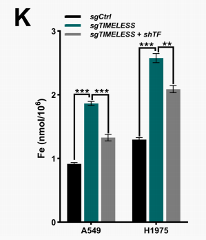
Fig. 5. TF silencing partially alleviates ferroptosis and restores tumor growth in TIMELESS knockout models. (K) Total iron content was quantified in A549 and H1975 cells from the sgCtrl, sgTIMELESS, and sgTIMELESS + shTF groups. (Hu et al., 2026)
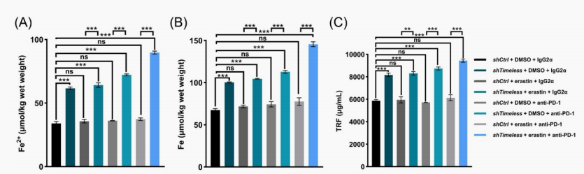
Supplementary Figure S13. Combined erastin and PD-1 blockade treatment induces ferroptosis and provides a favorable safety profile in Timeless-knockdown orthotopic lung tumors. (A-C) Fe2+ (A), total iron (B), and circulating TF (TRF) levels (C) in orthotopic tumors with or without Timeless knockdown (shTimeless) in response to combined erastin and PD-1 blockade treatment. (Hu et al., 2026)
Conclusion & Implications
This study demonstrates that ferroptosis is not only a tumor cell death mechanism but also a critical regulator of the tumor immune microenvironment. By targeting the TIMELESS/TF axis, ferroptosis induction enhances immune infiltration and improves responsiveness to anti-PD-1 immunotherapy, highlighting iron metabolism as an important therapeutic intervention point in cancer immunotherapy.
Related Resources
• Application Brochure: Cell Death Guide
• Experimental Videos: Cell Ferrous Iron Colorimetric Assay Kit Operation Guide Video
• Technical articles:
(1) How to Detect Ferroptosis Accurately Using Lipid ROS, Iron Assays, and GPX4 Validation
(2) Macrophage-Ferroptosis Crosstalk in Disease and Cancer
(3) Ferroptosis: Mechanisms, Disease Relevance, and Emerging Therapeutic Targets
Citation Note
All studies referenced on this page are published by independent research groups.
Figures and data are summarized or adapted for clarity. For full experimental details, please refer to the original publications.
Full Literature References
[1] Hu, C., Hu, F., Shao, C., He, Y., Su, L., Shi, D. & Yang, K. (2026). TIMELESS promotes LUAD growth via suppressing transferrin-mediated ferroptosis and reprograms the tumor microenvironment against anti-PD-1 immunotherapy. Cancer Communications.
[2] Chao, J., Liang, Y., Wang, H., Xun, Z., Wang, S., Xuan, Z. & Lu, L. (2025). FAD synthase confers ferroptosis resistance and restrains CD8+ T cell recruitment in hepatocellular carcinoma. Nature Communications, 16(1), 9547.
[3] Xiang, B., Zhang, M., Li, K., Zhang, Z., Liu, Y., Gao, M. & Zhang, J. (2025). The epitranscriptional factor PCIF1 orchestrates CD8+ T cell ferroptosis and activation to control antitumor immunity. Nature Immunology, 26(2), 252-264.
[4] Wang, Y., Li, Y., Ren, G., Zhou, J., Chen, W., Zhang, K. & Liu, Z. (2026). Targeting c-Myc-p300-CARM1 complex induces ferroptosis and reduces CD8+ T cell exhaustion in esophageal squamous cell carcinoma. International Journal of Biological Sciences, 22(3), 1266.
[5] Chen, X., Li, J., Tang, B., Wang, X., & Huang, Y. (2026). Deubiquitinating Enzyme UCHL1 Modulates FHL2 to Block Ferroptosis and Counteract CD8+ T Cell Anti‐Tumour Immunity in Lung Adenocarcinoma. Immunology, 177(2), 384-397.
[6] Zhang, C., Cao, S., Zeng, G., Dong, Y., Li, H., Ma, X. & Huang, Y. (2026). APR-246 drives ROS-dependent ferroptosis and apoptosis and enhances anti–PD-1 efficacy in bladder cancer. Scientific Reports.
[7] Zhou, X., Zou, L., Liao, H., Luo, J., Yang, T., Wu, J. & Mao, X. (2022). Abrogation of HnRNP L enhances anti-PD-1 therapy efficacy via diminishing PD-L1 and promoting CD8+ T cell-mediated ferroptosis in castration-resistant prostate cancer. Acta Pharmaceutica Sinica B, 12(2), 692-707.
[8] Zheng, P., Hu, Z., Shen, Y., Gu, L., Ouyang, Y., Duan, Y. & Xu, D. (2025). PSAT1 impairs ferroptosis and reduces immunotherapy efficacy via GPX4 hydroxylation. Nature Chemical Biology, 21(9), 1420-1432.

