

Adenosine triphosphate (ATP) is the central energy currency governing diverse cellular processes, ranging from biosynthetic reactions to immune cell activation. Accurate measurement of ATP levels relies on a spectrum of techniques, including the gold-standard luciferin-luciferase bioluminescence assays, real-time bioenergetic profiling via extracellular flux analyzers, and genetically encoded biosensors that permit live-cell imaging of subcellular dynamics. Beyond its canonical role in metabolism, ATP homeostasis serves as a critical determinant of cell fate; a precipitous drop in ATP shifts cell death from apoptosis to necrosis, particularly under pathological conditions such as ischemia and chemotherapy. In immunometabolism, fluctuations in the ATP/ADP ratio and metabolic reprogramming dictate T cell differentiation, influencing whether cells adopt an effector or memory phenotype.
In this context, we outline the essential methodological considerations for precise ATP quantification and highlight the molecule’s pivotal role as a biomarker of cellular stress and a metabolic checkpoint in health and disease.
Table of Contents
1. What is ATP and why is it essential for cellular energy?
2. How is ATP formed during cellular respiration?
3. Mitochondrial ATP production and bioenergetic homeostasis
4. ATP as a metabolic regulator of immune cell function
5. ATP depletion as a biomarker of cellular stress and cell death
6. Experimental methods for measuring intracellular ATP levels
01 What is ATP and why is it essential for cellular energy?
Adenosine triphosphate (ATP) is the universal energy currency of living cells, driving nearly all energy-requiring biochemical processes. Beyond bioenergetics, ATP plays pivotal roles in signal transduction, nucleic acid synthesis, enzyme catalysis, ion channel regulation, and protein phosphorylation[1,2]. Structurally comprising adenine, ribose, and three phosphate groups, ATP stores energy in high-energy phosphoanhydride bonds. Hydrolysis to ADP and inorganic phosphate releases approximately 30.5 kJ/mol under standard conditions, fueling anabolic processes, active transport, muscle contraction, and cellular motility[1].
The dynamic interconversion between ATP and ADP underpins cellular energy metabolism. In healthy cells, cytosolic ATP concentrations are tightly maintained at 2.5–4.1 mM, with mitochondrial levels slightly lower[3]. This homeostasis reflects a balance between ATP consumption and generation. The ATP/ADP ratio, sensed by regulators such as AMPK, serves as a critical indicator of cellular energy status, orchestrating metabolic adaptations. By sustaining ionic gradients, biomolecular synthesis, and mechanical work, ATP remains indispensable for life[2].
02 How is ATP formed during cellular respiration?
Cellular respiration in mitochondria integrates sequential catabolic reactions to drive ATP synthesis. Glycolysis in the cytosol converts glucose to pyruvate, yielding two ATP and two NADH. Pyruvate then enters the mitochondrial matrix to form acetyl-CoA, which fuels the tricarboxylic acid (TCA) cycle. This cycle generates reducing equivalents (NADH and FADH₂), GTP, and CO₂[2,4].
The conversion of ADP to ATP culminates in oxidative phosphorylation. NADH and FADH₂ transfer electrons to the electron transport chain (ETC), driving proton pumping across the inner membrane to establish the mitochondrial membrane potential (ΔΨm)[5]. This proton motive force powers F₁F₀-ATP synthase (Complex V), catalyzing bulk ATP formation. Ultimately, complete glucose oxidation yields approximately 30–36 ATP, with oxidative phosphorylation accounting for the majority of this output[1,5].
03 Mitochondrial ATP production and bioenergetic homeostasis
Mitochondria serve as the central hubs of mitochondrial ATP production, orchestrating pathways that sustain bioenergetic homeostasis. Beyond energy synthesis, they supply biosynthetic intermediates such as citrate and α-ketoglutarate while maintaining redox balance[2]. The rate of mitochondrial ATP production is dynamically matched to cellular demand via the ATP/ADP ratio, which modulates respiratory flux. The adenine nucleotide translocase couples synthesis to consumption by exchanging matrix ATP for cytosolic ADP[1,5].
To ensure stable ATP output, mitochondria oxidize diverse substrates including pyruvate, fatty acids, and amino acids based on nutrient availability. Specific transporters, namely the mitochondrial pyruvate carrier and carnitine palmitoyltransferase 1, govern substrate entry[2]. The efficiency of mitochondrial ATP production, quantified by the P/O ratio and modulated by uncoupling proteins, reflects the tight coupling of oxidative phosphorylation essential for cellular viability[5].
04 ATP as a metabolic regulator of immune cell function
Immune cell activation triggers profound metabolic remodeling, placing ATP function in cells at the core of immunometabolism. Naïve T cells maintain quiescence through oxidative phosphorylation and fatty acid oxidation to support survival. Upon activation, T cells undergo rapid metabolic reprogramming, upregulating aerobic glycolysis to meet the biosynthetic and energetic demands of clonal expansion and effector functions[6,7].
This switch is driven by signaling pathways that enhance glucose uptake and activate glycolytic enzymes. Transcriptional regulators coordinate the expression of glycolytic and glutaminolytic genes to ensure sufficient ATP and precursor supply. Notably, glycolytic flux directly regulates effector functions, as certain glycolytic enzymes also control cytokine translation[6]. Distinct T cell subsets exhibit specialized metabolic profiles; effector T cells rely on glycolysis, while regulatory T cells favor oxidative metabolism. Memory T cells develop enhanced mitochondrial capacity to enable rapid recall responses[6–8].
The AMP/ATP ratio serves as a critical metabolic checkpoint, with sensors promoting oxidative metabolism during stress to sustain immune responses. These metabolic dependencies offer therapeutic opportunities, as targeted modulation of glycolytic or glutaminolytic pathways presents promising strategies for treating cancer, autoimmunity, and chronic infections[6,8].

Fig. 1 Human CD3+ T cells were isolated from PBMCs using a sorting kit and cultured in vitro. The experimental group was stimulated with CD3/CD28 activation beads, while the control group remained unstimulated. After several days of culture, cellular ATP levels were measured (E-BC-F201). Results showed that ATP content was higher in activated T cells than in the control group (The data are provided by Elabscience).
05 ATP depletion as a biomarker of cellular stress and cell death
Intracellular ATP levels serve as a decisive switch between apoptosis and necrosis, with ATP depletion acting as both a hallmark and driver of cellular injury. Apoptosis is an energy-dependent process; once ATP falls below a critical threshold, cells shift from programmed cell death to necrosis. This mechanism underlies the prevalence of necrosis in pathologies involving energy crisis, such as ischemia, hypoxia, and metabolic poisoning[9].
The execution of apoptosis relies on ATP at multiple stages, including caspase activation, nuclear transport of effectors, and formation of apoptotic bodies. Severe ATP loss blocks these cascades entirely, redirecting cellular fate toward necrosis. Conversely, necrosis proceeds passively following catastrophic energy failure. This ATP-dependent bifurcation carries significant pathophysiological implications. In solid tumors, hypovascular regions trigger ATP depletion, fostering necrosis, inflammation, and heterogeneity. Similarly, acute ATP collapse during stroke or myocardial infarction exacerbates tissue damage. Additionally, hyperactivation of PARP in response to DNA damage can drain NAD⁺ and ATP reserves, directly linking genotoxic stress to necrotic cell death[9]. Consequently, monitoring ATP levels provides a critical biomarker for evaluating cellular viability, cytotoxicity, and therapeutic efficacy in clinical and preclinical settings.
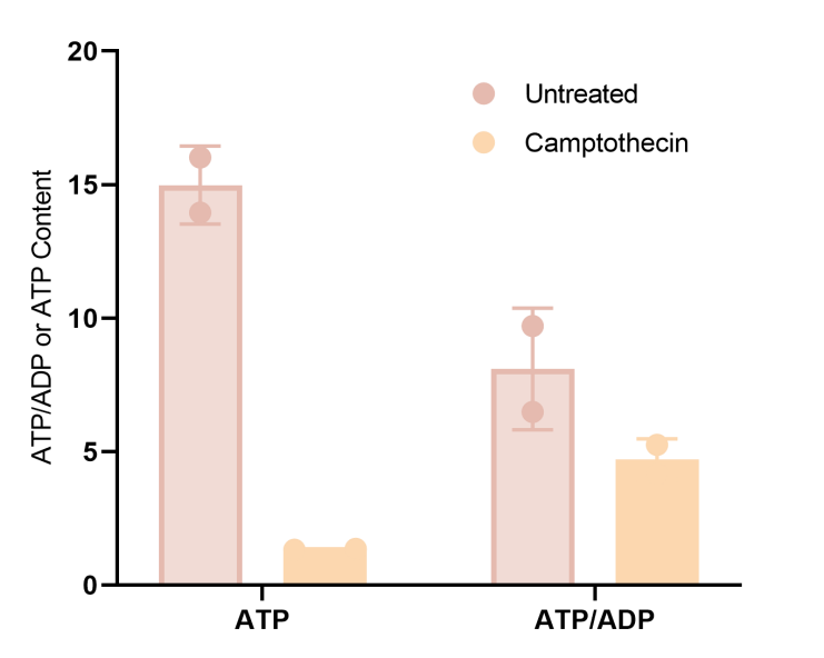
Fig. 2 After treatment with camptothecin apoptosis reagent, the ATP/ADP ratio (E-BC-F004) in 293T cells was decreased compared with untreated cells (The data are provided by Elabscience).
06 Experimental methods for measuring intracellular ATP levels
Accurate quantification of ATP is fundamental to deciphering cellular bioenergetics and evaluating cytotoxic or pharmacological effects. A diverse suite of methodologies supports these measurements, balancing sensitivity, throughput, and experimental context.
The gold-standard ATP assay utilizes the luciferin-luciferase bioluminescence reaction, which forms the basis of most commercial ATP detection kit formats. While offering exceptional sensitivity suitable for high-throughput screening, this approach requires cell lysis, precluding longitudinal studies. To overcome this, genetically encoded biosensors such as the BRET-based BTeam enable ratiometric quantification of cytosolic and mitochondrial ATP in live cells without excitation interference. Complementary fluorescence-based tools, including FRET sensors and AIE probes like TPE-Py-FP, facilitate subcellular imaging, with specific probes allowing real-time visualization of mitochondrial ATP dynamics[3,10,11].
For functional bioenergetic profiling, the Seahorse XF and Oroboros O2k systems quantify oxygen consumption and extracellular acidification. These data can be transformed into rates of cellular respiration in mitochondria and glycolysis, elucidating the contribution of each pathway to the total ATP pool under basal or stressed conditions. The selection of an ATP detection method ultimately depends on the required spatial resolution, temporal scale, and biological question[1,10,12].
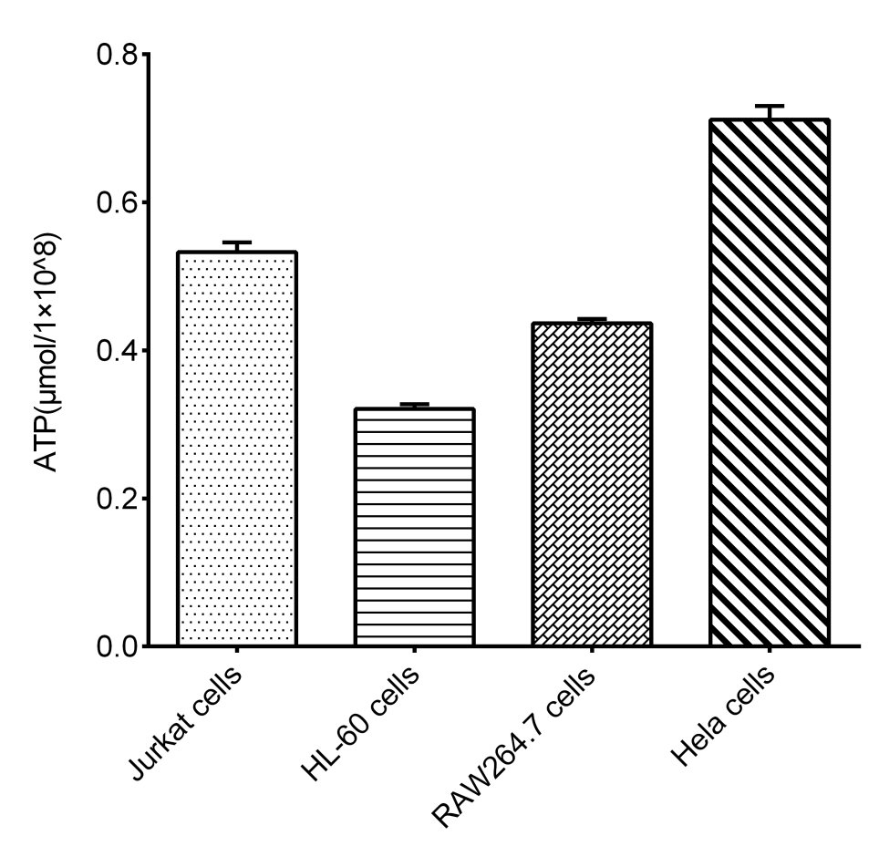
Fig. 3 ATP levels measured in different cell lines by ATP Chemiluminescence Assay Kit (E-BC-F300) (The data are provided by Elabscience).
Elabscience® Quick Overview of Popular Products:
Table 1. Assay Kits for energy metabolism
|
Cat. No. |
Product Name |
|
E-BC-F300 |
ATP Chemiluminescence Assay Kit (Double Reagent) |
|
E-BC-F004 |
ATP/ADP Ratio Chemiluminescence Assay kit |
|
E-BC-F201 |
Enhanced ATP Chemiluminescence Assay Kit |
|
E-BC-F070 |
Enhanced Oxygen Consumption Rate (OCR) Fluorometric Assay Kit |
|
E-BC-F069 |
Extracellular Acidification Rate (ECAR) Fluorometric Assay Kit |
|
E-BC-F084 |
Glycolysis Stress Fluorometric Assay Kit |
|
E-BC-K784-M |
Fatty Acid Oxidation (FAO) Colorimetric Assay Kit |
|
E-CK-A301 |
Mitochondrial Membrane Potential Assay Kit (with JC-1) |
|
E-BC-K153-M |
Mitochondrial Complex Ⅴ Activity Assay Kit |
|
E-BC-K149-M |
Mitochondrial Complex I (NADH-CoQ Reductase) Activity Assay Kit |
|
E-BC-K838-M |
Mitochondrial Complex Ⅴ Activity Assay Kit |
References:
[1] Handel, M.E., et al., The Whys and Hows of Calculating Total Cellular ATP Production Rate. Trends in Endocrinology & Metabolism, 2019. 30(7):412-416.
[2] Spinelli, J.B. and Haigis, M.C., The Multifaceted Contributions of Mitochondria to Cellular Metabolism. Nature Cell Biology, 2018. 20(7):745-754.
[3] Yoshida, T., et al., BTeam, a Novel BRET-based Biosensor for the Accurate Quantification of ATP Concentration Within Living Cells. Scientific Reports, 2016. 6(1):39618.
[4] Cliff, T.S. and Dalton, S., Metabolic switching and cell fate decisions: implications for pluripotency, reprogramming and development. Current Opinion in Genetics & Development, 2017. 46(1):44-49.
[5] Hill, B.G., et al., Mitochondrial Electron Transport Chain: Oxidative Phosphorylation, Oxidant Production, and Methods of Measurement. Redox Biology, 2020. 37:101674.
[6] Chang, C.-H. and Pearce, E.L., Emerging Concepts of T Cell Metabolism as a Target of Immunotherapy. Nature Immunology, 2016. 17(4):364-368.
[7] Madden, M.Z., et al., The Complex Integration of T-cell Metabolism and Immunotherapy. Cancer Discovery, 2021. 11(7): p. 1636-1643.
[8] Klein Geltink, R.I., et al., Unraveling the Complex Interplay Between T Cell Metabolism and Function. Annual Review of Immunology, 2018. 36(1):461-488.
[9] Tsujimoto, Y., Apoptosis and Necrosis: Intracellular ATP Level as a Determinant for Cell Death Modes. Cell Death & Differentiation, 1997. 4(6):429-434.
[10] Manfredi, G., Measurements of ATP in Mammalian Cells. Methods, 2002. 26(4):317-326.
[11] Yang, B., et al., Tetraphenylethylene Fluorophore Based AIE-Fluorescent Probe for Detection of ATP in Mitochondria. Dyes and Pigments, 2023. 215:111295.
[12] Lee, M.-S., et al., Intracellular ATP Assay of Live Cells Using PTD-Conjugated Luciferase. Sensors, 2012. 12(11):15628-15637.

