

Cytokines are core soluble signaling molecules that orchestrate immune cell activation, differentiation, and intercellular crosstalk, serving as critical biomarkers for reflecting immune homeostasis, inflammatory status, and disease progression. Accurate, sensitive, and standardized quantification of cytokine expression is therefore fundamental to immunological mechanism exploration, disease biomarker screening, immunotherapy evaluation, and vaccine immunogenicity assessment. Enzyme-linked immunosorbent assay (ELISA) has long been regarded as the most classic and widely applied technique for cytokine detection due to its high specificity, convenient operation, and reliable quantitative performance. Nevertheless, the continuous advancement of immunological research and the growing demand for detecting trace level cytokines and multiplex immune signatures have driven the iterative upgrading of ELISA-related technologies. In recent years, conventional single-plex ELISA has gradually evolved toward high-throughput ELISA multiplex platforms, ultrasensitive single molecule detection, automated standardized systems, and digital quantitative modalities, greatly overcoming the limitations of traditional assays in sensitivity, throughput, sample consumption, and reproducibility. Furthermore, continuously optimized ELISA systems have been extensively verified in DC-T cell co-culture functional experiments, enabling comprehensive phenotypic and mechanistic interpretation of complex immune responses.
This review article systematically summarizes the technical principles, advantages and limitations of conventional cytokine ELISA assay, elaborates the evolutionary trajectory from single-plex detection to high density multiplex profiling, and highlights emerging high sensitivity ELISA strategies, automated high-throughput platforms, and state of the art digital ELISA technologies. Meanwhile, it illustrates the broad applications of ELISA based cytokine detection in functional immunological studies, aiming to provide an in depth understanding of technical advancements and guide standardized, high precision cytokine profiling for future immunological and translational research.
Table of Contents
1. What Are Cytokine ELISA Assays and Why Are They Widely Used in Immunology Research?
2. Evolution of Cytokine Detection: From Single-plex ELISA to High-Density Multiplex Platforms
3. High-Sensitivity ELISA for Low-Abundance Cytokine Detection in Serum and Plasma
4. Advances in Automated ELISA Platforms for High-Throughput Screening
5. Next-Generation ELISA: Digital ELISA and Ultra-Sensitive Detection Technologies
6. Using Cytokine ELISA for Immune Cell Co-Culture and Functional Assays
01 What Are Cytokine ELISA Assays and Why Are They Widely Used in Immunology Research?
Cytokine ELISA is a robust protein quantification technique based on the double antibody sandwich principle. This workflow uses pre-coated capture antibodies to immobilize target cytokines in biological samples, followed by enzyme-conjugated detection antibodies to form immune sandwich complexes. Subsequent colorimetric reactions enable quantitative cytokine measurement, with the assay achieving high sensitivity at the picogram level. It is applicable to common biological matrices, including serum, plasma, and cell culture supernatants[1-3]. Cytokine ELISA kit supported ELISA has become a mainstream tool in immunological research, largely attributable to its superior specificity, rapid detection, and simplified operational procedures relative to traditional cytokine bioactivity assays[1,4]. Its excellent analytical performance and accessibility support extensive standardized applications in immunology: it serves as a key tool for detecting cytokine biomarkers in infectious, autoimmune, and malignant diseases to assess disease activity and prognosis; enables longitudinal cytokine dynamic monitoring in immunotherapy and vaccine clinical trials; and facilitates pharmacokinetic analysis of recombinant cytokine drugs[1,5,6].Nevertheless, this technique has inherent methodological pitfalls that may compromise data reliability. Variations in antibody pairs and calibration standards among commercial kits can cause substantial measurement discrepancies for identical samples. Pre analytical variables, including sample types, anticoagulants, storage conditions, and freeze-thaw cycles, greatly affect cytokine stability. Moreover, endogenous sample interferents such as soluble binding receptors and heterophilic antibodies may induce false detection results[1,3,4,6]. Strict quality control, including spike and recovery tests, dilution parallelism verification, and international standard calibration, is therefore required to ensure reliable and comparable ELISA data across studies.
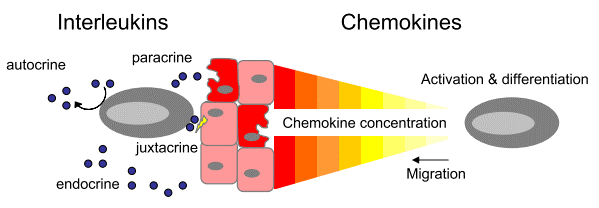
Fig. 1 General function of cytokines. When secreted cytokines (interleukins) can act on cells or tissue that secrete them (autocrine), surrounds them (paracrine), travel to distant cells (endocrine) or remain cell bound and activate neighboring cells (juxtacrine). On the other hand chemokines are regulatory proteins which induce migration, activation and differentiation of cells to inflamed or damaged tissue[4].
02 Evolution of Cytokine Detection: From Single-plex ELISA to High-Density Multiplex Platforms
Advancements in cytokine detection technologies mark a systematic transition from conventional single-plex ELISA to high-density multiplex analytical platforms. As the long standing gold standard for single analyte quantification, traditional ELISA delivers robust specificity and picogram level sensitivity. However, it suffers from critical limitations: only one target cytokine can be measured per assay run, it requires large sample volumes (≥100 μL), and it involves lengthy incubation procedures with substantial reagent costs[3,4]. To dissect the intricate and redundant cytokine regulatory networks, researchers have developed a series of alternative detection strategies. These include sequential ELISA, which splits a single sample across multiple ELISA plates for separate measurements[2]; fluorescent coded microsphere suspension arrays (e.g., Luminex xMAP) and electrochemiluminescence planar arrays (e.g., MSD)[7]; and protein microarrays exemplified by nucleic acid programmable protein chips[8]. Label free optical biosensors based on silicon photonic microring resonators have also been established to enable near real time multiplex kinetic monitoring of cytokines[9]. Multiplex cytokine assay platforms possess prominent analytical merits. They require merely microliter quantities of sample to quantify dozens to hundreds of cytokines simultaneously, with an extended dynamic range covering 3 to 5 orders of magnitude. Such systems drastically boost testing throughput and cut cumulative expenses compared with repeated single-plex ELISA measurements. Nevertheless, multiplexed cytokine detection still faces unresolved technical hurdles, including antibody cross reactivity, incompatible antibody pair combinations, and inconsistent absolute quantification values across distinct instrumental platforms[10]. The technical shift from single-plex ELISA to high-density multiplex systems reduces both sample consumption and experimental turnaround time. More importantly, it transforms analytical paradigms from single biomarker assessment to multi cytokine signature profiling, laying an essential technical foundation for precision immunodiagnostics and mechanistic investigations of complex diseases[7,8].
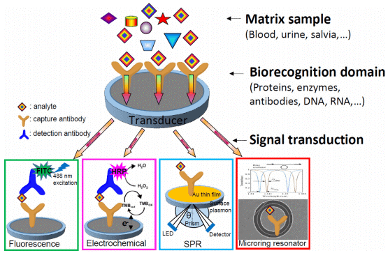
Fig. 2 The general scheme of an immunosensor which includes matrix sample, biorecognition domain and signal transduction. Four important signal transduction approaches are schemed for cytokine detection, such as fluorescence immunosensing, electrochemical immunosensing, surface plasmon resonance (SPR) based and microring resonator based immunosensing[10].
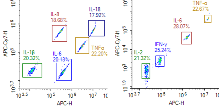
Fig. 3 The Presentation of multiplex cytokine assay data. (All experimental data were provided by Elabscience®)
03 High-Sensitivity ELISA for Low-Abundance Cytokine Detection in Serum and Plasma
Quantification of low-abundance cytokines (e.g., IL-6, IL-10, IFN-γ) in serum and plasma poses substantial analytical challenges, as their physiological concentrations often fall at pg/mL or even fg/mL levels, far below the limit of detection (LOD) of conventional ELISA. To address this technical bottleneck, multiple high-sensitivity detection strategies have been developed. Single Molecule Counting (SMC) generates digital readouts from laser excited fluorophore labeled antibody–antigen immune complexes. This technology achieves limits of quantification (LOQ) of 0.05 pg/mL for IL-17A and 0.15 pg/mL for IL-17F, enabling clear discrimination of serum cytokine profiles between psoriasis patients and healthy volunteers, whereas standard ELISA fails to detect these analytes in identical specimens[11]. Additional SMC based work has established the first reference intervals and biological variability datasets of circulating IL-6, TNF-α and IL-17A in healthy populations, establishing critical benchmarks for clinical data interpretation[12]. As a core detection tool for inflammatory biomarkers, IL-6 elisa kit enables precise quantification of trace IL-6 in peripheral blood, supporting the assessment of systemic inflammatory levels in various diseases.Nucleic acid linked immunosorbent assay (NULISA), another cutting edge modality, employs a dual capture and release mechanism for immune complexes to suppress background signals by over 10,000 fold. This platform attains attomolar (10^-¹⁸ M) sensitivity for IL-4 with a 7 log dynamic range, while retaining equivalent analytical performance within a 200-plex multiplex panel[13]. Digital bead based ELISA (e.g., Simoa) sequesters individual immune complexes in femtoliter sized microwells to enable single molecule enumeration; its sensitivity for HIV p24 detection is approximately 10 fold higher than that of conventional ELISA. Various signal amplification approaches also push detection sensitivity down to the fg/mL range, including nanomaterial functionalized sensing surfaces, enzymatic signal enhancement (e.g., HRP mediated 4 chloro 1 naphthol precipitation), and cascaded DNA amplification systems such as hybridization chain reaction and CRISPR/Cas13a collateral cleavage[6]. In conclusion, reliable high-sensitivity profiling of low-abundance cytokines relies on three core pillars: digital single molecule counting or background suppressed immunoassay architectures, complementary signal amplification via nanomaterials or enzymatic reactions, and fully standardized pre analytical protocols. Combined, these elements facilitate robust translational research from laboratory discovery to clinical implementation.
04 Advances in Automated ELISA Platforms for High-Throughput Screening
Automated ELISA platforms integrate the full analytical workflow, including sample handling, incubation, washing and signal readout, substantially boosting assay throughput and experimental standardization. Conventional manual ELISA is labor intensive and time consuming, with substantial inter-batch deviations arising from inconsistent operator performance. By contrast, automated systems equipped with high precision liquid handlers and integrated washers/plate readers cut hands on operational time by 6–10 fold and raise daily analytical throughput from fewer than 200 data points to over 1,300[14]. In terms of standardization, automation eliminates manual pipetting errors and inconsistent timing control, stabilizing intra-assay and inter-assay coefficients of variation (CV) at approximately 10% and 12%, respectively. Preset standardized scripts further guarantee uniform execution across all experimental steps[15,16]. Additionally, automated instruments support flexible sample dilution schemes and parallel multi plate processing, and enable traceable quantification when calibrated against ISO/WHO international reference materials[17]. Self-contained microfluidic or centrifugal force driven systems further miniaturize hardware footprint and cut overall costs to less than one-fortieth of conventional automated ELISA workstations[18,19]. These technical advances have rendered automated ELISA no longer exclusive to well-resourced central laboratories; the technology is increasingly adopted in routine clinical diagnostic facilities and resource limited regions[18, 20]. It provides a high-throughput, standardized technical foundation for a wide range of applications, including cytokine signature profiling, therapeutic antibody potency assessment, and serological surveillance of infectious diseases[14,16].
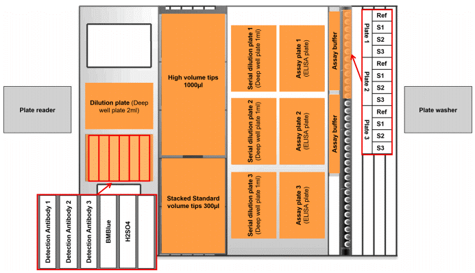
Fig. 4 Layout of the automated liquid handler deck for the automated potency ELISA. The “occupied” positions are orange. The red rectangle and arrow on the right show the positions of the reference standard (Ref) and samples (S1 - S3) for three ELISA plates. The red rectangles and arrow on the left indicate the position of HRP substrate (BMBlue), HRP stop solution (H2SO4), and detection antibodies for assay plates 1, 2 and 3. Plate reader and plate washer are positioned outside and accessible by the plate transport arm of the liquid handler[15].
05 Next-Generation ELISA: Digital ELISA and Ultra-Sensitive Detection Technologies
Digital ELISA marks a fundamental paradigm shift in immunoassays from analog to digital signal readout. Its core innovation lies in converting continuous analog signals of conventional ELISA into discrete counting of individual immune complexes, enabling ultrasensitive quantification at fg/mL or even ag/mL concentrations[21]. This technology operates on Poisson distribution statistics. When the number of target molecules is far lower than the number of microreactors (microdroplets or microwells), each microreactor accommodates at most one analyte molecule. Positive microreactors (digital “1”) and negative microreactors (digital “0”) are distinctly discriminated via accumulated enzymatic fluorescent products, and absolute quantification is achieved by enumerating all positive compartments[22,23]. A typical commercial platform, Quanterix Simoa single molecule array, adopts femtoliter scale microwell chips and delivers approximately 1,000 fold higher sensitivity than standard ELISA. It enables robust measurement of plasma Alzheimer’s disease biomarkers including p-tau and NfL, creating new avenues for non invasive early diagnosis[24]. Nevertheless, widespread deployment of digital ELISA remains hindered by considerable obstacles. Multiplex detection suffers false positive signals originating from optical crosstalk and substrate diffusion; bulky instruments and prohibitive costs also limit its accessibility in resource limited settings[21,25]. Multiple optimization strategies have therefore been proposed: combined magnetic and capillary forces boost bead loading efficiency above 60% to mitigate Poisson noise; rolling circle amplification (RCA) and tyramide signal amplification (TSA) support microchamber free digital detection with nearly 100% bead utilization; portable systems integrated with smartphone imaging and centrifugal droplet generation cut per sample testing costs below $0.5[21]. Future development of digital ELISA will center on three key directions. First, integration with CRISPR/Cas signal amplifiers allows direct detection of RNA viruses without pre-amplification of nucleic acids[23]. Second, artificial intelligence based image recognition algorithms facilitate spatial encoding and automated decoding of multiple analytes to strengthen multiplexing capacity. Third, fully integrated, miniaturized point-of care testing (POCT) devices will transfer ultrasensitive protein detection from central laboratories to community clinics and at home self-testing. Sustained technological breakthroughs in digital ELISA are poised to push protein detection sensitivity toward the single molecule limit, offering revolutionary analytical tools for early tumor screening, presymptomatic surveillance of neurodegenerative disorders, and longitudinal monitoring of immunotherapy responses.
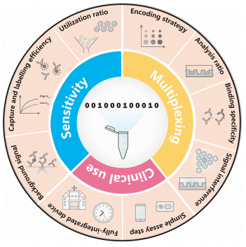
Fig. 5 Schematic summary of the key points of digital immunoassay[21].
06 Using Cytokine ELISA for Immune Cell Co-Culture and Functional Assays
ELISA serves as a core analytical tool for measuring functional cytokine secretion in co-culture systems of immune cells, and is extensively adopted to investigate crosstalk between dendritic cells (DCs) and T cells, as well as interactions between T and B lymphocytes. In DC–T co-culture models, ELISA quantifies secreted cytokines including IFN-γ, IL-17, IL-22 and TGF-β within culture supernatants to evaluate antigen specific T cell activation and differentiation fates. In one representative study, Marten et al. co-cultured cytokine induced immune effector cells with DCs pulsed with CEA or CAP-1 peptides for seven days. ELISA was applied to quantify supernatant IFN-γ release, and combined with ELISpot assay and flow cytometry to confirm effector cell activation and antigen specific TCR expression[26]. Similarly, Cools et al. subjected autologous CD8+ T cells to five rounds of restimulation with mature DCs loaded with HPV-16 E7 peptides. ELISA measured IFN-γ secretion validated E7 specific T cell clones, demonstrating the capacity of DCs to activate cytotoxic T lymphocytes via presentation of MHC class I restricted epitopes[27]. ELISA is also well suited to characterize the function of antibody secreting cells in T–B co-cultures. Piersma et al. isolated porcine peripheral blood mononuclear cells (PBMCs) from animals immunized with tetanus toxoid, followed by ex vivo antigen recall stimulation. ELISA detected tetanus toxoid specific antibodies and IFN-γ in culture supernatants; paired with antibody ELISpot for enumerating antibody producing B cells, this approach verified humoral immune responses driven by antigen specific T–B cell cooperation[28]. In another mechanistic study, Hall et al. co-cultured activated CD4+ T cells with hepatocellular carcinoma cell lines expressing the full HCV genome. ELISA quantification of supernatant TGF-β and IFN-γ uncovered a mechanism whereby HCV-infected hepatocytes trigger regulatory T cell differentiation through TGF-β to suppress Th1 immune responses[29]. Loubaki et al. further employed ELISA to measure IL-17, IL-22, IL-6, IL-1β and TGF-β in co-cultures of bronchial fibroblasts from asthmatic patients and CD4+ T cells. Combined with flow cytometry and western blotting, their work systematically delineated how fibroblast derived local microenvironments sustain and amplify Th17 mediated inflammatory cascades[30]. Collectively, these published works confirm that ELISA is an indispensable assay for functional immunology research using immune cell co-cultures. It not only characterizes lymphocyte activation and differentiation but also dissects molecular mechanisms underlying intercellular immune crosstalk. For rigorous experimental design, negative controls (unstimulated cells or irrelevant antigen), positive controls (PHA or anti-CD3/CD28 co-stimulation), and neutralizing antibody blockade groups are strongly recommended to validate antigen specificity and signal authenticity. Moreover, multiplexed readouts integrating ELISpot, flow cytometry and qPCR enable comprehensive profiling of immune responses across protein secretion, cellular phenotype and transcriptional levels, generating pivotal evidence for evaluating vaccine immunogenicity, developing tumor immunotherapies, and unravelling pathogenic pathways of inflammatory disorders.
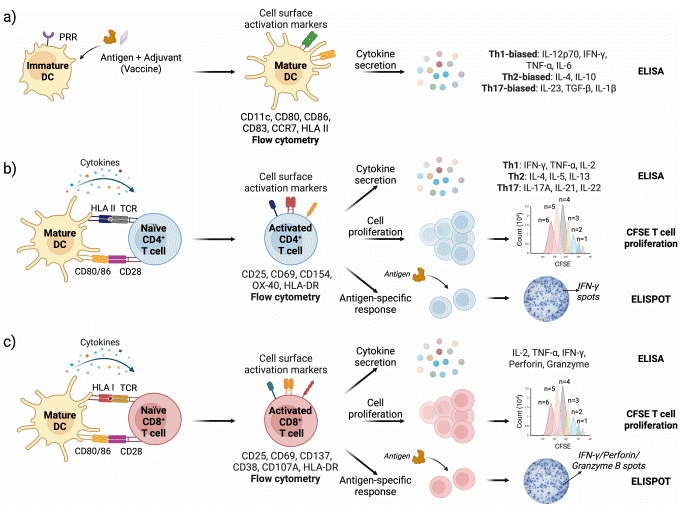
Fig. 6 Schematic representation of DC and T cell activation assays with readouts. (a) Vaccine stimulation activates DCs leading to upregulation of cell surface markers (assessed by flow cytometry) and cytokine secretion (measured by ELISA); (b) DC-CD4+ T cell and (c) DC-CD8+ T cell interface assays are used to assess T cell activation, proliferation, and antigen specific immune responses. Cell surface activation markers and cytokine response are analyzed by flow cytometry and ELISA, respectively. T cell proliferation is quantified by tracking CFSE dye dilution in cells using flow cytometry. Antigen specific responses are measured via ELISPOT, specifically detecting IFN-g (for both CD4+ and CD8+ T cells), and Granzyme B and Perforin specifically for CD8 T cells[31].
In summary, In summary, double antibody sandwich ELISA is a specific, routine cytokine quantification assay for serum, plasma and cell supernatants, widely used in biomarker screening, immunotherapy monitoring and pharmacokinetic studies. Yet single-plex ELISA features low throughput, large sample consumption and kit dependent bias, which necessitates rigorous quality control to offset matrix and pre analytical interference. High-density multiplex platforms such as Luminex and MSD were developed to address these deficits, enabling simultaneous detection of dozens of cytokines with tiny sample volumes and wider dynamic ranges, though cross reactivity and inter platform quantification discrepancies remain unresolved. For trace low-abundance cytokines, ultrasensitive approaches including single molecule counting, NULISA and digital Simoa ELISA reach fg/mL to attomolar sensitivity via signal amplification and single molecule enumeration. Automated ELISA reduces manual deviations and stabilizes assay CVs, while miniaturized microfluidic variants cut costs for under resourced clinical facilities. Digital ELISA realizes a shift from analog to digital signal readout, nearly achieving single molecule detection limits. Further optimization incorporating CRISPR amplification and AI decoding will facilitate portable POCT detection. Functionally, ELISA reliably characterizes DC–T and T–B cell crosstalk in immune co-cultures, revealing inflammatory and immune regulatory mechanisms when combined with flow cytometry, ELISpot and qPCR. Collectively, the technical evolution from manual single-plex ELISA to automated, multiplexed and digital ultrasensitive systems provides powerful tools for basic immunology, tumor immunotherapy, inflammatory research and early clinical diagnosis.
Elabscience® Quick Overview of Popular Products:
Table 1. Research Tools for Biomarker Detection
|
Cat. No. |
Product Name |
Features |
|
E-HSEL-H0003 |
High Sensitivity Human IL-6 (Interleukin 6) ELISA Kit |
Sensitivity: 0.49 pg/mL Citations:539 |
|
E-HSEL-H0005 |
High Sensitivity Human IL-10 (Interleukin 10) ELISA Kit |
Sensitivity: 0.49 pg/mL Citations:266 |
|
E-HSEL-H0006 |
High Sensitivity Human IL-17A (Interleukin 17A) ELISA Kit |
Sensitivity: 0.49 pg/mL Citations:58 |
|
E-EL-0162 |
TGF-β1(Transforming Growth Factor Beta 1) ELISA Kit |
Citations:307 |
|
E-EL-H0108 |
Human IFN-γ(Interferon Gamma) ELISA Kit |
Citations:217 |
|
E-EL-H0101 |
Human IL-4(Interleukin 4) ELISA Kit |
Citations:79 |
|
E-EL-H0109 |
Human TNF-α(Tumor Necrosis Factor Alpha) ELISA Kit |
Citations:875 |
|
CQH011 |
CellaQuant™ Human IL-22 (Interleukin 22) ELISA Kit |
Cell samples Citations:1 |
|
ESP-H0002 |
Human IFN-γ (Interferon Gamma) ELISPOT Kit |
Single-cell cytokine detection
|
|
MPA014 |
Aptplex™ Human Th1/Th2/Th17 12-Plex Panel |
Multiplex cytokine detection
|
References:
[1] Banks, R.E., Measurement of Cytokines in Clinical Samples Using Immunoassays: Problems and Pitfalls. 2000. 37(2): 131-182.
[2] Chiswick, E.L., et al., Detection and Quantification of Cytokines and Other Biomarkers, in Leucocytes. 2012. p. 15-30.
[3] WHITESIDE, T.L., Cytokine Measurements and Interpretation of Cytokine Assays in Human Disease. 1994. 14(6): 327-339.
[4] Keustermans, G.C.E., et al., Cytokine assays: An assessment of the preparation and treatment of blood and tissue samples. Methods, 2013. 61(1): p. 10-17.
[5] Amsen, D., K.E. Visser, and T. Town, Approaches to Determine Expression of Inflammatory Cytokines, in Inflammation and Cancer. 2009. p. 107-142.
[6] Liu, C., et al., Cytokines: From Clinical Significance to Quantification. Advanced Science, 2021. 8(15): 327-339.
[7] Litteljohn, D. and S. Hayley, Cytokines as Potential Biomarkers for Parkinson’s Disease: A Multiplex Approach, in Psychoneuroimmunology. 2012. p. 121-144.
[8] Taghavian, O., et al., Antibody Profiling by Proteome Microarray with Multiplex Isotype Detection Reveals Overlap between Human and Aotus nancymaae Controlled Malaria Infections. Proteomics, 2018. 18(2): 1700277.
[9] ROBISON, H.M., MULTIPLEXED IMMUNOASSAY DEVELOPMENT FOR PRECISION MEDICINE DIAGNOSTICS AND PROTEIN CHARACTERIZATION USING SILICON PHOTONIC MICRORING RESONATORS. 2017.
[10] Liu, G., et al., Recent advances in cytokine detection by immunosensing. Biosensors and Bioelectronics, 2016. 79: p. 810-821.
[11] Soderstrom, C., et al., Ultra-Sensitive Measurement of IL-17A and IL-17F in Psoriasis Patient Serum and Skin. The AAPS Journal, 2017. 19(4): p. 1218-1222.
[12] Todd, J., et al., Reference range and short- and long-term biological variation of interleukin (IL)-6, IL-17A and tissue necrosis factor-alpha using high sensitivity assays. Cytokine, 2013. 64(3): p. 660-665.
[13] Feng, W., et al., NULISA: a proteomic liquid biopsy platform with attomolar sensitivity and high multiplexing. Nature Communications, 2023. 14(1): 7238.
[14] Tighe, P.J., et al., ELISA in the multiplex era: Potentials and pitfalls. PROTEOMICS – Clinical Applications, 2015. 9(3-4): p. 406-422.
[15] Rey, G., et al., Automated ELISA for potency measurements of therapeutic antibodies and antibody fragments. Journal of Pharmaceutical and Biomedical Analysis, 2024. 245: 116141.
[16] Xin, X., et al., Revolutionizing ELISA development: The transformative impact of automation on monoclonal antibody potency assays. Journal of Pharmaceutical and Biomedical Analysis, 2026. 271.
[17] Filchtinski, D., et al., Development and performance evaluation of a novel, fully automated, high-throughput GAD65 autoantibody assay on the Phadia platform. Journal of Immunological Methods, 2025. 542: 113885.
[18] Aalizadeh, M., Automated ELISA and Machine Learning-Enhanced Accuracy in Biosensing and Colorimetric Measurements. 2025.
[19] Wang, Y., et al., Advancing Microfluidic Immunity Testing Systems: New Trends for Microbial Pathogen Detection. Molecules, 2024. 29(14): 3322.
[20] Hui Cai, M.G., Akshit Raj Gupta, William Grimm, Andrea Sease, Richard Rodriguez, Nesredin Mussa, and Z.J. Li, Automation of ELISAs & evaluation of emerging technologies for highthroughput quantitation of protein impurities. 2015.3(7):427-441.
[21] Zhang, Y., H. Gu, and H. Xu, Recent progress in digital immunoassay: how to achieve ultrasensitive, multiplex and clinical accessible detection? Sensors & Diagnostics, 2024. 3(1): p. 9-27.
[22] Noji, H., Y. Minagawa, and H. Ueno, Enzyme-based digital bioassay technology – key strategies and future perspectives. Lab on a Chip, 2022. 22(17): p. 3092-3109.
[23] Fan, W., et al., Emerging digital platforms for precise and ultrasensitive biosensing. npj Biosensing, 2026. 3(1).
[24] Duffy, D.C., Digital detection of proteins. Lab on a Chip, 2023. 23(5): p. 818-847.
[25] Su, K., et al., Emerging Trends in Integrated Digital Microfluidic Platforms for Next-Generation Immunoassays. Micromachines, 2024. 15(11): 1358.
[26] Märten, T.G.C.A., Generation of activated and antigen-specific T cells with cytotoxic activity after co-culture with dendritic cells. Cancer Immunology, Immunotherapy, 2002. 51(1): p. 25-32.
[27] Cools, N., et al., Sensitive detection of human papillomavirus type 16 E7-specific T cells by ELISPOT after multiple in vitro stimulations of CD8+ T cells with peptide-pulsed autologous dendritic cells. Molecular Cancer, 2006. 5(1): 49.
[28] Piersma, S., et al., An in vitro immune response model to determine tetanus toxoid antigen (vaccine) specific immunogenicity: Selection of sensitive assay criteria. Vaccine, 2006. 24(16): p. 3076-3083.
[29] Unutmaz, D., et al., HCV+ Hepatocytes Induce Human Regulatory CD4+ T Cells through the Production of TGF-β. PLoS ONE, 2010. 5(8): e12154.
[30] Cormier, Stephania A., et al., Co-Culture of Human Bronchial Fibroblasts and CD4+ T Cells Increases Th17 Cytokine Signature. PLoS ONE, 2013. 8(12): e81983.
[31] Bowley, T.Y., et al., Methods integrating innate and adaptive immune responses in human in vitro immunization assays. Frontiers in Immunology, 2025. 16: 1584852.

