

Ferroptosis is a non-apoptotic form of regulated cell death driven by iron-dependent lipid peroxidation, which has been implicated in a broad spectrum of pathological conditions including neurodegenerative diseases, ischemia-reperfusion injury, and cancer. Accurate ferroptosis detection requires a multi-parametric approach that encompasses the quantification of lipid reactive oxygen species (ROS), measurement of intracellular ferrous iron (Fe²⁺), assessment of glutathione peroxidase 4 (GPX4) activity, and analysis of downstream lipid peroxidation end-products. This review provides a comprehensive overview of experimental strategies currently employed for ferroptosis detection, including the C11-BODIPY oxidation assay for lipid ROS (a key ROS assay), ferrous iron-specific fluorescent probes (cell ferrous assay), immunofluorescence-based GPX4 activity validation, system Xc⁻ (SLC7A11) inhibition assays, detection of malondialdehyde (MDA) and 4-hydroxynonenal (4-HNE), and flow cytometry ROS-based multi-parametric approaches. Emphasis is placed on method validation, reagent specificity, and the integration of orthogonal techniques to establish definitive ferroptosis signatures.
Table of Contents
1. Lipid ROS quantification using C11-BODIPY oxidation assay
2. Intracellular ferrous iron (Fe²⁺) measurement methods
3. GPX4 activity level validation by immunofluorescence
4. System Xc⁻ (SLC7A11) inhibition assays to induce ferroptosis signatures
5. Lipid peroxidation end-products (MDA, 4-HNE) detection in ferroptosis models
6. Flow cytometry-based ferroptosis detection using ROS and iron probes
01 Lipid ROS quantification using C11-BODIPY oxidation assay
Lipid peroxidation constitutes the central biochemical hallmark of ferroptosis. Its reliable detection is essential for distinguishing this cell death modality from apoptosis, necroptosis, and other forms of regulated cell death. The C11-BODIPY⁵⁸¹/⁵⁹¹ probe is a fluorescent fatty acid analog extensively validated for monitoring lipid peroxidation in living cells and model membrane systems[1]. This probe represents a widely used ROS assay for lipid-specific oxidation. The principle is straightforward: the intact probe exhibits bright red fluorescence (emission ~595 nm). Upon free radical-induced oxidation of its conjugated diene bridge, the emission shifts to 520 nm (green fluorescence), enabling ratiometric imaging[1].
Drummen et al. provided a comprehensive (micro)spectroscopic characterization of C11-BODIPY⁵⁸¹/⁵⁹¹[1]. They demonstrated key properties: the probe is sensitive to oxy-radicals (hydroxyl, alkoxyl, peroxyl) and peroxynitrite, but not to superoxide, nitric oxide, or transition metal ions. Crucially, it exhibits minimal cytotoxicity up to 50 µM in Rat-1 fibroblasts, resists β-oxidation, and shows negligible sensitivity to pH or solvent polarity changes[1]. Once oxidized, C11-BODIPY⁵⁸¹/⁵⁹¹ remains lipophilic and does not leak from the bilayer, ensuring signal retention.
In ferroptosis research, Yang et al. demonstrated that erastin treatment of BJeLR cells caused a marked increase in C11-BODIPY oxidation by flow cytometry ROS analysis, while non-ferroptotic lethal compounds did not. von Krusenstiern et al.[2] further refined its application, revealing through confocal and Raman microscopy that the endoplasmic reticulum (ER) is the primary initial site of lipid peroxidation during ferroptosis, followed by the plasma membrane. Collectively, these studies solidify C11-BODIPY⁵⁸¹/⁵⁹¹ as the gold-standard probe for lipid ROS detection.
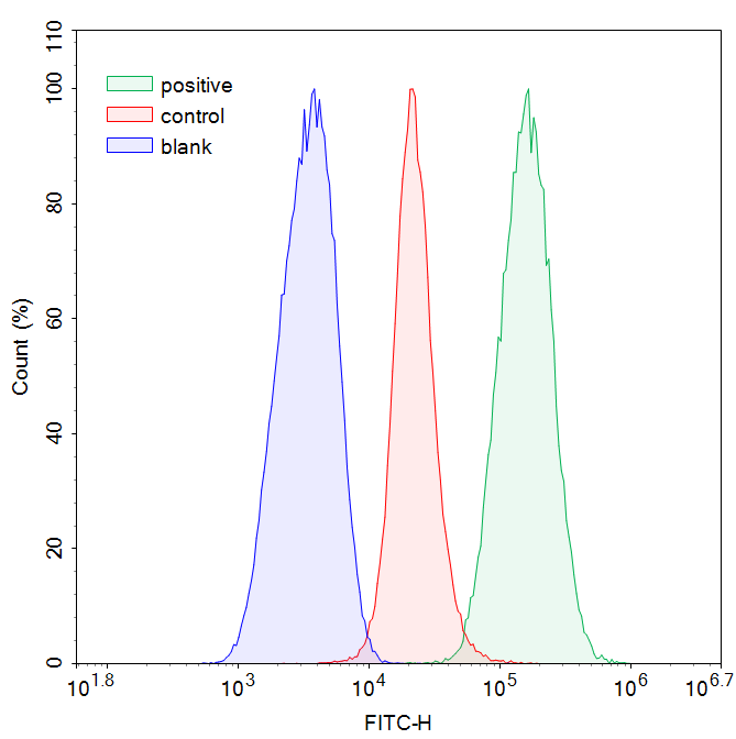
Fig. 1 Detection of lipid peroxidation levels in control and positive groups using C11-BODIPY⁵⁸¹/⁵⁹¹. 293T cells were treated with a lipid peroxidation-inducing agent (positive group).
02 Intracellular ferrous iron (Fe²⁺) measurement methods
The dependence of ferroptosis on iron is enshrined in its name, making the detection of labile Fe²⁺, the catalytically active form driving Fenton chemistry, critical. Several elegant fluorescent probes have been developed for organelle-specific Fe²⁺ detection, constituting a reliable cell ferrous assay. There are a series of Fe (II)-selective turn-on probes based on N-oxide chemistry: Ac-MtFluNox (mitochondria-targeted), Lyso-RhoNox (lysosome-targeted), and ER-SiRhoNox (ER-targeted)[3]. All exhibit similar reaction kinetics and off/on contrasts, enabling simultaneous multi-color imaging of labile Fe²⁺ in living cells.
Using a cocktail of these probes, Hirayama et al. showed that erastin treatment of HT1080 cells elevated labile Fe²⁺ in lysosomes and the ER prior to cell death, while mitochondrial Fe²⁺ remained unchanged. The iron chelator deferoxamine (DFO) abolished the fluorescence increase, confirming the specificity[3]. This organelle-resolution analysis revealed that ferritinophagy, the autophagic degradation of ferritin in lysosomes, liberates Fe²⁺, which then seeds lipid peroxidation.
In addition to microscopy, flow cytometric quantification of Fe²⁺ using probes like FerroOrange or Phen Green SK is widely adopted[4]. Colorimetric iron assay kits provide an alternative endpoint ferroptosis assay for tissue lysates[4]. The choice of method depends on the experimental question: subcellular resolution requires microscopy, while population-level quantification is suited to flow cytometry ROS platforms or plate-reader assays.
%20fluorometric%20assay%20kit%20(E-BC-F108)_.png)
Fig. 2 Measurement of ferrous iron levels in Jurkat cells using a cell ferrous (Fe²⁺) fluorometric assay kit (E-BC-F108). Jurkat cells were treated with ferric ammonium sulfate (FAS) alone or in combination with 2,2'-bipyridyl (BPY). The results showed that BPY suppressed the FAS-induced increase in ferrous iron. (The data are provided by Elabscience)
03 GPX4 activity level validation by immunofluorescence
Glutathione peroxidase 4 (GPX4) is the central regulator of ferroptosis. This selenoenzyme catalyzes the reduction of phospholipid hydroperoxides using glutathione (GSH) as a cofactor[5,6]. The seminal work by Yang et al.[5] established that all 12 ferroptosis-inducing compounds converge on GPX4 inhibition, either through GSH depletion (e.g., erastin) or direct covalent inhibition of its selenocysteine active site (e.g., RSL3). Consequently, GPX4 overexpression confers resistance, while knockdown sensitizes cells.
Generated knock-in mice expressing a cysteine (Cys) variant of GPX4 (Gpx4ᶜʸˢ/ᶜʸˢ). Using immunofluorescence and immunohistochemistry with a GPX4-specific antibody, they demonstrated that GPX4-Cys-expressing cells are exquisitely sensitive to peroxide-induced ferroptosis due to irreversible overoxidation of the catalytic Cys residue[6]. Therefore, immunofluorescence staining for GPX4, particularly when combined with lipid peroxidation markers, provides a robust validation strategy.
Seibt et al.[7] reviewed GPX4's role and emphasized that detecting GPX4 protein levels by western blotting or immunofluorescence should be complemented by functional activity assays. A GPX4 activity assay, using phosphatidylcholine hydroperoxide (PCOOH) as a substrate, measures NADPH oxidation coupled to glutathione reductase; loss of activity is observed upon erastin or RSL3 treatment[5,7]. This functional readout is an essential component of any comprehensive ferroptosis detection panel. Importantly, as a prelude to the next section, this loss of GPX4 activity is most commonly triggered by depletion of its essential cofactor, GSH.
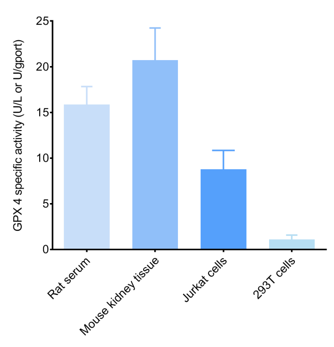
Fig. 3 GPX4 specific activity measured in four sample types: Rat serum, Mouse liver tissue, Jurkat cells, and 293T cells use Glutathione Peroxidase 4 (GPX4) Activity Assay Kit. (The data are provided by Elabscience)
04 System Xc⁻ (SLC7A11) inhibition assays to induce ferroptosis signatures
System Xc⁻ is a cystine/glutamate antiporter composed of the light chain SLC7A11 (xCT) and the heavy chain SLC3A2. It imports cystine, the rate-limiting precursor for GSH biosynthesis, in exchange for intracellular glutamate. Thus, pharmacological inhibition of system Xc⁻ by erastin (or its potent analogue erastin-2) leads to GSH depletion, subsequent GPX4 inactivation, and ultimately ferroptotic cell death.
Murray et al.[8] detailed a protocol for distinguishing ferroptosis using system Xc⁻ inhibitors. In this protocol, erastin-2 (0.005-10 µM) and RSL3 are used alongside specific inhibitors: ferrostatin-1 (1 µM, a radical-trapping antioxidant) or deferoxamine (50 µM, an iron chelator) to validate specificity. Cell viability is assessed by CellTiter-Glo luminescence or the Scalable Time-lapse Analysis of Cell Death Kinetics (STACK) method using mKate2 nuclear labeling and SYTOX Green dead-cell staining[8].
Chen et al.[4] summarized that the system Xc⁻/GSH/GPX4 pathway is the most well-characterized regulatory axis in ferroptosis. Quantification of the GSH/GSSG ratio, alongside SLC7A11 expression analysis by quantitative real-time polymerase chain reaction (qPCR) or western blotting, provides complementary evidence for system Xc⁻ inhibition[4]. Notably, SLC7A11 expression is positively regulated by activating transcription factor 4 (ATF4) under oxidative stress and repressed by tumor protein p53 (p53), linking ferroptosis detection to the cellular stress response.
05 Lipid peroxidation end-products (MDA, 4-HNE) detection in ferroptosis models
Malondialdehyde (MDA) and 4-hydroxynonenal (4-HNE) are reactive aldehydes generated as secondary decomposition products of lipid hydroperoxides. Their accumulation is a well-established, stable biochemical signature of ferroptosis. Commercially available lipid peroxidation assay kit options, such as those for MDA and 4-HNE, provide convenient and reproducible endpoints. MDA and 4-HNE levels have been widely used as endpoint markers in both cell culture and animal models of ferroptosis, often showing correlation with the extent of lipid peroxidation and the efficacy of ferroptosis inhibitors.
The thiobarbituric acid reactive substances (TBARS) assay is the most common method for MDA assay. Here, MDA reacts with thiobarbituric acid to produce a chromophore with absorbance at 532 nm, measurable spectrophotometrically or fluorometrically[4]. For 4-HNE assay, the aldehyde group reacts with 2, 4-dinitrophenylhydrazine (DNPH), and the resulting hydrazone is measured by HPLC or ELISA. Because MDA and 4-HNE are chemically stable end-products, they are preferable to the direct measurement of unstable primary hydroperoxides. Thus, incorporating an MDA assay or 4-HNE assay alongside a lipid peroxidation assay kit is recommended for validating ferroptosis in complex biological samples.
%20using%20enhanced%20cell%20malondialdehyde%20(MDA)%20colorimetric%20assay%20kit_.png)
Fig. 4 MDA content was measured in different cell lines (Molt-4, HepG2, 293T, and A549) using enhanced cell malondialdehyde (MDA) colorimetric assay kit. (The data are provided by Elabscience)
06 Flow cytometry-based ferroptosis detection using ROS and iron probes
Flow cytometry offers a high-throughput, quantitative approach to detect multiple ferroptosis-associated parameters at the single-cell level. This platform enables simultaneous execution of a ROS assay and a cell ferrous assay, greatly enhancing experimental efficiency. The combination of C11-BODIPY⁵⁸¹/⁵⁹¹ (for lipid ROS) with Fe²⁺-selective probes enables powerful multi-parametric analysis within a single experiment. Intracellular ROS can be detected using DCFH-DA or dihydroethidium (DHE)[1]. However, these lack specificity for lipid peroxidation. In contrast, C11-BODIPY⁵⁸¹/⁵⁹¹, detected in the FL1 (green, oxidized) and FL2 (red, non-oxidized) channels, provides a ratiometric readout largely independent of probe loading variations. This approach is a prime example of flow cytometry ROS application in ferroptosis research.
Murray et al. described a flow cytometry-compatible STACK protocol. In simpler terms, this quantifies the proportion of cells that have lost the live-cell nuclear marker (mKate2) and gained the dead-cell stain (SYTOX Green). Ferroptosis was distinguished from apoptosis by the lack of caspase-3 cleavage and by its suppression with ferrostatin-1, but not with the pan-caspase inhibitor Q-VD-OPh[8].
For iron quantification by flow cytometry, Lyso-RhoNox and FerroOrange can detect labile Fe²⁺, with specificity confirmed by DFO co-treatment[2]. Recent advances in nanoflow cytometry have even enabled the detection of ferrous ions at the single-extracellular-vesicle level, opening new avenues for biomarker discovery.
Summary and Recommended Workflow
In summary, a definitive ferroptosis detection strategy should integrate multiple orthogonal markers. A complete ferroptosis assay panel may include the following; we propose a tiered approach:
For initial screening: C11-BODIPY ROS assay + cell ferrous assay + ferrostatin-1 rescue experiment
For mechanistic validation: Add functional GPX4 activity assay & immunofluorescence staining + System Xc⁻ function assessment (GSH/GSSG ratio & SLC7A11 expression level)
For tissue samples: Prioritize MDA assay and 4-HNE assay (or a general commercial lipid peroxidation assay kit) + immunohistochemistry for GPX4 and SLC7A11
For high-throughput single-cell analysis: Flow cytometry ROS detection combined with viability and iron probes
The integration of at least three orthogonal markers (evidence of lipid peroxidation, iron dependency, and GPX4 pathway involvement) is recommended to definitively establish ferroptosis.
Quick Overview of Popular Products:
Table 1. Reagents for Ferroptosis detection
|
Cat. No. |
Product Name |
|
E-BC-F003 |
Lipid Peroxide (LPO) Fluorometric Assay Kit |
|
E-BC-K025-M |
Malondialdehyde (MDA) Colorimetric Assay Kit (TBA Method) |
|
E-BC-K814-M |
Enhanced Cell Malondialdehyde (MDA) Colorimetric Assay Kit |
|
E-EL-0128 |
4-HNE(4-Hydroxynonenal) ELISA Kit |
|
E-BC-F108 |
Cell Ferrous (Fe2+) Fluorometric Assay Kit |
|
E-BC-K881-M |
Cell Ferrous Iron Colorimetric Assay Kit |
|
E-BC-K138-F |
Reactive Oxygen Species (ROS) Fluorometric Assay Kit (Green) |
|
E-BC-F005 |
Reactive Oxygen Species (ROS) Fluorometric Assay Kit (Red) |
|
E-BC-F066 |
Cystine Uptake Fluorometric Assay Kit |
|
E-BC-F045 |
Total Glutathione (T-GSH) And Reduced Glutathione (GSH) Assay Kit |
|
E-BC-K903-M |
Glutamic Acid (Glu) Colorimetric Assay Kit |
|
E-BC-K883-M |
Glutathione Peroxidase 4 (GPX4) Activity Assay Kit |
|
E-BC-K771-M |
Lactate Dehydrogenase (LDH) Cytotoxicity Colorimetric Assay Kit |
|
E-CK-A362 |
Enhanced Cell Counting Kit 8 (WST-8/CCK8) |
References:
[1] Drummen, G.P.C., et al., C11-BODIPY581/591, an Oxidation-Sensitive Fluorescent Lipid Peroxidation Probe: (Micro)spectroscopic Characterization and Validation of Methodology. Free Radical Biology and Medicine, 2002. 33(4):473-490.
[2] von Krusenstiern, A.N., et al., Identification of Essential Sites of Lipid Peroxidation in Ferroptosis. Nature Chemical Biology, 2023. 19(6):719-730.
[3] Hirayama, T., A. Miki, and H. Nagasawa, Organelle-Specific Analysis of Labile Fe(II) during Ferroptosis by Using a Cocktail of Various Colour Organelle-Targeted Fluorescent Probes. Metallomics, 2019. 11(1):111-117.
[4] Chen, Z., et al., The Application of Approaches in Detecting Ferroptosis. Heliyon, 2024. 10(1):e23507.
[5] Yang, Wan S., et al., Regulation of Ferroptotic Cancer Cell Death by GPX4. Cell, 2014. 156(1):317-331.
[6] Ingold, I., et al., Selenium Utilization by GPX4 Is Required to Prevent Hydroperoxide-Induced Ferroptosis. Cell, 2018. 172(3):409-422.e21.
[7] Seibt, T.M., B. Proneth, and M. Conrad, Role of GPX4 in Ferroptosis and Its Pharmacological Implication. Free Radical Biology and Medicine, 2019. 133:144-152.
[8] Murray, M.B., et al., Protocol for Detection of Ferroptosis in Cultured Cells. STAR Protocols, 2023. 4(3):102457.

