| Form |
Liquid |
| Clone No. |
G4.18(See Other Available Formats) |
| Host |
Mouse |
| Isotype |
Mouse IgG3, κ |
| Isotype Control |
PerCP/Cyanine5.5 Mouse IgG3, κ Isotype Control[A112-3] [Product E-AB-F09753J] |
| Reactivity |
Rat |
| Application |
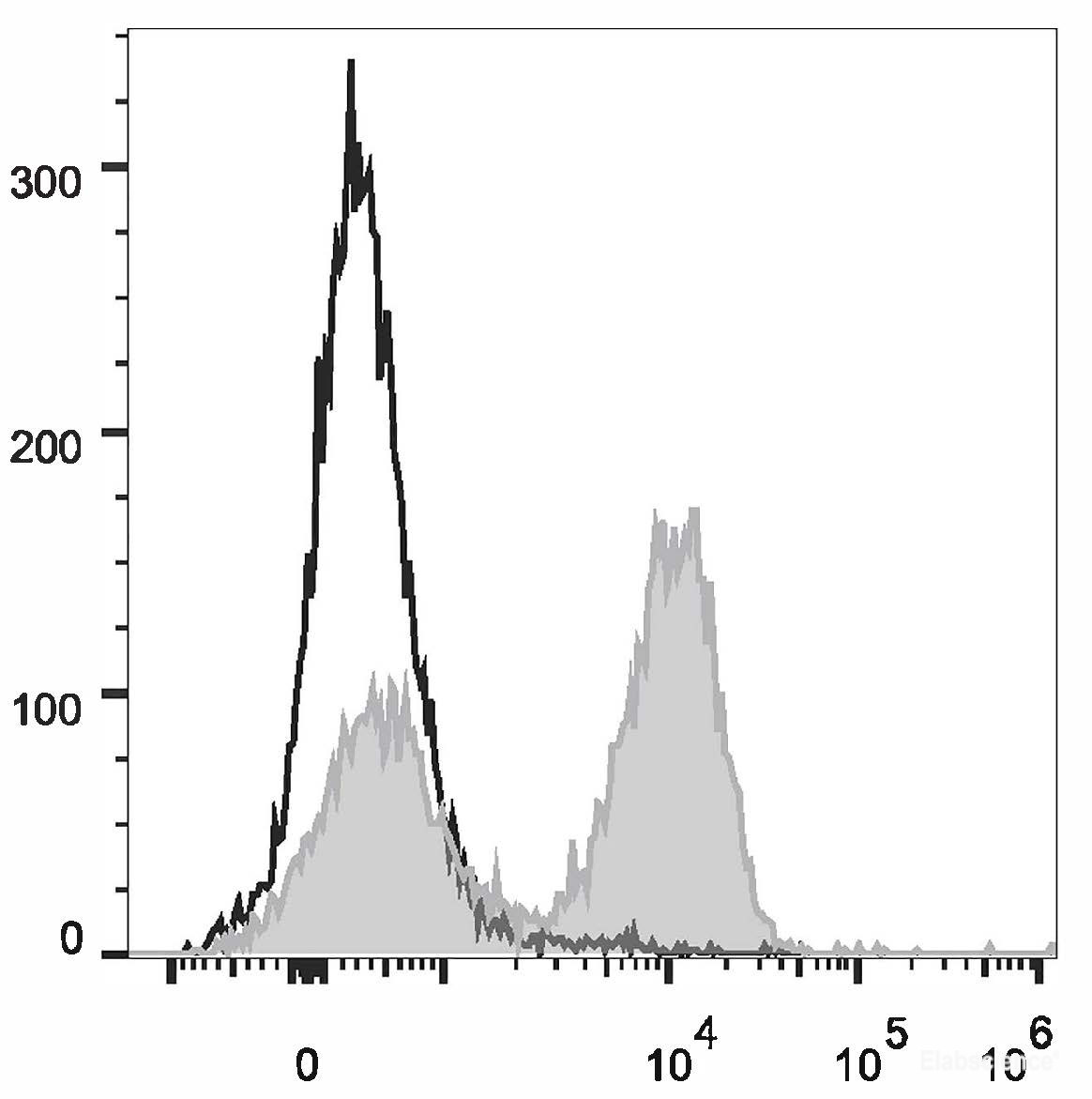
FCM (See the Application Description) |
| Storage Buffer |
Phosphate buffered solution, pH 7.2, containing 0.09% stabilizer and 1% protein protectant. |
| Recommended Use |
Each lot of this antibody is quality control tested by flow cytometric analysis. Please check your vial before the experiment. Since applications vary, the appropriate dilutions must be determined for individual use. We suggest each investigator should titrate the reagent to obtain optimal results [The recommended concentration is 0.1-1 μg/106 cells in 100 μL volume]. |
| Shipping |
Biological ice pack at 4℃ |
| Stability & Storage |
Keep as concentrated solution.
Store at 2~8°C and protected from prolonged exposure to light. Do not freeze.
Centrifuge before opening to ensure complete recovery of vial contents.
This product is guaranteed up to one year from purchase. |
| Conjugation |
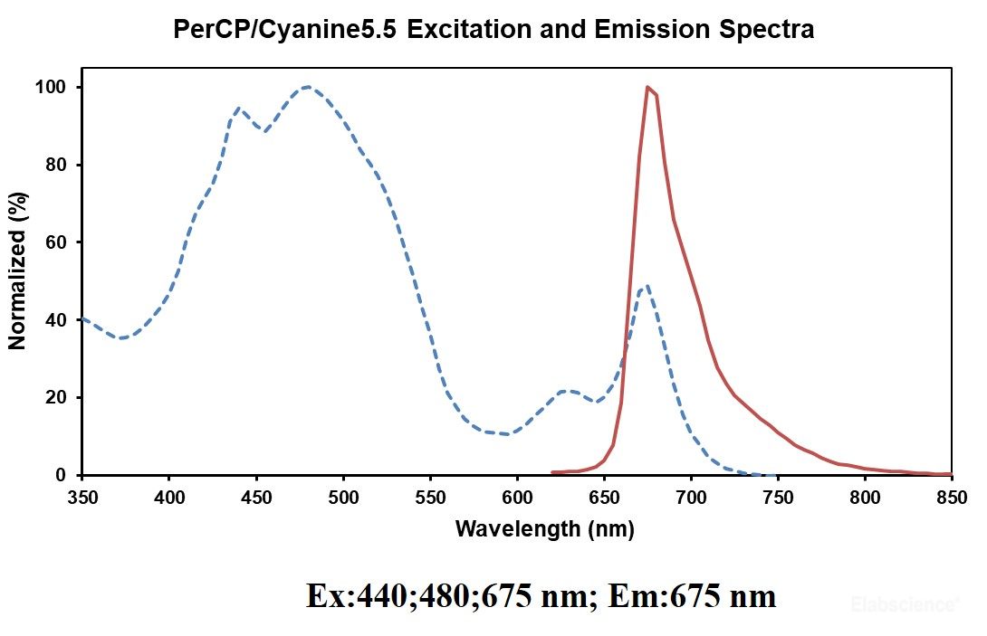
PerCP/Cyanine5.5 |
|
PerCP/Cyanine5.5 is designed to be excited by the blue laser (488 nm) and detected using an optical filter centered near 675 nm (e.g., a 690/50 nm bandpass filter). |
|
|